. 2017 Jan;6(1):223-237.
doi: 10.5966/sctm.2016-0044. Epub 2016 Aug 8.

Enhanced Immunosuppression of T Cells by Sustained Presentation of Bioactive Interferon-γ Within Three-Dimensional Mesenchymal Stem Cell Constructs

Affiliations

Enhanced Immunosuppression of T Cells by Sustained Presentation of Bioactive Interferon-γ Within Three-Dimensional Mesenchymal Stem Cell Constructs

Joshua A Zimmermann et al. Stem Cells Transl Med. 2017 Jan.

Abstract

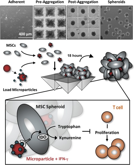
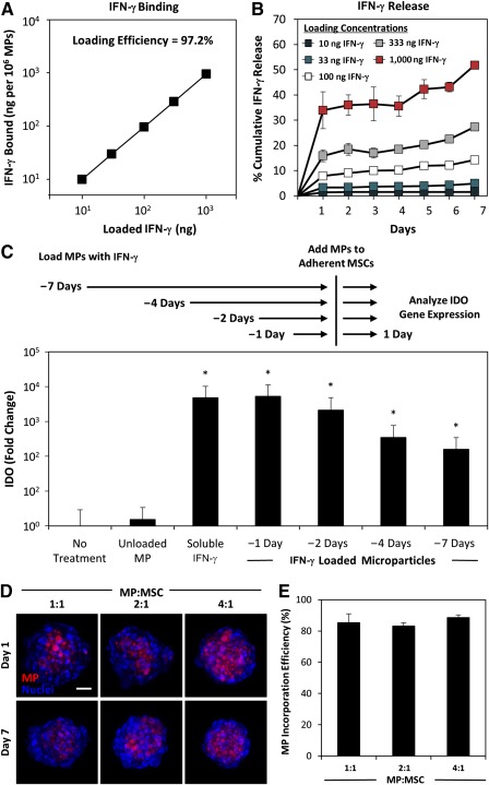
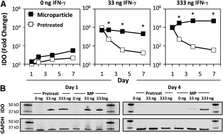
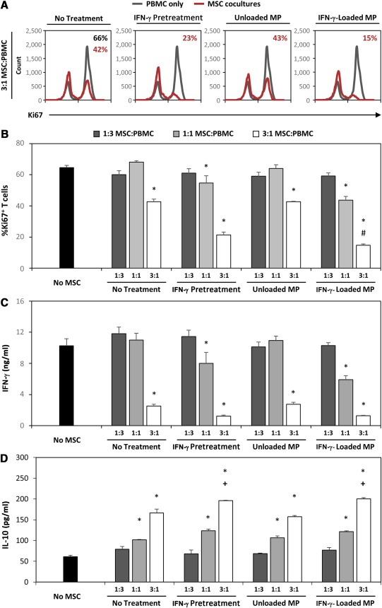
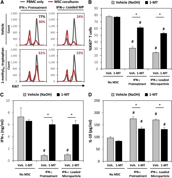
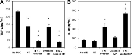
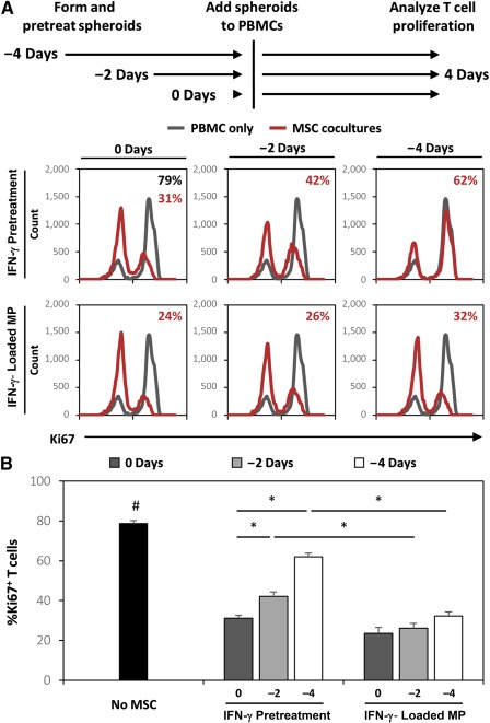
The immunomodulatory activity of mesenchymal stem/stromal cells (MSCs) to suppress innate and adaptive immune responses offers a potent cell therapy for modulating inflammation and promoting tissue regeneration. However, the inflammatory cytokine milieu plays a critical role in stimulating MSC immunomodulatory activity. In particular, interferon-γ (IFN-γ)-induced expression of indoleamine 2,3-dioxygenase (IDO) is primarily responsible for MSC suppression of T-cell proliferation and activation. Although pretreatment with IFN-γ is commonly used to prime MSCs for immunomodulatory activity prior to transplantation, the transient effects of pretreatment may limit the potential of MSCs to potently modulate immune responses. Therefore, the objective of this study was to investigate whether microparticle-mediated presentation of bioactive IFN-γ within three-dimensional spheroidal MSC aggregates could precisely regulate and induce sustained immunomodulatory activity. Delivery of IFN-γ via heparin-microparticles within MSC aggregates induced sustained IDO expression during 1 week of culture, whereas IDO expression by IFN-γ-pretreated MSC spheroids rapidly decreased during 2 days. Furthermore, sustained IDO expression induced by IFN-γ-loaded microparticles resulted in an increased and sustained suppression of T-cell activation and proliferation in MSC cocultures with CD3/CD28-activated peripheral blood mononuclear cells. The increased suppression of T cells by MSC spheroids containing IFN-γ-loaded microparticles was dependent on induction of IDO and supported by affecting monocyte secretion from pro- to anti-inflammatory cytokines. Altogether, microparticle delivery of IFN-γ within MSC spheroids provides a potent means of enhancing and sustaining immunomodulatory activity to control MSC immunomodulation after transplantation and thereby improve the efficacy of MSC-based therapies aimed at treating inflammatory and immune diseases. Stem Cells Translational Medicine 2017;6:223-237.

Keywords: Cellular; Immunomodulation; Indoleamine 2,3-dioxygenase; Mesenchymal stromal cells; Spheroids.


Figures

Figure 1
Figure 1
Microparticle delivery of interferon (IFN)‐γ within mesenchymal stem/stromal cell (MSC) spheroids. To form aggregates, we dissociated MSCs from adherent culture and mixed them with IFN‐γ‐loaded microparticles. Spheroids were subsequently formed via forced aggregation of cells and particles within microwell arrays to entrap cytokine‐loaded microparticles within the spheroidal construct to provide sustained local presentation of IFN‐γ within the MSC aggregates. Abbreviations: IFN, interferon; MSC, mesenchymal stem/stromal cell; IDO, indoleamine 2,3‐dioxygenase.
Figure 2
Figure 2
Heparin microparticles bind and present bioactive interferon (IFN)‐γ and incorporate stably within mesenchymal stem/stromal cell (MSC) aggregates. (A): Heparin microparticles bound >97% of IFN‐γ from solution over the ranges tested. (B): Although an initial burst release of IFN‐γ from the particles was observed, little IFN‐γ was subsequently released from the particles during 7 days. (C): Particle‐bound IFN‐γ remained bioactive and capable of inducing MSC expression of indoleamine 2,3‐dioxygenase during 7 days. (D): Fluorescently labeled microparticles were incorporated within MSC aggregates at various doses and remained stably incorporated for at least 7 days of culture. Maximal intensity projection, scale bar = 50 μm. (E): Approximately 85% of microparticles added during aggregate formation were incorporated within MSC spheroids within the range of microparticle‐to‐cell ratios tested, allowing for precise control of microparticle and loaded cytokine dose. ∗p < .05, in comparison with no treatment. Abbreviations: IDO, indoleamine 2,3‐dioxygenase; IFN, interferon; MP, microparticle; MSC, mesenchymal stem/stromal cell.
Figure 3
Figure 3
Interferon (IFN)‐γ‐loaded heparin microparticles induce sustained IDO expression. (A): IDO gene expression rapidly decreased after initial pretreatment of mesenchymal stem/stromal cell (MSC) spheroids with IFN‐γ, whereas IDO gene expression was sustained during 7 days when IFN‐γ was delivered by microparticles within MSC spheroids. ∗p < .05, comparing pretreatment and microparticle treatment on the same day of culture. (B): IDO protein expression was induced by IFN‐γ but was only expressed by MSC spheroids after 4 days when IFN‐γ was delivered by microparticles. Abbreviations: GAPDH, glyceraldehyde‐3‐phosphate dehydrogenase; IDO, indoleamine 2,3‐dioxygenase; IFN, interferon; MP, microparticle.
Figure 4
Figure 4
Microparticle delivery of interferon (IFN)‐γ enhances mesenchymal stem/stromal cell (MSC) spheroid suppression of T‐cell activation and proliferation. (A): Representative histograms of Ki67+ T‐cells in coculture with MSC spheroids at 3:1 MSC:peripheral blood mononuclear cell (PBMC) ratios demonstrate increased suppression of T‐cell proliferation when treated with IFN‐γ via pretreatment or microparticle delivery. (B): Coculture of activated PBMCs with IFN‐γ‐loaded microparticle‐treated MSC spheroids resulted in the greatest suppression of T‐cell proliferation at a 3:1 MSC:PBMC ratio. Only MSCs treated with IFN‐γ (via pretreatment or microparticle) suppressed T‐cell proliferation at a 1:1 MSC:PBMC ratio. (C): MSC spheroids suppressed expression of the effector cytokine IFN‐γ, and the greatest suppression was observed in cocultures with MSCs treated with IFN‐γ. (D): IL‐10 expression was increased in cocultures with MSC spheroids and was greatest when MSCs were treated with IFN‐γ via pretreatment or microparticle delivery. ∗, p < .05 in comparison with No‐MSC cultures; #, p < .05 in comparison with IFN‐γ‐pretreated MSC spheroids at the same MSC:PBMC ratio; +, p < .05 in comparison with No Treatment and Unloaded Microparticle MSC spheroid cocultures. Abbreviations: IFN, interferon; IL, interleukin; MP, microparticle; MSC, mesenchymal stem/stromal cell; PBMC, peripheral blood mononuclear cell.
Figure 5
Figure 5
Increased suppression of T‐cells by microparticle delivery of interferon (IFN)‐γ is dependent on induction of indoleamine 2,3‐dioxygenase (IDO) expression. (A): Representative histograms of Ki67+ T‐cells in coculture with IFN‐γ‐pretreated or IFN‐γ‐loaded microparticle‐ treated MSC spheroids with or without the IDO inhibitor 1‐methylDL‐tryptophan (1‐MT). (B): Addition of 1‐MT to mesenchymal stem/stromal cell (MSC)‐peripheral blood mononuclear cell (PBMC) coculture (3:1 MSC:PBMC ratio) significantly inhibited the ability of IFN‐γ‐primed MSCs to inhibit T‐cell proliferation in both pretreated and microparticle‐treated spheroids. Inhibition of IDO activity with 1‐MT increased T‐cell expression of IFN‐γ (C) and reduced the induction of interleukin‐10 expression in PBMC cocultures (D). ∗, p < .05 in comparison with groups denoted by bar. #, p < .05 in comparison with No‐MSC cultures. Abbreviations: IFN, interferon; IL‐10, interleukin‐10; MSC, mesenchymal stem/stromal cell; PBMC, peripheral blood mononuclear cell; Veh., vehicle; 1‐MT, 1‐methylDL‐tryptophan.
Figure 6
Figure 6
Microparticle delivery of interferon (IFN)‐γ increases mesenchymal stem/stromal cell (MSC) spheroid polarization of monocytes to anti‐inflammatory phenotypes. (A): Monocyte expression of tumor necrosis factor‐α, a proinflammatory cytokine, was decreased in all cocultures with MSC spheroids but was greatest in cultures with IFN‐γ pretreated or microparticle‐treated MSCs. (B): Conversely, interleukin‐10 production, an anti‐inflammatory cytokine, was greatest in cocultures with IFN‐γ‐loaded microparticle‐treated MSCs and not increased in cocultures with untreated MSCs. Polarization of monocytes to anti‐inflammatory phenotypes by MSCs treated with IFN‐γ‐loaded microparticles can aid in the suppression of T‐cell proliferation and activation in peripheral blood mononuclear cell cocultures. ∗, p < .05 in comparison with No‐MSC group; #, p < .05 in comparison with IFN‐γ‐pretreated MSC spheroid group; +, p < .05 in comparison with No‐Treatment group. Abbreviations: IFN, interferon; IL‐10, interleukin‐10; MSC, mesenchymal stem/stromal cell; MP, microparticle; NT, no treatment; TNF‐α, tumor necrosis factor‐α.
Figure 7
Figure 7
Microparticle delivery of interferon (IFN)‐γ enhances mesenchymal stem/stromal cell (MSC) suppression of T‐cells for more than 1 week. (A): Representative histograms of Ki67+ T‐cells in coculture with MSC spheroids treated with IFN‐γ or IFN‐γ‐loaded microparticles 0, 2, and 4 days prior to addition to peripheral blood mononuclear cell coculture. (B): IFN‐γ‐loaded microparticles sustained the enhanced MSC suppression of T‐cell proliferation in response to IFN‐γ for up to 8 days, whereas the effects of pretreating MSC spheroids with IFN‐γ were transient and resulted in decreased ability to suppress T‐cell proliferation over time. ∗, p < .05 in comparison with groups denoted by bars; #, p < .05 in comparison with No‐MSC group. Abbreviations: IFN, interferon; MP, microparticle; MSC, mesenchymal stem/stromal cell; PBMC, peripheral blood mononuclear cell.

References

    1. Di Nicola M, Carlo‐Stella C, Magni M et al. Human bone marrow stromal cells suppress T‐lymphocyte proliferation induced by cellular or nonspecific mitogenic stimuli. Blood 2002;99:3838–3843. - PubMed
    1. Krampera M, Glennie S, Dyson J et al. Bone marrow mesenchymal stem cells inhibit the response of naive and memory antigen‐specific T cells to their cognate peptide. Blood 2003;101:3722–3729. - PubMed
    1. Corcione A, Benvenuto F, Ferretti E et al. Human mesenchymal stem cells modulate B‐cell functions. Blood 2006;107:367–372. - PubMed
    1. Németh K, Leelahavanichkul A, Yuen PST et al. Bone marrow stromal cells attenuate sepsis via prostaglandin E(2)‐dependent reprogramming of host macrophages to increase their interleukin‐10 production. Nat Med 2009;15:42–49. - PMC - PubMed
    1. Nauta AJ, Kruisselbrink AB, Lurvink E et al. Mesenchymal stem cells inhibit generation and function of both CD34+‐derived and monocyte‐derived dendritic cells. J Immunol 2006;177:2080–2087. - PubMed

Publication types

MeSH terms