

Wavelength-Dependent Second Harmonic Generation Circular Dichroism for Differentiation of Col I and Col III Isoforms in Stromal Models of Ovarian Cancer Based on Intrinsic Chirality Differences
- PMID: 28170263
- PMCID: PMC5494177
- DOI: 10.1021/acs.jpcb.6b06822
Wavelength-Dependent Second Harmonic Generation Circular Dichroism for Differentiation of Col I and Col III Isoforms in Stromal Models of Ovarian Cancer Based on Intrinsic Chirality Differences
Abstract
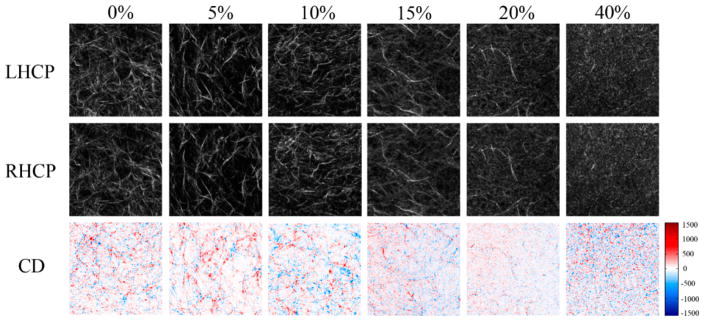
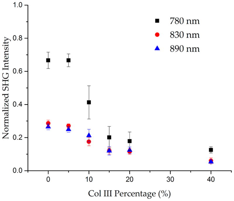
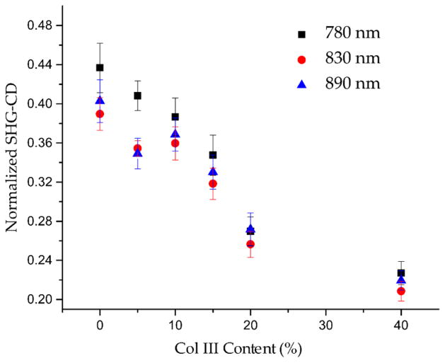
Extensive remodeling of the extracellular matrix (ECM) occurs in many epithelial cancers. For example, in ovarian cancer, upregulation of collagen isoform type III has been linked to invasive forms of the disease, and this change may be a potential biomarker. To examine this possibility, we implemented wavelength-dependent second harmonic generation circular dichroism (SHG-CD) imaging microscopy to quantitatively determine changes in chirality in ECM models comprised of different Col I/Col III composition. In these models, Col III was varied between 0 and 40%, and we found increasing Col III results in reduced net chirality, consistent with structural biology studies of Col I and III in tissues where the isoforms comingle in the same fibrils. We further examined the wavelength dependence of the SHG-CD to both optimize the response and gain insight into the underlying mechanism. We found using shorter SHG excitation wavelengths resulted in increased SHG-CD sensitivity, where this is consistent with the electric-dipole-coupled oscillator model suggested previously for the nonlinear chirality response from thin films. Moreover, the sensitivity is further consistent with the wavelength dependency of SHG intensity fit to a two-state model of the two-photon absorption in collagen. We also provide experimental calibration protocols to implement the SHG-CD modality on a laser scanning microscope. We last suggest that the technique has broad applicability in probing a wide range of diseased states with changes in collagen molecular structure.
Conflict of interest statement
The authors declare no competing financial interest.
Figures
References
-
- Ricciardelli C, Rodgers RJ. Extracellular Matrix of Ovarian Tumors. Semin Reprod Med. 2006;24:270–82. - PubMed
-
- Nelson AR, Fingleton B, Rothenberg ML, Matrisian LM. Matrix Metalloproteinases: Biologic Activity and Clinical Implications. J Clin Oncol. 2000;18:1135–49. - PubMed
-
- Pupa SM, Menard S, Forti S, Tagliabue E. New Insights into the Role of Extracellular Matrix During Tumor Onset and Progression. J Cell Physiol. 2002;192:259–267. - PubMed
-
- Sherman-Baust CA, Weeraratna AT, Rangel LB, Pizer ES, Cho KR, Schwartz DR, Shock T, Morin PJ. Remodeling of the Extracellular Matrix through Overexpression of Collagen Vi Contributes to Cisplatin Resistance in Ovarian Cancer Cells. Cancer Cell. 2003;3:377–86. - PubMed
Publication types
MeSH terms
Substances
Grants and funding
LinkOut - more resources
Full Text Sources
Other Literature Sources
Medical
