. 2016 Nov 15;25(22):4898-4910.
doi: 10.1093/hmg/ddw313.

The human RHOX gene cluster: target genes and functional analysis of gene variants in infertile men

Affiliations

The human RHOX gene cluster: target genes and functional analysis of gene variants in infertile men

Jennifer Borgmann et al. Hum Mol Genet. .

Abstract

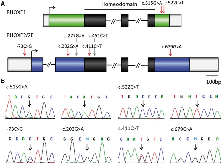
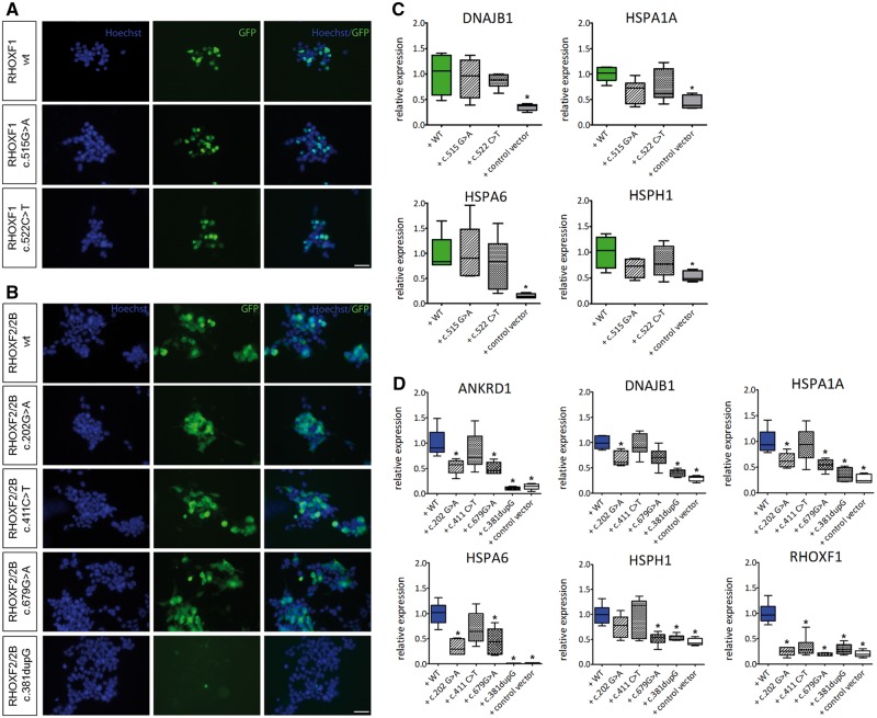
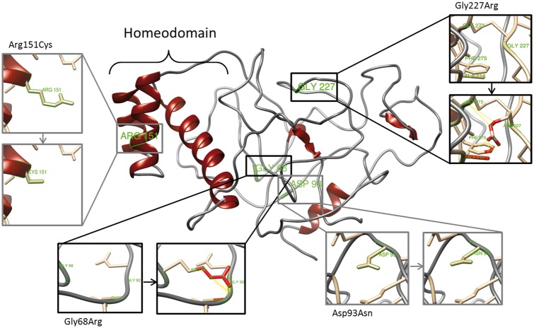
The X-linked reproductive homeobox (RHOX) gene cluster encodes transcription factors preferentially expressed in reproductive tissues. This gene cluster has important roles in male fertility based on phenotypic defects of Rhox-mutant mice and the finding that aberrant RHOX promoter methylation is strongly associated with abnormal human sperm parameters. However, little is known about the molecular mechanism of RHOX function in humans. Using gene expression profiling, we identified genes regulated by members of the human RHOX gene cluster. Some genes were uniquely regulated by RHOXF1 or RHOXF2/2B, while others were regulated by both of these transcription factors. Several of these regulated genes encode proteins involved in processes relevant to spermatogenesis; e.g. stress protection and cell survival. One of the target genes of RHOXF2/2B is RHOXF1, suggesting cross-regulation to enhance transcriptional responses. The potential role of RHOX in human infertility was addressed by sequencing all RHOX exons in a group of 250 patients with severe oligozoospermia. This revealed two mutations in RHOXF1 (c.515G > A and c.522C > T) and four in RHOXF2/2B (-73C > G, c.202G > A, c.411C > T and c.679G > A), of which only one (c.202G > A) was found in a control group of men with normal sperm concentration. Functional analysis demonstrated that c.202G > A and c.679G > A significantly impaired the ability of RHOXF2/2B to regulate downstream genes. Molecular modelling suggested that these mutations alter RHOXF2/F2B protein conformation. By combining clinical data with in vitro functional analysis, we demonstrate how the X-linked RHOX gene cluster may function in normal human spermatogenesis and we provide evidence that it is impaired in human male fertility.


Figures

Figure 1.
Figure 1.
RHOX expression in transiently transfected HEK293 cells. (A) Immunofluorescent imaging of HEK293 cells 48h after transfection. GFP signal reflects RHOX expression while nuclei were counterstained with Hoechst. Scale bar: 10 µm (right row). (B) RHOX expression after 24h and 48h of transfection (n = 8). Total RNA was isolated, reverse transcribed, and amplified with specific primers. Values were normalized to endogenous GAPDH. ***P < 0.001. (C) Western Blot analysis of RHOX after 24h and 48h of transfection.
Figure 2.
Figure 2.
Identification of genes regulated by the RHOX gene cluster. Heat map of RHOXF1 (A) and RHOXF2/2B (B) differentially expressed genes (DEGs) after 48h of transfection. Each column represents individual samples (S: RHOX transfected samples, C: control vector transfected samples), each row shows a single gene and expression levels were indicated by a color scale (red: high expression, green: low expression). (C) Analysis of DEGs by qRT-PCR. After different transfections with RHOX, mRNA expression levels of MSH5 (n = 15), ANKRD1 (n = 15), DNAJB1 (n = 15), HSPA1A (n = 8), HSPA2 (n = 6), HSPA6 (n = 15), HSPH1 (n = 8) and RHOXF1 (n = 15) were measured and normalized to GAPDH. The data are represented as median and interquartile range. **P < 0.01, ***P < 0.001. (D) Immunohistological analysis of RHOX in men with normal spermatogenesis. Pachytene spermatocytes and round spermatids showed positive staining for RHOXF1 (D.1–D.3), while type B spermatogonia and preleptotene – zygotene spermatocytes were predominantly stained with RHOXF2/2B specific antibody (D.5–D.7). IgG controls were negative (D.4, D.8). B, type B spermatogonia; P, pachytene spermatocytes; Pl-Z, preleptotene – zygotene spermatocytes; RS, round spermatids. Scale bar: 50 μm (D.1, D.4, D.5, D.8) and 25 μm (D.2, D.3, D.6, D.7). (E) Expression of RHOX regulated genes in testis with normal spermatogenesis (grey, n = 3) and in testis with SCO (white; n = 3). Data are presented as mean with SEM.
Figure 3.
Figure 3.
Genetic variants identified in RHOX genes in men with severe oligozoospermia. (A) Location of identified variants within RHOX gene structure. The homeodomain is shown in black, mutations are marked with red arrows and SNPs with grey arrows. (B) Chromatograms obtained from Sanger sequencing validate the mutations found by IonTorrent sequencing. For RHOXF1 c.515G > A and c.522C > T wild type sequence (left) and mutated sequence (right) are shown. In case of mutations -73C > G, c.202G > A, c.411C > T, and c.679G > A only one of the two RHOXF2 copies is affected resulting in a double peak at these positions. N = ambiguous call.
Figure 4.
Figure 4.
Functional analysis of RHOX mutations. (A) Immunofluorescent staining of HEK293 cells after 48h of transfection with RHOXF1 c.515G > A, c.522C > T and (B) RHOXF2/2B c.202G > A, c.411C > T, c.679G > A, and c.381dupG expression vectors. GFP signal reflects RHOX expression, nuclei were counterstained with Hoechst. Scale bar: 50 μm. (C) Gene expression levels of RHOXF1 DEGs after transfection with c.515G > A and c.522C > T expression vectors. (D) Gene expression levels of RHOXF2/2B DEGs after transfection with c.202G > A, c.411C > T, c.679G > A, and c.381dupG expression vectors. Transformed data are represented as whiskers with 5–95 percentiles. *Results are significant after Bonferroni correction for multiple testing (P < 0.01).
Figure 5.
Figure 5.
Effect of non-synonymous mutations on RHOXF2/2B protein structure. Diagram of the top predicted tertiary structure model of RHOXF2/2B using Phyre2. The homeodomain region was modeled with template structures of homeodomain containing proteins and the remaining residues ab initio. The model was colored by secondary structure using UCSF Chimera software: helix structures are shown in red and coiled structures are shown in grey. Each insert shows the identified non-synonymous mutations (grey) and SNPs (black) as sticks. Predicted clashes/contacts for c.202G > A (p.Gly68Arg) and c.679G > A (p.Gly227Arg) are marked with yellow lines (14 and 5, respectively) and atoms involved in the potential contacts are highlighted in red (10 and 8, respectively). No clashes/contacts were predicted for c.277G > A (p.Asp93Asn) and c.451C > T (p.Arg151Cys).

References

    1. Tüttelmann F., Gromoll J., Kliesch S. (2008) Genetics of male infertility. Der. Urol., 47, 1561–1562. 1564–1567. - PubMed
    1. Tüttelmann F., Gromoll J. (2010) Novel genetic aspects of Klinefelter’s syndrome. Mol. Hum. Reprod., 16, 386–395. - PubMed
    1. Bansal S.K., Jaiswal D., Gupta N., Singh K., Dada R., Sankhwar S.N., Gupta G., Rajender S. (2016) Gr/gr deletions on Y-chromosome correlate with male infertility: an original study, meta-analyses, and trial sequential analyses. Sci. Rep., 6, 19798.. - PMC - PubMed
    1. Wang P.J., McCarrey J.R., Yang F., Page D.C. (2001) An abundance of X-linked genes expressed in spermatogonia. Nat. Genet., 27, 422–426. - PubMed
    1. Mueller J.L., Skaletsky H., Brown L.G., Zaghlul S., Rock S., Graves T., Auger K., Warren W.C., Wilson R.K., Page D.C. (2013) Independent specialization of the human and mouse X chromosomes for the male germ line. Nat. Genet., 45, 1083–1087. - PMC - PubMed

Publication types