Review
. 2017 Feb 6;40(3):221-233.
doi: 10.1016/j.devcel.2017.01.001.

Macrophage Functions in Tissue Patterning and Disease: New Insights from the Fly

Affiliations
Review

Macrophage Functions in Tissue Patterning and Disease: New Insights from the Fly

Will Wood et al. Dev Cell. .

Abstract

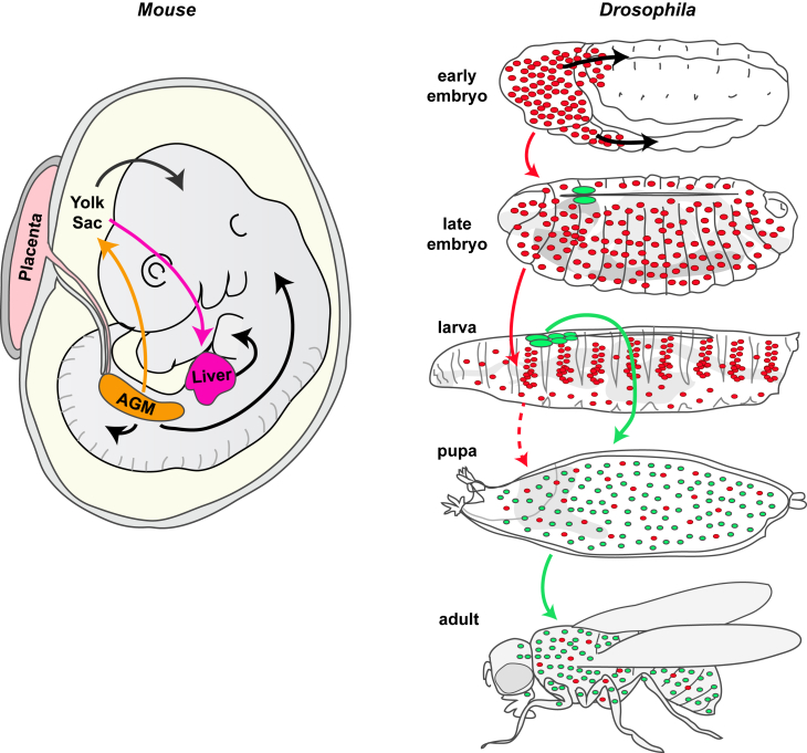
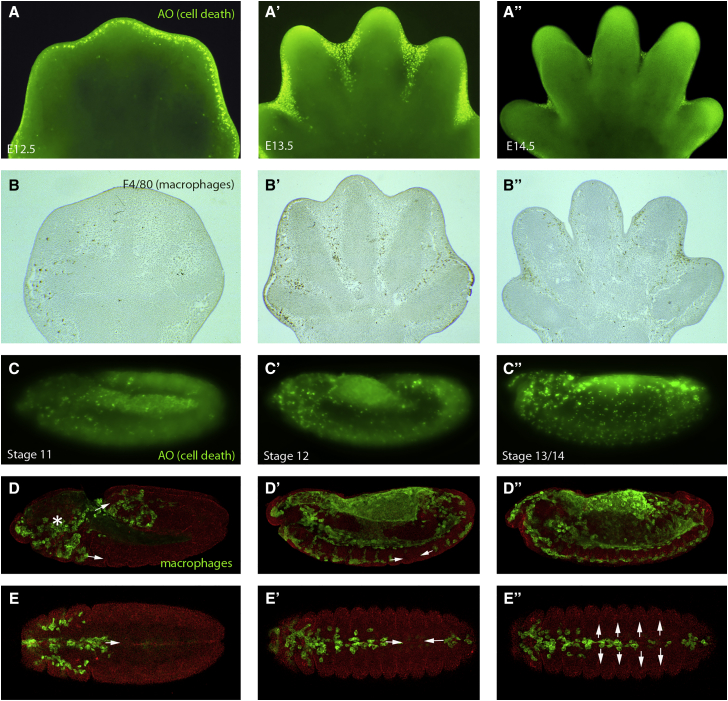
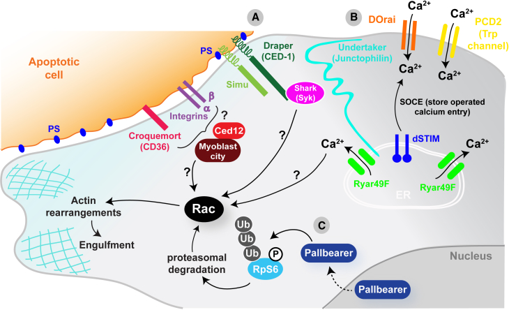
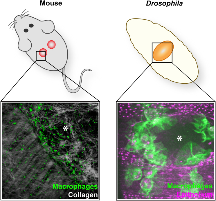
Macrophages are multifunctional innate immune cells that seed all tissues within the body and play disparate roles throughout development and in adult tissues, both in health and disease. Their complex developmental origins and many of their functions are being deciphered in mammalian tissues, but opportunities for live imaging and the genetic tractability of Drosophila are offering complementary insights into how these fascinating cells integrate a multitude of guidance cues to fulfill their many tasks and migrate to distant sites to either direct developmental patterning or raise an inflammatory response.

Keywords: Drosophila; apoptosis; development; disease; immunity; inflammation; macrophage; migration; wound.


Figures

Figure 1
Figure 1
Hematopoiesis in Mouse and Fly A schematized, limb bud stage mouse embryo with arrows indicating the flow of macrophage progenitors, which are all initially derived from the yolk sac and aorta-gonad-mesonephros (AGM), but with some populations moving directly onto their eventual tissues and others bypassing and differentiating further in the liver. In Drosophila (right), as in vertebrates, hematopoiesis occurs in two waves. The first during early embryogenesis gives rise to embryonic macrophages (red) that disperse throughout the embryo and later populate the larva organizing into sessile patches and circulating blood cells; these can be considered the fly equivalent of tissue macrophages. A second population arise from the larval lymph gland (green); these cells are released during pupal development, make up most of the population of blood cells in both the pupa and the adult, and can be considered the fly equivalent of bone marrow-derived macrophages.
Figure 2
Figure 2
Macrophages Clear Developmental Apoptosis during Development in the Mouse and Fly Acridine orange (AO) staining of mouse embryo footplates between 12.5 and 14.5 days of development reveals cell death (bright green) in the interdigital tissue of the developing limb (A–A″). Corresponding stage limbs stained with F4/80 reveal macrophages (brown) in the same location as they engulf the resulting apoptotic corpses (B–B″). AO staining in the Drosophila embryo (bright green in C–C″) or expressing GFP in macrophages (green in D–D″) reveals a similarly tight correlation between position of developmental cell death and macrophages throughout development in the fly embryo. Fly macrophages are born in the head (asterisk in D) and migrate through two routes, one into the extended germband and one along the ventral midline (arrows in D). (E)–(E′) show ventral views of Drosophila embryos at stages corresponding to those in (C)–(C′), highlighting the developmental migration of macrophages (green) along the ventral midline (arrows in E′). This is then followed by a rapid lateral migration from the midline (arrows in E″).
Figure 3
Figure 3
Apoptotic Recognition and Clearance Signaling in the Fly Several transmembrane proteins have been identified that allow detection of apoptotic corpses in the fly (A) including Croquemort (homolog of vertebrate CD36 scavenger receptor), the CED-1 homolog Draper, Six-microns under (Simu), and βv/αPS3 integrin heterodimers. Draper and Simu have been shown to bind phosphatidylserine (PS) on the surface of apoptotic cells, but how activation of any of these receptors upon binding to their ligands leads to the activation of Rac and the subsequent actin rearrangements required for engulfment remains largely unknown. In the case of Draper, activation of Rac could be through the Syk kinase homolog shark and in other cases will likely involve ELMO/Ced12 and Myoblast city. Two signaling cascades have been identified in the fly macrophage downstream of apoptotic engagement. The first (B) involves a calcium signaling pathway driven by intracellular store operated calcium entry (SOCE) downstream of Draper. A junctophilin (undertaker), an ER calcium sensor (Dstim), a calcium release activated channel (DOrai), and a TRP channel (Pkd2) are all required for this calcium signaling event. Again, how this calcium signaling leads to the activation of Rac remains unknown. The second (C) involves an F-box protein that acts as an E3 ubiquitin ligase called Pallbearer. This interacts with phosphorylated ribosomal protein S6 (RpS6) to promote its ubiquitylation and proteasomal degradation, which leads to Rac activation and subsequent actin remodeling required for engulfment.
Figure 4
Figure 4
Wounding Triggers a Recruitment of Macrophages in the Mouse and Fly Right: F480 immunostaining of a wound made to the back skin of an adult mouse with multiphoton second harmonics revealing collagen (white) to reveal the wound margin running from top left to bottom right of the field of view. Macrophages (green) are clustered at the wound edge. Left: similarly, laser ablation wounds made in the epithelium of a fly embryo trigger a rapid chemotactic response from macrophages (green), which are recruited to the wound within minutes and remain at the wound site throughout closure. Wounds are marked with an asterisk. Mouse wound image courtesy of Jenna Cash, and fly image courtesy of Helen Weavers.
Figure 5
Figure 5
A Three-Part Signaling System Drives the Inflammatory Response in the Fly (1) In Drosophila, macrophages are initially primed to respond to a wound by engulfing an apoptotic corpse. The process of engulfment triggers a calcium signaling event in the macrophage which, through activation of the JNK pathway, leads to upregulation of the damage receptor draper and makes these cells “primed” for response to a subsequent wound. (2) Upon wounding, hydrogen peroxide (H2O2) is rapidly released from the wound site diffusing at approximately 84,000 μm/min, acting as a “permissive signal” for macrophage migration to wounds by activating Src-dependent phosphorylation of Draper on its ITAM domain, which in turn recruits the downstream kinase shark. (3) A third unknown directional signal (signal X) is also produced upon wounding and diffuses away from the wound at a speed of approximately 200 μm/min. This signal operates as an attractive cue to pull the macrophage to the wound and could be detected by Draper or unknown damage receptors (receptor X).

References

    1. Abram C.L., Lowell C.A. The ins and outs of leukocyte integrin signaling. Annu. Rev. Immunol. 2009;27:339–362. - PMC - PubMed
    1. Agaisse H., Petersen U.M., Boutros M., Mathey-Prevot B., Perrimon N. Signaling role of hemocytes in Drosophila JAK/STAT-dependent response to septic injury. Dev. Cell. 2003;5:441–450. - PubMed
    1. Allen J.E., Sutherland T.E. Host protective roles of type 2 immunity: parasite killing and tissue repair, flip sides of the same coin. Semin. Immunol. 2014;26:329–340. - PMC - PubMed
    1. Anderl I., Vesala L., Ihalainen T.O., Vanha-Aho L.M., Ando I., Ramet M., Hultmark D. Transdifferentiation and proliferation in two distinct hemocyte lineages in Drosophila melanogaster larvae after wasp infection. PLoS Pathog. 2016;12:e1005746. - PMC - PubMed
    1. Antunes M., Pereira T., Cordeiro J.V., Almeida L., Jacinto A. Coordinated waves of actomyosin flow and apical cell constriction immediately after wounding. J. Cell Biol. 2013;202:365–379. - PMC - PubMed

Publication types

LinkOut - more resources