. 2016 Oct 15;25(20):4369-4375.
doi: 10.1093/hmg/ddw267.

Vitamin B12 ameliorates the phenotype of a mouse model of DiGeorge syndrome

Affiliations

Vitamin B12 ameliorates the phenotype of a mouse model of DiGeorge syndrome

Gabriella Lania et al. Hum Mol Genet. .

Erratum in

Abstract

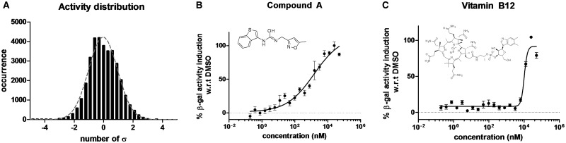
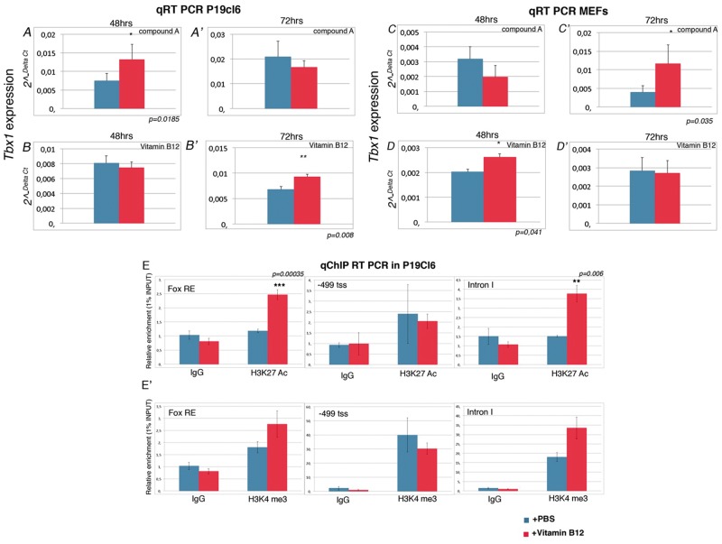
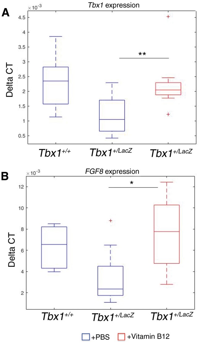
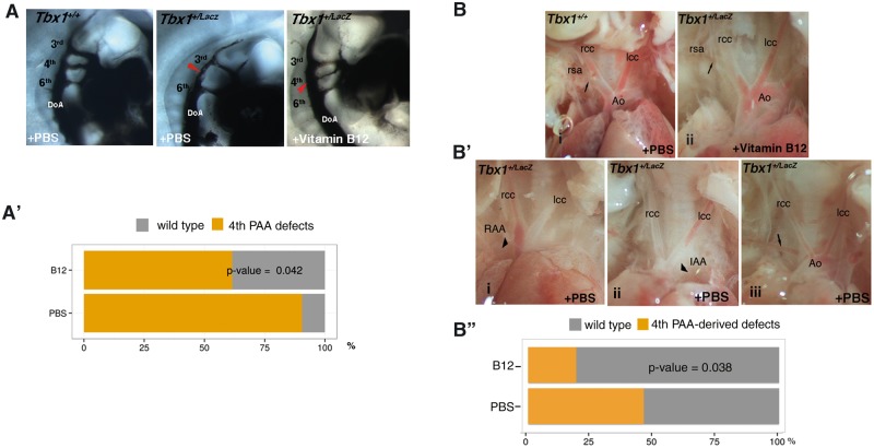
Pathological conditions caused by reduced dosage of a gene, such as gene haploinsufficiency, can potentially be reverted by enhancing the expression of the functional allele. In practice, low specificity of therapeutic agents, or their toxicity reduces their clinical applicability. Here, we have used a high throughput screening (HTS) approach to identify molecules capable of increasing the expression of the gene Tbx1, which is involved in one of the most common gene haploinsufficiency syndromes, the 22q11.2 deletion syndrome. Surprisingly, we found that one of the two compounds identified by the HTS is the vitamin B12. Validation in a mouse model demonstrated that vitamin B12 treatment enhances Tbx1 gene expression and partially rescues the haploinsufficiency phenotype. These results lay the basis for preclinical and clinical studies to establish the effectiveness of this drug in the human syndrome.


Figures

Figure 1.
Figure 1.
High throughput screening results. Occurrence distribution of compound screening activity (A). The bin width was set to +3 standard deviations. The grey dotted line represents the best fitted Gaussian distribution. Dose response of the β-gal activity for compound A (B) and vitamin B12 (C). The percentage of induction was calculated with relative to the average DMSO levels (0% induction).
Figure 2.
Figure 2.
Compound A and vitamin B12 enhance Tbx1 expression in P19Cl6 and MEF cells. (A-D') Quantitative RT-PCR evaluation of Tbx1 expression in P19Cl6 cells treated for 48h and 72h with the compound A (A-A’) and vitamin B12 (B-B’). Quantitative RT-PCR evaluation of Tbx1 expression in MEFs cells treated for 48h and 72h with chemical Compound A (C-C’) and vitamin B12 (D-D’). (E-E') Quantitative ChIP (qChIP) results for three loci, Fox responsive element (FOX-RE) (mm9 chr16:18601587-18601697), promoter (-499 fro transcription start site; mm9 chr16:18587346-18587459), and intron I of Tbx1 gene (mm9 chr16:18587346-18587459). qChIP assays were performed on P19Cl6 cells with and without vitamin B12 treatment. The histograms represent the mean of 6 independent experiments. Data are presented as mean ± SEM. *P < 0.05; ** P < 0.01; *** P < 0.001.
Figure 3.
Figure 3.
Tbx1 and Fgf8 gene expression is upregulated by vitamin B12 in vivo. Box plots of qRT-PCR evaluation of Tbx1 (A) and Fgf8 (B) gene expression in E9.5 embryos (n = 11 for each point), with or without vitamin B12 treatment. The error bars represent the minimal and maximal values of relative expression. *** P = 0.0045; * P = 0.031.
Figure 4.
Figure 4.
Vitamin B12 treatment partially rescues the Tbx1 haploinsufficiency phenotype. (A) Pharyngeal arch arteries of E10.5 embryos visualised by ink injection. Lateral view of PBS-treated WT (left panel), PBS-treated Tbx1lacZ/+ (centre), and vitamin B12-treated Tbx1lacZ/+ (right) embryos. (A’) Graphic representation of the percentage of embryos with 4th PAA defects (PBS sample n = 21; vitamin B12-treated sample n =  26). (B) Representative immages of aortic arch and great vessels of E17.5 fetuses. (i) WT pattern; (ii) normal pattern in a vitamin B12-treated Tbx1lacZ/+ fetus; (B’) Examples of aortic arch patterning defects in Tbx1lacZ/+ fetuses: (i) right aortic arch (RAA), the arrowhead indicates the retropositioned arch; (ii) interrupted arch aortic (IAA) type B, the arrowhead indicates the interruption; (iii) retroesophageal right subclavian artery. Arrows indicate the right subclavian artery. Ao: aorta; lcc: left common carotid artery; rcc: right common carotid artery; rsa: right subclavian artery. (B'') Graphic representation of the percentage of fetuses with aortic arch patterning abnormalities derived from 4th PAA defects (PBS sample n = 26; vitamin B12-treated sample n =  26).

References

    1. Yagi H., Furutani Y., Hamada H., Sasaki T., Asakawa S., Minoshima S., Ichida F., Joo K., Kimura M., Imamura S., et al. (2003) Role of TBX1 in human del22q11.2 syndrome. Lancet, 362, 1366–1373. - PubMed
    1. Paylor R., Glaser B., Mupo A., Ataliotis P., Spencer C., Sobotka A., Sparks C., Choi C.H., Oghalai J., Curran S., et al. (2006) Tbx1 haploinsufficiency is linked to behavioral disorders in mice and humans: implications for 22q11 deletion syndrome. Proc. Natl. Acad. Sci. U. S. A, 103, 7729–7734. - PMC - PubMed
    1. Swillen A., McDonald-McGinn D. (2015) Developmental trajectories in 22q11.2 deletion. Am. J. Med. Genet. C Semin. Med. Genet., 169, 172–181. - PMC - PubMed
    1. Vitelli F., Lania G., Huynh T., Baldini A. (2010) Partial rescue of the Tbx1 mutant heart phenotype by Fgf8: genetic evidence of impaired tissue response to Fgf8. J. Mol. Cell. Cardiol., 49, 836–840. - PMC - PubMed
    1. Caprio C., Baldini A. (2014) p53 suppression partially rescues the mutant phenotype in mouse models of DiGeorge syndrome. Proc. Natl. Acad. Sci. U. S. A, 111, 13385–13390. - PMC - PubMed

Publication types