

Effects of Angelicae dahuricae Radix on 2, 4-Dinitrochlorobenzene-Induced Atopic Dermatitis-Like Skin Lesions in mice model
- PMID: 28173791
- PMCID: PMC5297152
- DOI: 10.1186/s12906-017-1584-8
Effects of Angelicae dahuricae Radix on 2, 4-Dinitrochlorobenzene-Induced Atopic Dermatitis-Like Skin Lesions in mice model
Abstract
Background: Atopic dermatitis (AD) is an inflammatory, chronically relapsing, and intensively pruritic skin disease that affect 10-30% of the global population. Angelicae dahuricae Radix (ADR) has been reported to be anti-inflammatory in Korean Medicine. In the present study, we investigated whether ADR suppresses the progression of AD in animal model.
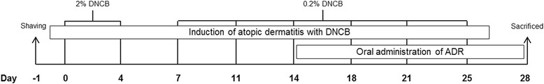
Methods: AD was induced by 2, 4-Dinitrochlorobenzene (DNCB). ADR was orally administered to mice to study the effect of ADR on AD. Histological Analysis, immunohistochemistry, blood analysis, RT-PCR, and ELISA assay were performed.
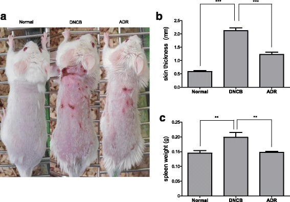
Results: ADR significantly suppressed AD-like symptoms in BALB/c mice: ADR decreased skin thickness and spleen weight of mice. ADR reduced infiltration of mast cells, inflammatory cells and CD4+ cells into mouse skin. ADR lowered the number of WBCs in the blood of mice. ADR reduced the levels of IgE, IL-6, IL-10 and IL-12 in mice serum. ADR down-regulated mRNA expression of IL-4, IL-6 and TNF-α in mouse skin tissue.
Conclusion: Our present study clearly indicates that ADR suppresses the progression of AD induced by DNCB in BALB/c mice. This suggests that ADR might be a useful drug for the treatment of AD.
Keywords: 2, 4-Dinitrocholrlbenzene; Angelicae dahuricae Radix; Atopic dermatitis; BALB/c mice; Cytokine; Inflammation.
Figures


Similar articles
-
The prevention of 2,4-dinitrochlorobenzene-induced inflammation in atopic dermatitis-like skin lesions in BALB/c mice by Jawoongo.BMC Complement Altern Med. 2018 Jul 13;18(1):215. doi: 10.1186/s12906-018-2280-z. BMC Complement Altern Med. 2018. PMID: 30005655 Free PMC article.
-
Topical application of Taglisodog-eum inhibits the development of experimental atopic dermatitis.J Ethnopharmacol. 2013 Jan 30;145(2):536-46. doi: 10.1016/j.jep.2012.11.026. Epub 2012 Dec 2. J Ethnopharmacol. 2013. PMID: 23211659
-
Suppression of dust mite extract and 2,4-dinitrochlorobenzene-induced atopic dermatitis by the water extract of Lindera obtusiloba.J Ethnopharmacol. 2011 Sep 1;137(1):802-7. doi: 10.1016/j.jep.2011.06.043. Epub 2011 Jul 5. J Ethnopharmacol. 2011. PMID: 21762765
-
Management of Atopic Dermatitis Via Oral and Topical Administration of Herbs in Murine Model: A Systematic Review.Front Pharmacol. 2022 May 24;13:785782. doi: 10.3389/fphar.2022.785782. eCollection 2022. Front Pharmacol. 2022. PMID: 35685636 Free PMC article.
-
Kampo therapy for adult atopic dermatitis by dieting and herbal medications: evaluating the disappearance of disease phases. Dieting and herbs for atopic dermatitis.Adv Exp Med Biol. 2004;546:297-310. doi: 10.1007/978-1-4757-4820-8_22. Adv Exp Med Biol. 2004. PMID: 15584383 Review. No abstract available.
Cited by
-
Inhibitory Effect of Bisdemethoxycurcumin on DNCB-Induced Atopic Dermatitis in Mice.Molecules. 2022 Dec 30;28(1):293. doi: 10.3390/molecules28010293. Molecules. 2022. PMID: 36615486 Free PMC article.
-
Anti-allergic actions of a Chinese patent medicine, huoxiangzhengqi oral liquid, in RBL-2H3 cells and in mice.Pharm Biol. 2021 Dec;59(1):672-682. doi: 10.1080/13880209.2021.1928242. Pharm Biol. 2021. PMID: 34078224 Free PMC article.
-
Beneficial Effects of Two Hydrogen Sulfide (H2S)-Releasing Derivatives of Dexamethasone with Antioxidant Activity on Atopic Dermatitis in Mice.Pharmaceutics. 2023 Jul 8;15(7):1907. doi: 10.3390/pharmaceutics15071907. Pharmaceutics. 2023. PMID: 37514093 Free PMC article.
-
Anti-inflammatory effects of amarogentin on 2,4-dinitrochlorobenzene-induced atopic dermatitis-like mice and in HaCat cells.Animal Model Exp Med. 2023 Jun;6(3):255-265. doi: 10.1002/ame2.12260. Epub 2022 Sep 21. Animal Model Exp Med. 2023. PMID: 36131559 Free PMC article.
-
Ethnobotanical Survey on Skin Whitening Prescriptions of Traditional Chinese Medicine in Taiwan.Front Pharmacol. 2021 Nov 30;12:736370. doi: 10.3389/fphar.2021.736370. eCollection 2021. Front Pharmacol. 2021. PMID: 34916932 Free PMC article.
References
-
- Vale S, Smith J, Paterson T, Bramah S, Butt C. Australasian Society of Clinical Immunology and Allergy (ASCIA) 26th Annual Conference, 9–12 September 2015, Adelaide, Australia. Intern Med J. 2015;45(Suppl 4):1–30. - PubMed
MeSH terms
Substances
LinkOut - more resources
Full Text Sources
Other Literature Sources
Medical
Research Materials
