. 2017 Feb 7;14(1):22.
doi: 10.1186/s12985-017-0694-8.

Identification of influenza A nucleoprotein body domain residues essential for viral RNA expression expose antiviral target

Affiliations

Identification of influenza A nucleoprotein body domain residues essential for viral RNA expression expose antiviral target

Alicia M Davis et al. Virol J. .

Abstract

Background: Influenza A virus is controlled with yearly vaccination while emerging global pandemics are kept at bay with antiviral medications. Unfortunately, influenza A viruses have emerged resistance to approved influenza antivirals. Accordingly, there is an urgent need for novel antivirals to combat emerging influenza A viruses resistant to current treatments. Conserved viral proteins are ideal targets because conserved protein domains are present in most, if not all, influenza subtypes, and are presumed less prone to evolve viable resistant versions. The threat of an antiviral resistant influenza pandemic justifies our study to identify and characterize antiviral targets within influenza proteins that are highly conserved. Influenza A nucleoprotein (NP) is highly conserved and plays essential roles throughout the viral lifecycle, including viral RNA synthesis.

Methods: Using NP crystal structure, we targeted accessible amino acids for substitution. To characterize the NP proteins, reconstituted viral ribonucleoproteins (vRNPs) were expressed in 293 T cells, RNA was isolated, and reverse transcription - quantitative PCR (RT-qPCR) was employed to assess viral RNA expressed from reconstituted vRNPs. Location was confirmed using cellular fractionation and western blot, along with observation of NP-GFP fusion proteins. Nucleic acid binding, oligomerization, and vRNP formation, were each assessed with native gel electrophoresis.

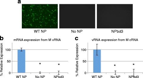
Results: Here we report characterization of an accessible and conserved five amino acid region within the NP body domain that plays a redundant but essential role in viral RNA synthesis. Our data demonstrate substitutions in this domain did not alter NP localization, oligomerization, or ability to bind nucleic acids, yet resulted in a defect in viral RNA expression. To define this region further, single and double amino acid substitutions were constructed and investigated. All NP single substitutions were functional, suggesting redundancy, yet different combinations of two amino acid substitutions resulted in a significant defect in RNA expression, confirming these accessible amino acids in the NP body domain play an important role in viral RNA synthesis.

Conclusions: The identified conserved and accessible NP body domain represents a viable antiviral target to counter influenza replication and this research will contribute to the well-informed design of novel therapies to combat emerging influenza viruses.

Keywords: Influenza; Nucleoprotein; RNA; Virus.


Figures

Fig. 1
Fig. 1
Components of the Viral Ribonucleoprotein and Nucleoprotein. a Graphical illustration of the viral Ribonucleoprotein (vRNP) complex, comprised of the viral polymerase subunits, PB1, PB2, and PA, bound to both the 5′ and 3′ ends of the viral RNA segment, and multiple copies of nucleoprotein (NP). b Domains of Nucleoprotein monomer crystal structure [32] using Deep View-Swiss-PdbViewer 4.0, with head domain, body domain, RNA pocket, and tail loop regions labeled. c. Nucleoprotein body domain substitutions of NPbd3 using Deep View-Swiss-PdbViewer 4.0 to analyze the accessible residues in the body domain of the NP monomer crystal structure [32]. In both b and c, residue color represents accessibility within the NP monomer as determined by the Deep View-Swiss-PdbViewer 4.0 “color” tool, with greatest to least accessible as follows: red, orange, yellow, green, light blue, and dark blue. Residues 289, 293, 294, 308, and 309 mutated in NPbd3 are highlighted in C. d Nucleoprotein (NP) sequence alignment from select influenza subtypes using CLS freeware and amino acids 281-310. Colors correlate with amino acid property. Asterisks indicate NPbd3 mutant
Fig. 2
Fig. 2
Reconstituted RNP Expression System. 293 T cells are transfected with plasmids to express mRNAs encoding the viral RdRP and NP or NP substitution mutants, and a specific vRNA or cRNA template. Cellular RNA polymerase II drives expression of viral mRNAs from pcDNA3 plasmid backbone while cellular RNA polymerase I drives expression of the cRNA or vRNA template from pHH21 plasmid backbone. vRNP function is assessed by gene expression from vRNA or cRNA templates
Fig. 3
Fig. 3
NPbd3 is defective for viral protein and RNA expression in reconstituted vRNPs. a Plasmids to express reconstituted vRNPs with GFP-M vRNA and either WT-NP, no NP, or NPbd3 were transfected into 293 T cells. 48 h post transfection cells were observed for GFP-M expression. WT NP represents the positive control while no NP is the negative control. GFP was visualized with a Nikon Eclipse TS100 (Nikon Intensilight C-HGFI for fluorescence) inverted microscope and images captured with the Nikon DS-Qi1Mc camera with NS Elements software. b and c RNA was purified from cells expressing reconstituted vRNPs (b) or cRNPs (c) as indicated. 1 μg was DNase treated and subject to reverse transcription with oligo dT (b) or vRNA specific primers (c) and quantitative PCR with M gene specific primers to calculate relative M RNA expression in each sample. Data are from triplicate trials; asterisks indicate p < 0.02
Fig. 4
Fig. 4
NPbd3 is expressed and localized as wild type NP. a Cells expressing reconstituted vRNPs were collected and fractionated with NP-40 non-ionic detergent to break open cellular plasma membrane. Microscopy was used to confirm disrupted plasma membranes and intact nuclei. Nuclei were pelleted by centrifugation and proteins isolated. Proteins were separated on a 10% SDS PAGE gel and transferred to nitrocellulose. Western was performed with anti-FLAG to detect WT NP and NPbd3 and anti-Hsp90 to detect Hsp90, a protein localized in the cytoplasm, which serves as confirmation of cellular fractionation. Expected size was confirmed by comparison with Fisher BioReagents EZ-Run protein standards. b NP-GFP and NPbd3-GFP fusion proteins or eGFP as indicated were expressed in cells grown on poly-L-Lysine cover slips. Cells were washed and fixed using a 1:1 methanol and acetone mixture. The coverslips were mounted onto glass slides using SouthernBiotech™ Dapi-Fluoromount-G™ Clear Mounting Media which stains the cell nucleus blue. Slides were observed on a Nikon ECLIPSE TE2000-U fluorescent microscope and images were captured with an Andor Clara DR-3446 camera using NIS-Elements AR software
Fig. 5
Fig. 5
Titration of NP and reconstituted vRNP activity. 293 T cells were transfected with plasmids to express reconstituted vRNPs with GFP-M vRNA and either WT-NP, no NP, or NPbd3 at concentrations indicated. a Total protein from titration samples was isolated at 48 h post transfection and run on a 10% SDS PAGE and transferred to nitrocellulose. The membrane was probed with FLAG to identify NP and Tubulin as the loading control. Expected size was confirmed by comparison with Fisher BioReagents EZ-Run protein standards. b Cells were observed for GFP-M expression 48 h post transfection. WT-NP expressed by transfection of 400 ng plasmid is the standard concentration used in our reconstituted vRNP experiments and serves as positive control, while no NP is the negative control. GFP was visualized with a Nikon Eclipse TS100 (Nikon Intensilight C-HGFI for fluorescence) inverted microscope and images captured with the Nikon DS-Qi1Mc camera with NS Elements software
Fig. 6
Fig. 6
NPbd3 maintains nucleic acid binding, oligmerization, and vRNP formation. a WT NP and NPbd3 were immuno purified using anti-FLAG antibody. Purified WT NP and NPbd3 were incubated with biotin labeled single stranded DNA and separated on an 8% PAGE TBE non denaturing gel before transfer to nitrocellulose. Biotin ssDNA was detected by interaction with Streptavidin-HRP and ECL reagents. No NP samples expressed no NP protein and serve as negative control. b Blue Native Gel Electrophoresis was utilized to migrate protein extracts of cells expressing NP and NPbd3 followed by western blot with anti-FLAG antibody to demonstrate NP oligomer formation. Asterisk represents Native Mark Unstained Protein standard at 480 kDa. c Blue Native Gel Electrophoresis was utilized to migrate protein extracts of cells expressing reconstituted vRNPs comprised of either NP or NPbd3 followed by western blot with anti-FLAG antibody to demonstrate vRNP formation. Asterisk represents Native Mark Unstained Protein standard at 1048 kDa
Fig. 7
Fig. 7
NP body domain single amino acid substitution mutants display near wild type activity in reconstituted vRNP assay. a Cells expressing reconstituted vRNPs with GFP-M vRNA were visualized for GFP expression with a Nikon Eclipse TS100 (Nikon Intensilight C-HGFI for fluorescence) inverted microscope and images captured with the Nikon DS-Qi1Mc camera with NS Elements software. Number indicates NP single residue mutated. b Total protein was isolated from cells expressing reconstituted vRNPs with GFP-M vRNA template and the indicated NP substitution mutant. Total protein extract was separated on 10% SDS PAGE and transferred to nitrocellulose. WT NP, NPbd3, and NP single mutants were detected with anti-FLAG. Anti-tubulin was used as loading control. Expected size was confirmed by comparison with Fisher BioReagents EZ-Run protein standards. c RNA from cells expressing reconstituted vRNPs with FLAG-M vRNA and either WT NP, no NP, or NPbd3, or the single amino acid substitution mutants were isolated and analyzed by 1% bleach/agarose gel electrophoresis. d One microgram of RNA was treated with DNase before being reverse transcribed with oligo dT (mRNA) and analyzed through quantitative PCR with primers targeting the M gene. qPCR reactions were carried out in triplicate. Significance was evaluated through t-test by comparing WT NP; asterisks indicate p-values <0.01
Fig. 8
Fig. 8
NP double amino acid substitution mutants display decreased activity in reconstituted vRNP assay. a Cells expressing reconstituted vRNPs with GFP-M vRNA were visualized for GFP expression with a Nikon Eclipse TS100 (Nikon Intensilight C-HGFI for fluorescence) inverted microscope and images captured with the Nikon DS-Qi1Mc camera with NS Elements software. Number indicates the two NP residues substituted with glycine. b Total protein was isolated from cells expressing reconstituted vRNPs with GFP-M vRNA template and the indicated NP mutant. Total protein extract was separated on 10% SDS PAGE and transferred to nitrocellulose. WT NP, NPbd3, and NP double mutants were detected with anti-FLAG. Anti-tubulin was used as loading control. Expected size was confirmed by comparison with Fisher BioReagents EZ-Run protein standards. c RNA from cells expressing reconstituted vRNPs with FLAG-M vRNA and either WT NP, no NP, or NPbd3, or the double amino acid mutants were isolated and analyzed by 1% bleach/agarose gel electrophoresis. d One microgram of RNA was treated with DNase before being reverse transcribed with oligo dT (mRNA) and analyzed through quantitative PCR with primers targeting the M gene. qPCR reactions were carried out in triplicate. Significance was evaluated through t-test by comparing WT NP; asterisks indicate p-values <0.02

Similar articles

Cited by

References

    1. Centers for Disease Control and Prevention. http://www.cdc.gov/flu/avianflu/avian-in-humans.htm. Accessed 15 June 2016.
    1. Centers for Disease Control and Prevention. http://www.cdc.gov/flu/about/viruses/transmission.htm. Accessed 15 June 2016.
    1. Neumann G, Noda T, Kawaoka Y. Emergence and pandemic potential of swine-origin H1N1 influenza virus. Nature. 2009;459:931–9. doi: 10.1038/nature08157. - DOI - PMC - PubMed
    1. Centers for Disease Control and Prevention. http://www.cdc.gov/flu/professionals/vaccination/effectiveness-studies.htm. Accessed 15 June 2016.
    1. Centers for Disease Control and Prevention. http://www.cdc.gov/media/releases/2016/s0622-laiv-flu.html. Accessed 15 July 2016.

MeSH terms