. 2017 Feb 7;12(1):14.
doi: 10.1186/s13024-017-0155-2.

Gene-environment interaction between lead and Apolipoprotein E4 causes cognitive behavior deficits in mice

Affiliations

Gene-environment interaction between lead and Apolipoprotein E4 causes cognitive behavior deficits in mice

Anna K Engstrom et al. Mol Neurodegener. .

Erratum in

Abstract

Background: Alzheimer's disease (AD) is characterized by progressive cognitive decline and memory loss. Environmental factors and gene-environment interactions (GXE) may increase AD risk, accelerate cognitive decline, and impair learning and memory. However, there is currently little direct evidence supporting this hypothesis.

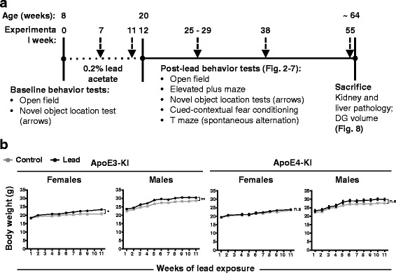
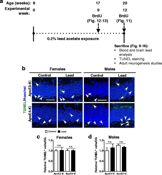
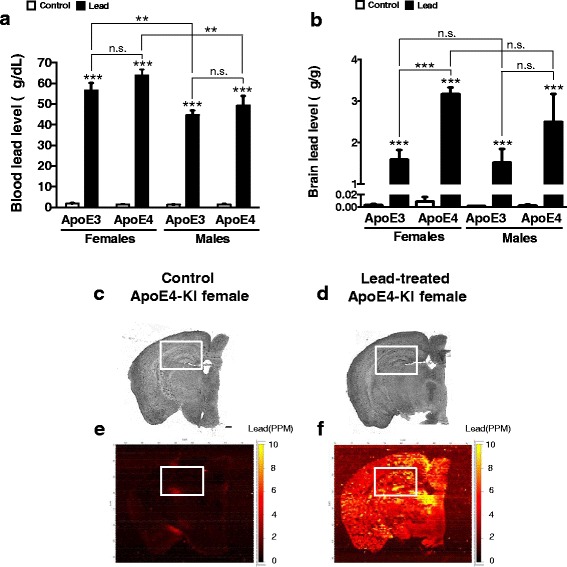
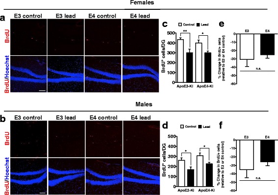
Methods: In this study, we assessed for a GXE between lead and ApoE4 on cognitive behavior using transgenic knock-in (KI) mice that express the human Apolipoprotein E4 allele (ApoE4-KI) or Apolipoprotein E3 allele (ApoE3-KI). We exposed 8-week-old male and female ApoE3-KI and ApoE4-KI mice to 0.2% lead acetate via drinking water for 12 weeks and assessed for cognitive behavior deficits during and after the lead exposure. In addition, we exposed a second (cellular) cohort of animals to lead and assessed for changes in adult hippocampal neurogenesis as a potential underlying mechanism for lead-induced learning and memory deficits.

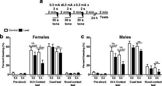
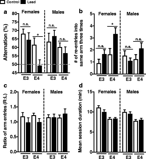
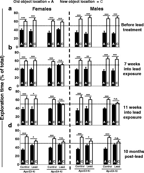
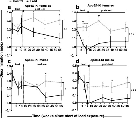
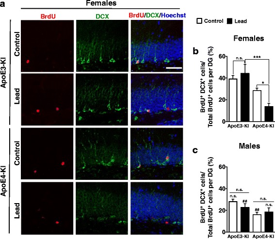
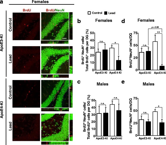
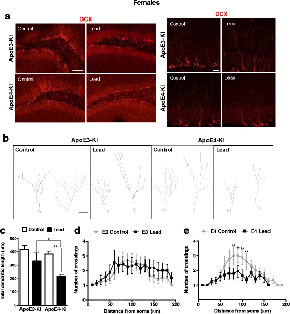
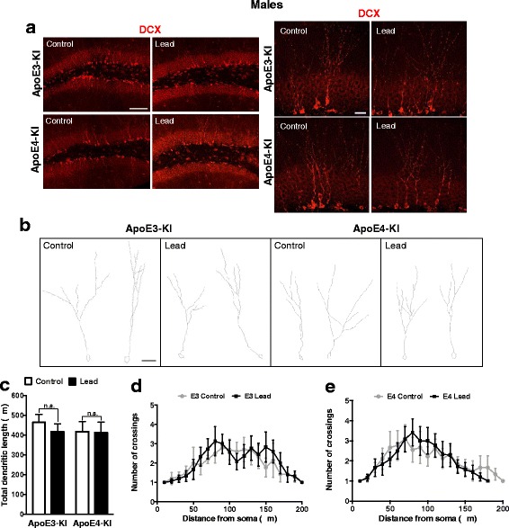
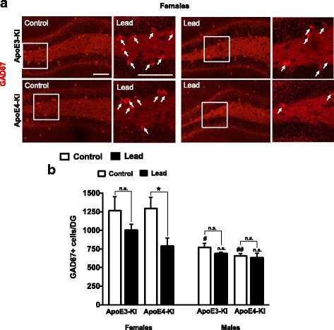
Results: In the behavior cohort, we found that lead reduced contextual fear memory in all animals; however, this decrease was greatest and statistically significant only in lead-treated ApoE4-KI females. Similarly, only lead-treated ApoE4-KI females exhibited a significant decrease in spontaneous alternation in the T-maze. Furthermore, all lead-treated animals developed persistent spatial working memory deficits in the novel object location test, and this deficit manifested earlier in ApoE4-KI mice, with female ApoE4-KI mice exhibiting the earliest deficit onset. In the cellular cohort, we observed that the maturation, differentiation, and dendritic development of adult-born neurons in the hippocampus was selectively impaired in lead-treated female ApoE4-KI mice.

Conclusions: These data suggest that GXE between ApoE4 and lead exposure may contribute to cognitive impairment and that impaired adult hippocampal neurogenesis may contribute to these deficits in cognitive behavior. Together, these data suggest a role for GXE and sex differences in AD risk.

Keywords: Adult hippocampal neurogenesis; Apolipoprotein E; Cognitive behavior; Lead; Learning and memory.


Figures

Fig. 1
Fig. 1
Adult male and female ApoE3-KI and ApoE4-KI mice do not exhibit weight loss upon exposure to 0.2% lead acetate for 12 weeks. a Experimental design and timeline for the behavior cohort. b Body weight was measured every 1–2 weeks during the lead exposure window. The lead-treated mice did not exhibit any weight loss relative to controls of the same sex and genotype. ApoE3-KI mice exposed to lead weighed slightly but significantly more at several time points than control mice during the lead exposure. Data are mean ± SEM with n = 8–13 per genotype/sex/treatment. Two-way ANOVA, significant main effect of treatment: n.s., not significant; * p < 0.05; ** p < 0.01
Fig. 2
Fig. 2
Lead-treated ApoE3-KI and ApoE4-KI male and female mice do not exhibit locomotor deficits in the open field test compared to controls. Mice were exposed to lead as previously described and the open field test was conducted after the cessation of the lead exposure. There was a significant main effect of lead treatment in female animals on floor plane distance, moving time, and average speed (Two-way ANOVA: floor plane distance, F(1,31) = 22.85, p < 0.0001; moving time, F(1,31) = 20.47, p < 0.0001; average speed, F(1,31) = 20.49, p < 0.0001). Post-hoc analyses found that lead-treated ApoE3-KI and/or ApoE4-KI females traveled a (a) greater distance, (b) spent the same or more time moving, and (c) traveled slightly faster than controls. There were no significant differences between lead and control ApoE3-KI and ApoE4-KI males or between lead-treated ApoE3-KI and ApoE4-KI mice (males or females) in any of the locomotor endpoints. Data are mean ± SEM with n = 8–13 per genotype/sex/treatment. Two-way ANOVA with Holm-Sidak post-hoc tests: n.s., not significant; * p < 0.05; *** p < 0.001
Fig. 3
Fig. 3
Male and female ApoE3-KI and ApoE4-KI mice exposed to lead do not exhibit overt anxiety in the open field test or elevated plus maze. The open field test was used to determine if lead treatment causes anxiety, measured as more time or distance in the margin, less time or distance in the center, or reduced center entries. There was a significant main effect of lead treatment in female mice on center distance and center entries (Two-way ANOVA: center distance, F(1,31) = 5.936, p = 0.0208; center entries, F(1,31) = 14.76, p = 0.0006). Post-hoc analyses found that there were no significant differences between lead-treated ApoE3-KI and ApoE4-KI mice (males and females) on the (a) time or (b) distance traveled in the margins, the (c) time or (d) distance traveled in the center, or the (e) number of center entries in the open field test. D, ApoE3-KI females treated with lead traveled a slightly greater distance in the center and E, made more center entries than controls. There were no significant differences between control and lead-treated ApoE4-KI females, ApoE3-KI males, or ApoE4-KI males in the open field test. f Representative open field track plots from female control and lead-treated ApoE3-KI and ApoE4-KI mice. In the elevated plus maze, both male and female ApoE3-KI and ApoE4-KI mice exposed to lead spent a (g) similar or greater amount of time and traveled a (h) similar or greater distance in the open arms of the maze compared to controls. Lead-treated males and females did not exhibit reduced (i) open arm or (j) open arm end entries compared to control animals. Data are mean ± SEM with n = 8–13 per genotype/treatment. Two-way ANOVA with Holm-Sidak post-hoc tests: n.s., not significant; * p < 0.05; *** p < 0.001
Fig. 4
Fig. 4
Lead impairs contextual fear memory in lead-treated ApoE4-KI females at 22–24 weeks of age. a Schematic of cued and contextual fear conditioning test performed post-lead exposure. b, c Female and male mice had low baseline freezing behavior (Pre-shock). In the 24 h Context test, there was a significant main effect of lead treatment on freezing behavior in both males and females (Two-way ANOVA: females, F(1,33) = 6.803, p =0.0136; males, F(1,42) = 4.435, p =0.0412). Although lead treatment reduced contextual memory in all animals (manifested as reduced freezing 24 h after fear conditioning) post-hoc tests revealed that this was only statistically significant between control and lead-treated ApoE4-KI females (Holm-Sidak post-hoc test: F(1,33) = 2.648, p = 0.0245). Auditory-cued (hippocampus-independent) fear memory was not affected (Cued test) in any lead-exposed animals. All animals did not freeze when placed into a novel, non-shock context (Novel context test). Data are mean ± SEM with n = 8–13 per sex/genotype/treatment. Two-way ANOVA with Holm-Sidak post-hoc test. n.s. not significant; * p < 0.05
Fig. 5
Fig. 5
Lead impairs spontaneous alternation and increases repetitive arm entry in lead-treated ApoE4-KI females. Spontaneous alternation was assessed using the T-maze at 12–13 months of age. a There was a significant main effect of genotype on spontaneous alternation in females (Two-way ANOVA: genotype, F(1,32) = 7.666, p = 0.0093). Lead-treated ApoE4-KI female mice exhibited reduced spontaneous alternation compared to female ApoE4-KI control mice (Holm-Sidak post-test: F(1,32) =2.356, p =0.0490). b There was a significant main effect of both genotype and treatment on repetitive arm entries in females (Two-way ANOVA: genotype, F(1,32) = 5.915, p =0.0208; treatment, F(1,32) = 6.164, p =0.0185). ApoE4-KI females exposed to lead exhibited significantly increased repetitive arm entries compared to control ApoE4-KI female mice (Holm-Sidak post-test: F(1,32) = 2.584, p 0.0289). There was no significant difference in spontaneous alternation or arm re-entries in lead-treated ApoE3-KI and ApoE4-KI male mice. c Female and male mice did not exhibit any arm preference. d There was a significant main effect of genotype on mean session duration in both the females and males, with ApoE4-KI mice completing the alternation task slightly faster than ApoE3-KI mice of the same sex (Two-way ANOVA: females, F(1,30) = 19.50, p = 0.0001; males F(1,41) =9.803, p = 0.0032). There was no significant difference in mean session duration between control and lead-treated animals of the same genotype and sex. Data are mean ± SEM with n = 8–13 per sex/genotype/treatment. n.s. not significant; * p < 0.05
Fig. 6
Fig. 6
The effect of lead on short-term spatial memory in the NOL test. The NOL test was performed before, during, and after the lead exposure to assess for spatial working memory deficits. The time animals spent investigating the object in the old (location A) vs. new (location C) locations was quantified. More time spent exploring the object in the novel vs. old location indicates memory for the old location. ( A ) All the animals had intact spatial memory prior to the lead exposure. ( B ) At 7 weeks into the lead exposure, only the lead-treated ApoE4-KI females did not discriminate between the old vs. new object locations (Two-tailed t-test (A vs. C): p = 1.000). ( C ) At 11 weeks, both the lead-treated ApoE4-KI females and males no longer discriminated between the object locations (females, p = 0.4042; males, p = 0.1959) while the ApoE3-KI females and males spent significantly more time exploring the novel object location (females, p = 0.0107; males, p = 0.0098). ( D ) Lead-treated ApoE4-KI females and males continued to exhibit a deficit in spatial working memory at 10 months post-lead exposure (females, p = 0.0961; males, p = 0.1855). Lead-treated ApoE3-KI mice still spent statistically significantly more time exploring the novel object location at 10 months post-lead exposure (females, p = 0.0186; males, p = 0.0320). Data are mean ± SEM with n = 8–13 per genotype/treatment. Two-tailed t–test: n.s. not significant, * p < 0.05; ** p < 0.01; *** p < 0.001
Fig. 7
Fig. 7
Genotype and sex differences in lead-induced reduction of discrimination index over time. The discrimination index in the NOL test was calculated by dividing the difference in exploration time between the novel (c) and familiar (a) locations by the total exploration time, and used to compare changes of spatial memory over time. There was a main effect of lead exposure on the discrimination index in all lead-exposed animals (Multiple mixed-effects linear regression: ApoE3-KI females, p = 0.001; ApoE4-KI females, p < 0.0001; ApoE3-KI males, p = 0.019; ApoE4-KI males, p < 0.0001). a There was no difference in the discrimination index between control and lead-treated ApoE3-KI females during the lead exposure (Two-tailed t-test: 7 week, p = 0.3046; 11 week, p = 0.9977). Lead-treated ApoE3-KI females had a significantly lower discrimination index at 3 and 10 months post-lead exposure (3 months, p = 0.0268; 10 months, p = 0.0156) and a non-significant decrease 6 months post-lead (p = 0.0901) compared to controls. b Lead-treated ApoE4-KI females had a significantly lower discrimination index than ApoE4-KI control mice starting at 7 weeks into the lead exposure and this persisted through 10 months post-lead (7 weeks, p = 0.0016; 11 weeks, p = 0.0002; 3 months, p = 0.0030; 10 months, p = 0.0269). The lead-treated ApoE4-KI discrimination index was lower than controls at 6 months post-lead but not statistically significant (p = 0.0713). c Lead-treated ApoE3-KI males had a significantly lower discrimination index at 6 and 10 months post-lead compared to controls (6 months, p = 0.0356; 10 months, p = 0.0418). d In contrast, lead-treated ApoE4-KI males had a significantly lower discrimination index than controls starting at 11 weeks into the lead exposure and this effect persisted through 10 months post-lead exposure (11 weeks, p = 0.0064; 4 months, p = 0.0433; 6 months, p = 0.0382; 10 months, p = 0.0275). Data are mean ± SEM with n = 8–13 per sex/genotype/treatment. Multi-level mixed-effects linear regression; significant effect of treatment: # p < 0.05; ## p < 0.01; ### p < 0.001. Two-tailed t-test; significant difference between control and lead: n.s., not significant; * p < 0.05; ** p < 0.01; *** p < 0.001
Fig. 8
Fig. 8
Adult lead does not cause significant liver or kidney toxicity or DG volume loss in aged ApoE3-KI and ApoE4-KI mice. 8-week-old female ApoE3-KI and ApoE4-KI mice were exposed to 0.2% lead acetate for 12 weeks, then switched to normal drinking water and sacrificed 43–45 weeks post-lead exposure (14.5–15 months old). H&E staining of a kidney and b liver sections and quantification of c kidney glomerular nephropathy and d liver cytoplasmic vacuolation in female ApoE3-KI and ApoE4-KI mice. e DG volume was measured in female ApoE3-KI and ApoE4-KI mice using ImageJ analysis of confocal images of coronal sections from one brain hemisphere. Data are mean ± SEM. n = 3–5 per genotype/treatment. Two-way ANOVA with Fisher’s LSD post-test: n.s., not significant. Scale bars, 100 μm
Fig. 9
Fig. 9
Adult-only lead exposure does not increase the number of TUNEL+ apoptotic cells in the DG of the hippocampus. a Experimental design and timeline for cellular studies (BrdU dosing) for Figs. 9, 10, 11, 12, 13, 14, 15 and 16. b Representative images of TUNEL+ (green) cells in the DG of control and lead-treated ApoE3-KI and ApoE4-KI males and females. Quantification of TUNEL+ cells per DG in ApoE3-KI and ApoE4-KI c females and d males. Data are mean ± SEM with n = 3–4 per genotype/sex/treatment. Two-way ANOVA with Fisher’s LSD post-test: n.s., not significant. Scale bar, 50 μm
Fig. 10
Fig. 10
Adult-only lead exposure results in elevated blood lead levels and lead deposition in the brain. 8-week-old ApoE3-KI and ApoE4-KI male and female mice were exposed to 0.2% lead acetate for 12 weeks and then sacrificed. Blood and one brain hemisphere were collected at sacrifice and (a) blood lead and (b) brain lead levels were measured using ICP-MS. Brightfield images of one brain hemisphere from a female (c) control and (d) lead-treated ApoE4-KI mouse after the 12 week lead exposure. Semi-quantitative measurement of lead in the brain of a (e) control and (f) lead-treated ApoE4-KI mouse using LA-ICP-MS. Two-way ANOVA with Fisher’s LSD post-test: n.s., not significant; ** p < 0.01; *** p < 0.001. Scale bars, 100 μm
Fig. 11
Fig. 11
Adult lead exposure decreases adult-born cell proliferation in the DG of the hippocampus of all mice. 8-week-old male and female ApoE3- and ApoE4-KI mice were exposed to 0.2% lead acetate for 12 weeks and then sacrificed. BrdU was administered 2 h prior to sacrifice (1 x 100 mg/kg). Representative images of BrdU (red) immunostaining in the DG of 20-week-old (a) female and (b) male ApoE3-KI and ApoE4-KI control and lead-treated mice. Quantification of the total BrdU+ cells per DG in (c) females and (d) males. Quantification of the percent change in total BrdU+ cells in lead-treated mice relative to ApoE3-KI or ApoE4-KI control (e) females and (f) males. Data are mean ± SEM with n = 3–5 per genotype/sex/treatment. Two-way ANOVA with Fisher’s LSD post-test: n.s., not significant; * p < 0.05; ** p < 0.01. Scale bars, 100 μm
Fig. 12
Fig. 12
Lead decreases adult-born immature neuron maturation in the DG of ApoE4-KI female mice. 8-week-old male and female ApoE3- and ApoE4-KI mice were exposed to 0.2% lead acetate for 12 weeks and then sacrificed. 100 mg/kg BrdU was administered 5 times in 1 day (every 2 h) 3 weeks prior to sacrifice (i.e., at 9 weeks into lead exposure). a Representative images of BrdU (red) and DCX (green) immunostaining in the DG of control and lead-treated female ApoE3-KI and ApoE4-KI animals. Quantification of the percent of total BrdU+ cells that are BrdU+DCX+ per DG in females (b) and males (c) respectively. Data are mean ± SEM with n = 4–5 per genotype/sex/treatment. Two-way ANOVA with Fisher’s LSD post-test: Comparison between control and lead-treated: n.s., not significant; * p < 0.05; *** p < 0.001. Comparison between males and females of the same genotype and treatment: n.s., not significant; ##, p < 0.01. Scale bar, 50 μm
Fig. 13
Fig. 13
Lead decreases adult-born neuron differentiation in the DG of ApoE4-KI female mice. 8-week-old male and female ApoE3-KI and ApoE4-KI mice were exposed to 0.2% lead acetate for 12 weeks and then sacrificed. 100 mg/kg BrdU was administered 5 times in 1 day (every 2 h) 3 weeks prior to sacrifice (i.e., at 9 weeks into lead exposure). a Representative images of BrdU (red) and NeuN (green) immunostaining in the DG of control and lead-treated female ApoE3-KI and ApoE4-KI animals. Quantification of the percent of total BrdU+ cells that are BrdU + NeuN+ per DG in b females and c males, respectively. Quantification of the total number of BrdU + NeuN+ cells in d females and e males, respectively. Data are mean ± SEM with n = 4–5 per genotype/sex/treatment. Two-way ANOVA with Fisher’s LSD post-test: n.s., not significant; * p < 0.05; ** p < 0.01. Scale bar, 50 μm
Fig. 14
Fig. 14
Lead decreases the dendritic complexity of immature neurons in the DG of ApoE4-KI female mice. a, Representative confocal images of DCX (red) immunostaining in the DG of 20-week-old female ApoE3-KI and ApoE4-KI control and lead-treated mice (left scale bar, 100 μm; right scale bar, 25 μm). b, Representative examples of DCX+ neurons from female ApoE3-KI and ApoE4-KI control and lead-treated mice traced using the ImageJ Simple Neurite Tracer plug-in (scale bar, 25 μm). c, Quantification of the total dendritic length of DCX+ neurons in the DG. Sholl analysis of DCX+ neurons from (d) ApoE3-KI and (e) ApoE4-KI female mice. Data are mean ± SEM with n = 4–5 per genotype/treatment. Two-way ANOVA with Fisher’s LSD post-test for analysis of total dendritic length; two-tailed t-test for within genotype comparisons of the number of crossings in control vs. lead-treated mice: * p < 0.05; ** p < 0.01
Fig. 15
Fig. 15
Lead does not impair the dendritic complexity of immature neurons in the DG of male mice. a, Representative confocal images of DCX (red) immunostaining in the DG of 20-week-old male ApoE3-KI and ApoE4-KI control and lead-treated mice (left scale bar, 100 μm; right scale bar, 25 μm). b, Representative examples of DCX+ neurons from male ApoE3-KI and ApoE4-KI control and lead-treated mice traced using the ImageJ Simple Neurite Tracer plug-in (scale bar, 25 μm). c, Quantification of the total dendritic length of DCX+ neurons in the DG. Sholl analysis of DCX+ neurons from d ApoE3-KI and e ApoE4-KI male mice. Data are mean ± SEM with n = 4–5 per genotype/treatment. Two-way ANOVA with Fisher’s LSD post-test: n.s., not significant
Fig. 16
Fig. 16
Lead decreases the total number of GAD67+ GABAergic interneurons in 20-week-old female ApoE4-KI mice. 8-week-old male and female ApoE3-KI and ApoE4-KI mice were exposed to 0.2% lead acetate for 12 weeks and sacrificed. a Representative images of GAD67 (red) immunostaining in the DG of 20-week-old female ApoE3-KI and ApoE4-KI control and lead-treated mice. b Quantification of the total number of GAD67+ cells in the DG of 20-week-old ApoE3-KI and ApoE4-KI females and males. Data are mean ± SEM with n = 3–5 per genotype/treatment. Two-way ANOVA with Fisher’s LSD post-test. Comparison between control and lead-treated: n.s., not significant; * p < 0.05. Comparison between males and females of the same genotype and treatment: n.s., not significant; #, p < 0.05; ##, p < 0.01. Scale bars, 100 μm

Similar articles

Cited by

References

    1. Ott A, Breteler MM, van Harskamp F, Claus JJ, van der Cammen TJ, Grobbee DE, Hofman A. Prevalence of Alzheimer’s disease and vascular dementia: association with education. The Rotterdam study. BMJ. 1995;310(6985):970–973. doi: 10.1136/bmj.310.6985.970. - DOI - PMC - PubMed
    1. Wu JF, Basha R, Zawia NH. The environment, epigenetics and amyloidogenesis. Journal of Molecular Neuroscience. 2008;34(1):1–7. doi: 10.1007/s12031-007-0009-4. - DOI - PubMed
    1. Gatz M, Fratiglioni L, Johansson B, Berg S, Mortimer JA, Reynolds CA, Fiske A, Pedersen NL. Complete ascertainment of dementia in the Swedish Twin Registry: the HARMONY study. Neurobiol Aging. 2005;26(4):439–447. doi: 10.1016/j.neurobiolaging.2004.04.004. - DOI - PubMed
    1. Raiha I, Kaprio J, Koskenvuo M, Rajala T, Sourander L. Alzheimer’s disease in twins. Biomed Pharmacother. 1997;51(3):101–104. doi: 10.1016/S0753-3322(97)86906-5. - DOI - PubMed
    1. Ketelaar ME, Hofstra EM, Hayden MR. What monozygotic twins discordant for phenotype illustrate about mechanisms influencing genetic forms of neurodegeneration. Clin Genet. 2012;81(4):325–333. doi: 10.1111/j.1399-0004.2011.01795.x. - DOI - PubMed

Publication types