Review
. 2017 Apr 15:234:58-73.
doi: 10.1016/j.virusres.2017.01.023. Epub 2017 Feb 6.

Nidovirus RNA polymerases: Complex enzymes handling exceptional RNA genomes

Affiliations
Review

Nidovirus RNA polymerases: Complex enzymes handling exceptional RNA genomes

Clara C Posthuma et al. Virus Res. .

Abstract

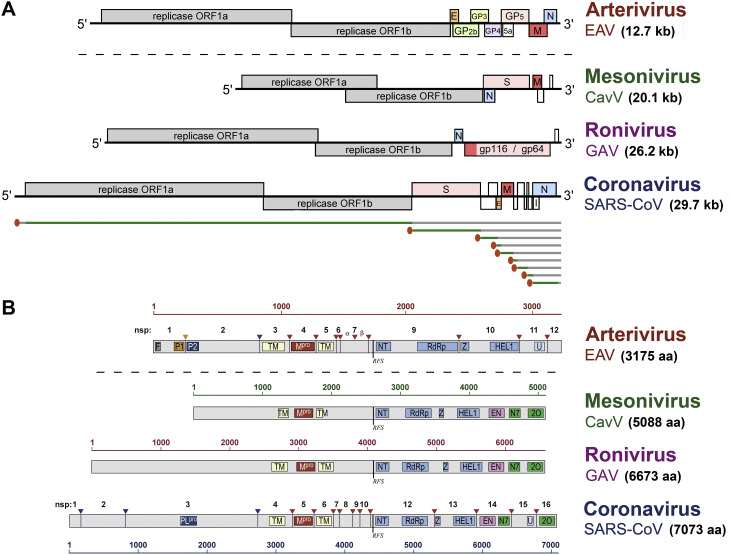
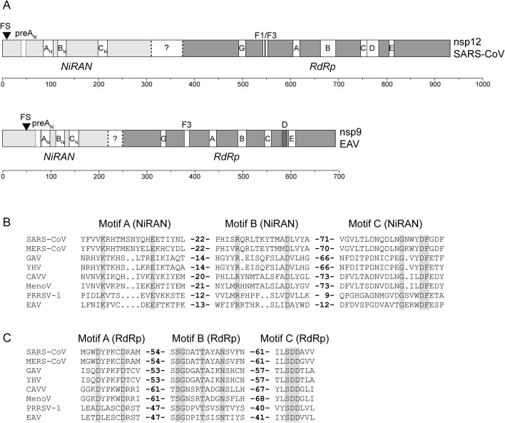
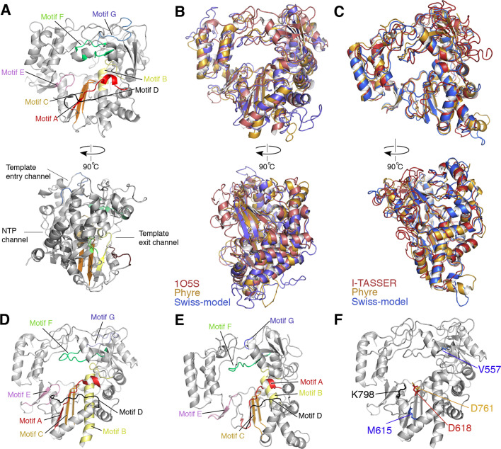
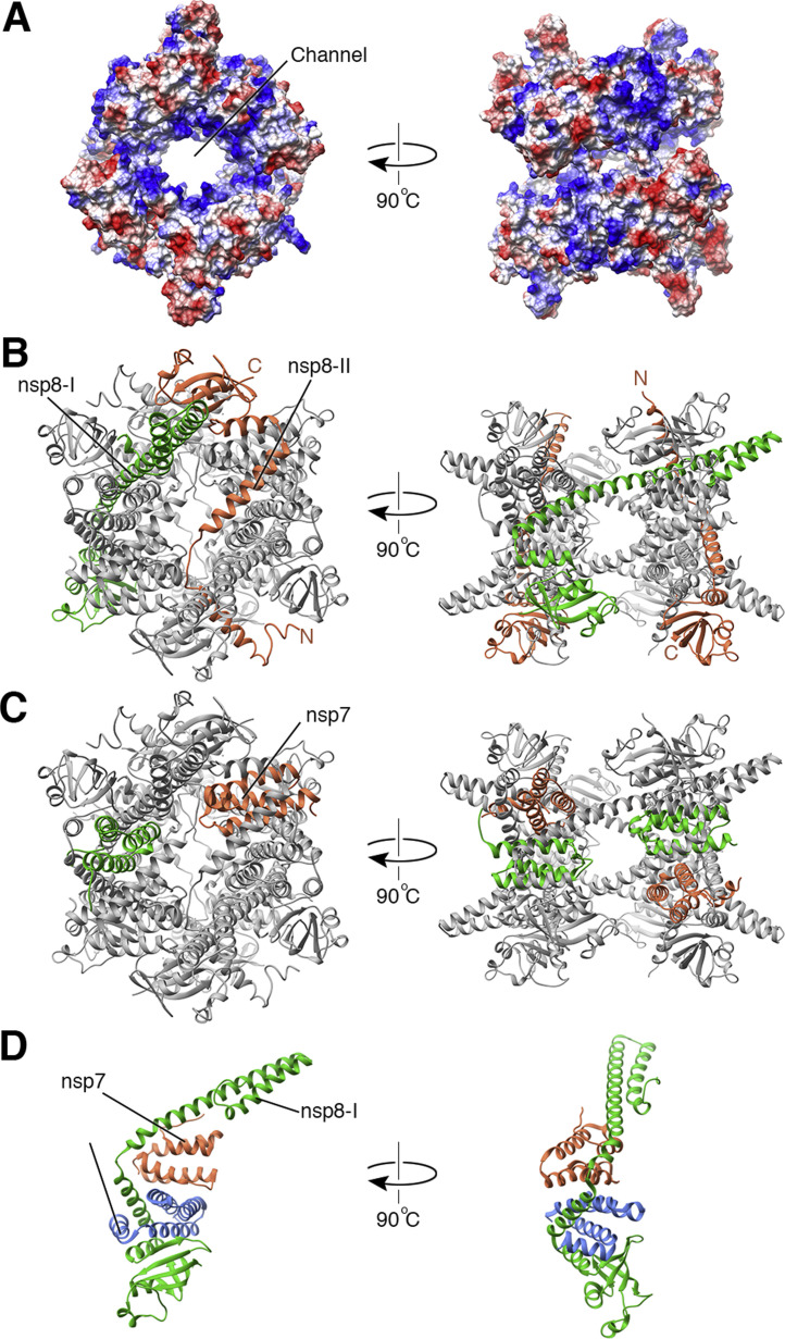
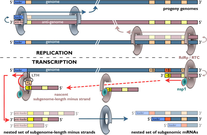
Coronaviruses and arteriviruses are distantly related human and animal pathogens that belong to the order Nidovirales. Nidoviruses are characterized by their polycistronic plus-stranded RNA genome, the production of subgenomic mRNAs and the conservation of a specific array of replicase domains, including key RNA-synthesizing enzymes. Coronaviruses (26-34 kilobases) have the largest known RNA genomes and their replication presumably requires a processive RNA-dependent RNA polymerase (RdRp) and enzymatic functions that suppress the consequences of the typically high error rate of viral RdRps. The arteriviruses have significantly smaller genomes and form an intriguing package with the coronaviruses to analyse viral RdRp evolution and function. The RdRp domain of nidoviruses resides in a cleavage product of the replicase polyprotein named non-structural protein (nsp) 12 in coronaviruses and nsp9 in arteriviruses. In all nidoviruses, the C-terminal RdRp domain is linked to a conserved N-terminal domain, which has been coined NiRAN (nidovirus RdRp-associated nucleotidyl transferase). Although no structural information is available, the functional characterization of the nidovirus RdRp and the larger enzyme complex of which it is part, has progressed significantly over the past decade. In coronaviruses several smaller, non-enzymatic nsps were characterized that direct RdRp function, while a 3'-to-5' exoribonuclease activity in nsp14 was implicated in fidelity. In arteriviruses, the nsp1 subunit was found to maintain the balance between genome replication and subgenomic mRNA production. Understanding RdRp behaviour and interactions during RNA synthesis and subsequent processing will be key to rationalising the evolutionary success of nidoviruses and the development of antiviral strategies.

Keywords: Arterivirus; Coronavirus; Polymerase fidelity; Processivity factors; Replication and transcription complex; Subgenomic mRNA synthesis.


Figures

Fig. 1
Fig. 1
Nidovirus genome organization and the core replicase domains. (A) Genome organization of representatives of the four major nidovirus lineages (CavV, Cavally virus; GAV, gill-associated virus; note that the EAV genome is drawn to a different scale). For each genome, the known open reading frames (ORFs) are indicated with the replicase ORFs 1a and 1b depicted in grey and structural protein genes depicted in different colours. ORFs encoding ‘accessory proteins’ (in SARS-CoV) or poorly characterized products are depicted in white. To illustrate the principle of subgenomic mRNA synthesis, as employed by all nidoviruses, the nested set structure and composition of the mRNAs is summarized for SARS-CoV, with the common 5′ leader sequence indicated in red and the translated part of the genome and each of the subgenomic mRNAs depicted in green. See main text for more details. (B) Domain organization of the pp1ab replicase polyprotein for the four major nidovirus lineages (note that the AV protein is drawn to a different scale). Proteolytic cleavages and non-structural protein numbering are indicated for EAV and SARS-CoV. The scheme highlights the conservation of the so-called nidovirus ‘core replicase’, consisting of the ORF1a-encoded main protease (Mpro) flanked by two transmembrane (TM) domains, followed by the ORF1b-encoded NiRAN nucleotidyl transferase (NT), RNA polymerase (RdRp), zinc binding domain (Z) and superfamily 1 helicase (HEL1). Accessory (papain-like) protease domains and their cleavage sites are indicated for EAV and SARS-CoV (P1, P2, PLpro). The zinc-finger domain (F) in EAV nsp1 that is crucial for subgenomic mRNA synthesis (see text) is also highlighted. The C-terminal part of pp1ab encodes a number of enzymatic domains that are not strictly conserved among all nidovirus lineages: U, endoribonuclease, conserved in vertebrate nidoviruses; EN, exoribonuclease (ExoN) conserved in nidoviruses with genome sizes >20 kb (see text); N7- and 2′-O methyl transferases (N7 and 2O) involved in cap modification (not identified in AVs).
Fig. 2
Fig. 2
Schematic representation of nsp12 from SARS-CoV and nsp9 from EAV. A) The position of NiRAN (light grey), the RdRp domain (dark grey) and the respective motifs (white boxes; subscript “N” was added to NiRAN motifs to dicriminate them from RdRp motifs) are indicated in the figure. The exact C-terminal border of NiRAN, as well as the N-terminal border of the RdRp are not defined yet and are indicated with dashed lines. The position of ribosomal frameshifting is indicated with a triangle; translation of the preceding ORF1a products terminates shortly downstream the frameshift site (SARS-CoV nsp11 within 4 amino acids, EAV nsp8 within 1 amino acid, not indicated in the figure). NiRAN and RdRp motifs are displayed as white boxes in the figure, based on (Lehmann et al., 2015a) for NiRAN motifs, and (Xu et al., 2003) (SARS-CoV) or (Beerens et al., 2007) (EAV) for the RdRp motifs. Note that Motif D was not defined for EAV nsp9 in Beerens et al. and that the approximate position is indicated as a dashed box. The rulers indicate amino acid positions in the proteins. Question mark indicates part of the nsp that may represent a linking domain or a domain with an additional (unknown) function. B) Alignment of NiRAN motifs A, B and C from eight representative nidoviruses from all 4 families (Modified from (Lehmann et al., 2015a)). C) Alignment of RdRp motifs A, B and C from the same nidoviruses. Completely conserved residues are indicated in grey boxes. SARS-CoV, SARS coronavirus Frankfurt 1 (AY291315; Coronaviridae); MERS-CoV, Middle East respiratory syndrome coronavirus EMC/2012 (JX869059.2; Coronaviridae); GAV, Gill-associated virus (AF227196; Roniviridae); YHV, yellow head virus (EU487200; Roniviridae); CAVV, Cavally virus (HM746600; Mesoniviridae); MenoV, Meno virus (JQ957873; Mesoniviridae); PRRSV-1, porcine reproductive and respiratory syndrome virus, European genotype (GU737264.2; Arteriviridae). EAV, Equine arteritis virus (DQ846750; Arteriviridae).
Fig. 3
Fig. 3
Models of the nidovirus RdRps. A) Structure of the FMDV RdRp (pdb 2E9R). In the top panel, conserved polymerase motifs A-F are indicated. In the bottom panel, the template entry, template exit and NTP entry channels are indicated. B) Models of the SARS-CoV nsp12 RdRp generated by Swiss-Model and Phyre2 superposed on the model 1O5S (Xu et al., 2003). All three models are based on the C-terminal polymerase domain and excluded the N-terminal NiRAN domain. All models show an overall similar fold. Differences exist in the fingers subdomain and surface loops of the thumb subdomain. C) Superposed models of the EAV nsp9 RdRp generated by use of I-TASSER, Swiss-Model and Phyre2. Overall, all three models show a very similar fold, with small differences in the fingers subdomain. D) Motifs of the polymerase domain of SARS-CoV nsp12 indicated on the structural model 1O5S (Xu et al., 2003). E) Motifs of the polymerase domain of EAV nsp9 that was generated using Phyre2. F) Key active site and fidelity residues indicated on the model of polymerase domain of SARS-CoV nsp12 1O5S.
Fig. 4
Fig. 4
The structure of the CoV nsp7-8 complex. A) The hollow hexadecameric ring of the SARS-CoV nsp7-8 complex has a positively charged channel (blue surface shading) that is likely important for RNA binding. The outside of the hexadecamer is predominantly negatively charged (red surface shading). B) The SARS-CoV nsp8 crystal structure (pdb 2AHM) resembles a ‘golf club' with a long stick at the N-terminus (N) and a head-like shape at the C-terminus (C). The nsp8 structure can adopt two conformations, here shaded green (nsp8-I) and orange (nsp8-II). C) In the hexadecamer each of the two nsp8 structures is present four times and complemented by eight nsp7 subunits that act as mortar. Two orientations of the nsp7 subunit are indicated (orange and green). D) The FCOV heterotrimeric nsp7-8 complex (pdb 3ub0) consists of one nsp8 (shaded green) subunit and two nsp7 subunits (orange and blue). The nsp8 subunit adopts an nsp8-I conformation.
Fig. 5
Fig. 5
Nidovirus RNA synthesis. Model for replication and transcription using a hypothetical genome encoding three sg mRNAs. The top half of the scheme depicts the replication of the genome from a full-length minus-strand intermediate (antigenome). The bottom half illustrates how minus-strand RNA synthesis can be attenuated at a body TRS (+B), after which the nascent minus strand, having a body TRS complement (−B) at its 3′ end, is redirected to the leader TRS (+L) near the 5′ end of the genome. Guided by a base-pairing interaction between the −B and +L sequences, RNA synthesis is resumed to add the anti-leader sequence to each nascent subgenome-length minus strand. Subsequently, the latter serves as template to produce a sg mRNA. The RdRp complexes engaged in replication and transcription may differ, as transcription-specific regulatory protein factors, like EAV nsp1, have been described (Nedialkova et al., 2010). For further details, see text. Adapted from (Snijder and Kikkert, 2013).

References

    1. Ahn D.G., Choi J.K., Taylor D.R., Oh J.W. Biochemical characterization of a recombinant SARS coronavirus nsp12 RNA-dependent RNA polymerase capable of copying viral RNA templates. Arch. Virol. 2012;157:2095–2104. - PMC - PubMed
    1. Alphonse S., Ghose R. Cystoviral RNA-directed RNA polymerases: regulation of RNA synthesis on multiple time and length scales. Virus Res. 2017;234:135–152. - PMC - PubMed
    1. Banerjee D., Reddy K.R. Review article: safety and tolerability of direct-acting anti-viral agents in the new era of hepatitis C therapy. Aliment. Pharmacol. Ther. 2016;43:674–696. - PubMed
    1. Beaucourt S., Vignuzzi M. Ribavirin: a drug active against many viruses with multiple effects on virus replication and propagation. Molecular basis of ribavirin resistance. Curr. Opin. Virol. 2014;8:10–15. - PMC - PubMed
    1. Becares M., Pascual-Iglesias A., Nogales A., Sola I., Enjuanes L., Zuniga S. Mutagenesis of Coronavirus nsp14 reveals its potential role in modulation of the innate immune response. J. Virol. 2016;90:5399–5414. - PMC - PubMed

Publication types