Review
. 2017 Mar 6;216(3):559-570.
doi: 10.1083/jcb.201610048. Epub 2017 Feb 7.

Patterned cell and matrix dynamics in branching morphogenesis

Affiliations
Review

Patterned cell and matrix dynamics in branching morphogenesis

Shaohe Wang et al. J Cell Biol. .

Abstract

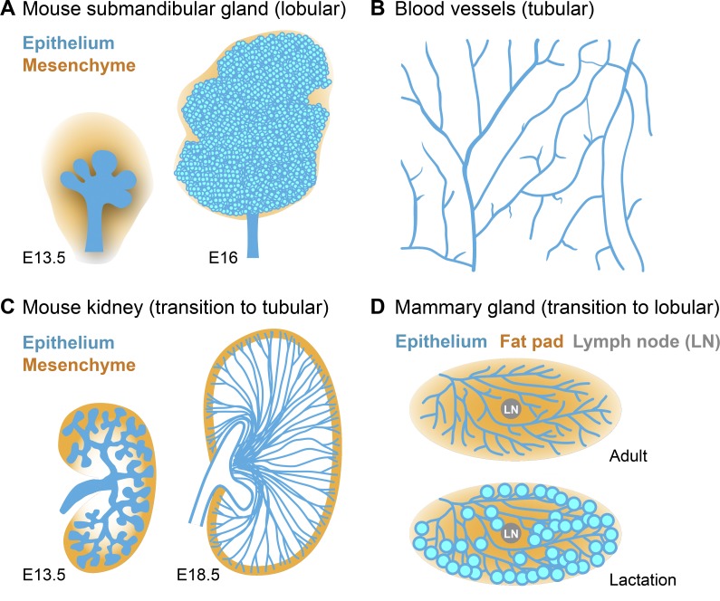
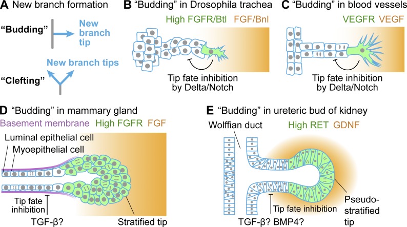
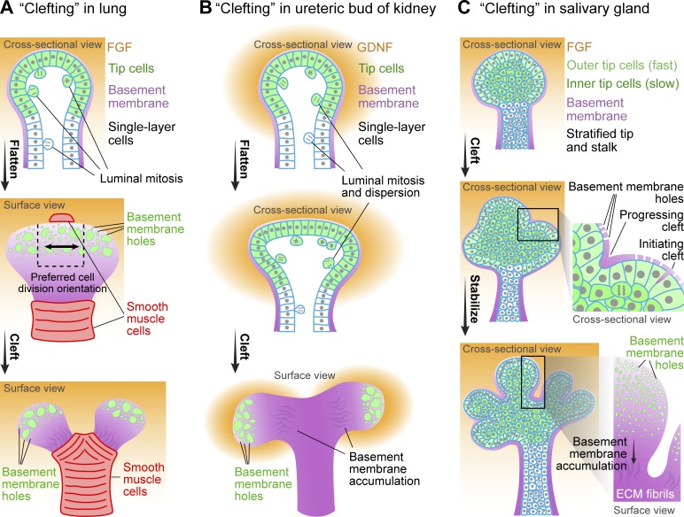
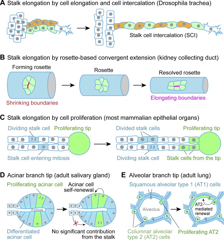
Many embryonic organs undergo branching morphogenesis to maximize their functional epithelial surface area. Branching morphogenesis requires the coordinated interplay of multiple types of cells with the extracellular matrix (ECM). During branching morphogenesis, new branches form by "budding" or "clefting." Cell migration, proliferation, rearrangement, deformation, and ECM dynamics have varied roles in driving budding versus clefting in different organs. Elongation of the newly formed branch and final maturation of the tip involve cellular mechanisms that include cell elongation, intercalation, convergent extension, proliferation, and differentiation. New methodologies such as high-resolution live imaging, tension sensors, and force-mapping techniques are providing exciting new opportunities for future research into branching morphogenesis.


Figures

Figure 1.
Figure 1.
Varying morphology of branched organs. (A) Schematics of mouse submandibular glands from embryonic day (E) 13.5 and E16 stage embryos. Submandibular gland morphology is predominantly lobular. (B) Schematic of blood vessels from a mouse retina. The branched vascular network is completely tubular. (C) Schematics of the ureteric bud of mouse kidney from E13.5 and E18.5 stage embryos. At E13.5, mouse kidney is relatively lobular. At E18.5, the elongated collecting ducts convert kidney morphology to predominantly tubular. (D) Schematics of mouse mammary gland at young adult stage and lactation stage. At the young adult stage, the mammary gland is a tubular network. During pregnancy and lactation, dramatic remodeling occurs in the mammary gland so that lactating alveoli form at branch tips, which transforms the structure to primarily lobular.
Figure 2.
Figure 2.
New branch formation by budding. (A) New branches can form through two geometrically distinct processes, budding or clefting. Schematics of budding in Drosophila trachea (B) and mouse retina blood vessels (C), where budding occurs by an invasive form of collective migration. The identity of the protrusive tip cell is specified by high RTK signaling. A Delta/Notch-mediated lateral inhibition mechanism prevents follower cells from becoming leader cells. Schematics of budding in mouse mammary gland (D) and the ureteric bud of mouse kidney (E), where budding occurs by a noninvasive form of collective migration and regionalized cell proliferation. The stratified tip of the mammary gland, or TEB, has high FGF receptor (FGFR) activity and higher proliferation rate than the stalk, which contains two cell layers. The tip of the ureteric bud at this early stage is pseudostratified, and its identity is specified by high RET signaling activity. EGFR, EGF receptor; VEGFR, VEGF receptor.
Figure 3.
Figure 3.
New branch formation by clefting. (A) Schematics of clefting (or terminal bifurcation) in mouse lung. The clefting tip of developing lung contains a single layer of cells that flattens before clefting. Within the tip, cells toward the center (dashed box in the center panel) divide preferentially parallel to the axis of flattening (black arrow in the center panel). Before clefting, smooth muscle cells differentiate at the future clefting site, which helps to deform the flattened tip to complete clefting. (B) Schematics of clefting in the ureteric bud of mouse kidney. The clefting tip of kidney contains a single layer of cells that also flattens before clefting. For cell proliferation, premitotic cells delaminate from the single-layered epithelium, complete cell division in the lumen, and then reinsert into the epithelium. (C) Schematics of clefting in mouse salivary gland. In these images, the clefting tip (bud) is stratified. The outer tip cells are more columnar and more regularly arranged than the inner tip cells. The outer tip cells also move much faster than the inner tip cells. Shallow clefts form stochastically with ECM invasion into the outer layer of epithelium, and they widen and stabilize to complete clefting. Clefting in all three systems is accompanied by microscopic perforations in the basement membrane toward the tip and accumulation of basement membrane components away from the tip. For each organ, a critically essential growth factor regulator is listed, although others contribute. GDNF, glial cell–derived neurotrophic factor.
Figure 4.
Figure 4.
Refining organ architecture by branch elongation and maturation. (A–C) Schematics of stalk elongation by cell elongation and intercalation in Drosophila trachea. (A) In response to the pulling force of the migrating tip cell, stalk cells elongate and intercalate, resulting in >2-fold increased stalk length. (B) Schematics of stalk elongation by rosette-based convergent extension in vertebrate kidney collecting duct. (C) Schematics of branch stalk elongation by cell proliferation in most mammalian epithelial organs. Some cells divide within the stalk, whereas some cells are deposited by the proliferating tip to elongate the stalk. (D) Schematics of the acinar branch tip in adult mouse salivary gland. During maturation, cells at the branch tip differentiate to become columnar cells that surround a relatively small cavity to form an acinus for saliva secretion. Homeostasis of the acinar cells is maintained by self-renewal with little contribution from the stalk. (E) Schematics of the alveolar branch tip in adult mouse lung. During maturation, lung cells at the branch tip differentiate into flat, squamous AT1 cells and columnar AT2 cells. AT2 cell–mediated self-renewal can be triggered by AT1 injury.

References

    1. Affolter M., and Caussinus E.. 2008. Tracheal branching morphogenesis in Drosophila: New insights into cell behaviour and organ architecture. Development. 135:2055–2064. 10.1242/dev.014498 - DOI - PubMed
    1. Affolter M., Zeller R., and Caussinus E.. 2009. Tissue remodelling through branching morphogenesis. Nat. Rev. Mol. Cell Biol. 10:831–842. 10.1038/nrm2797 - DOI - PubMed
    1. Akhtar N., and Streuli C.H.. 2013. An integrin-ILK-microtubule network orients cell polarity and lumen formation in glandular epithelium. Nat. Cell Biol. 15:17–27. 10.1038/ncb2646 - DOI - PMC - PubMed
    1. Akhtar N., Marlow R., Lambert E., Schatzmann F., Lowe E.T., Cheung J., Katz E., Li W., Wu C., Dedhar S., et al. . 2009. Molecular dissection of integrin signalling proteins in the control of mammary epithelial development and differentiation. Development. 136:1019–1027. 10.1242/dev.028423 - DOI - PMC - PubMed
    1. Akhtar N., Li W., Mironov A., and Streuli C.H.. 2016. Rac1 controls both the secretory function of the mammary gland and its remodeling for successive gestations. Dev. Cell. 38:522–535. 10.1016/j.devcel.2016.08.005 - DOI - PMC - PubMed

Publication types

MeSH terms

LinkOut - more resources