. 2017 Feb 21;114(8):E1500-E1508.
doi: 10.1073/pnas.1612991114. Epub 2017 Feb 7.

SGK3 sustains ERα signaling and drives acquired aromatase inhibitor resistance through maintaining endoplasmic reticulum homeostasis

Affiliations

SGK3 sustains ERα signaling and drives acquired aromatase inhibitor resistance through maintaining endoplasmic reticulum homeostasis

Yuanzhong Wang et al. Proc Natl Acad Sci U S A. .

Abstract

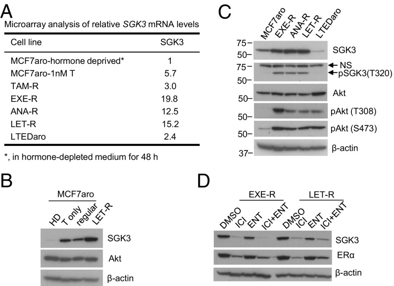
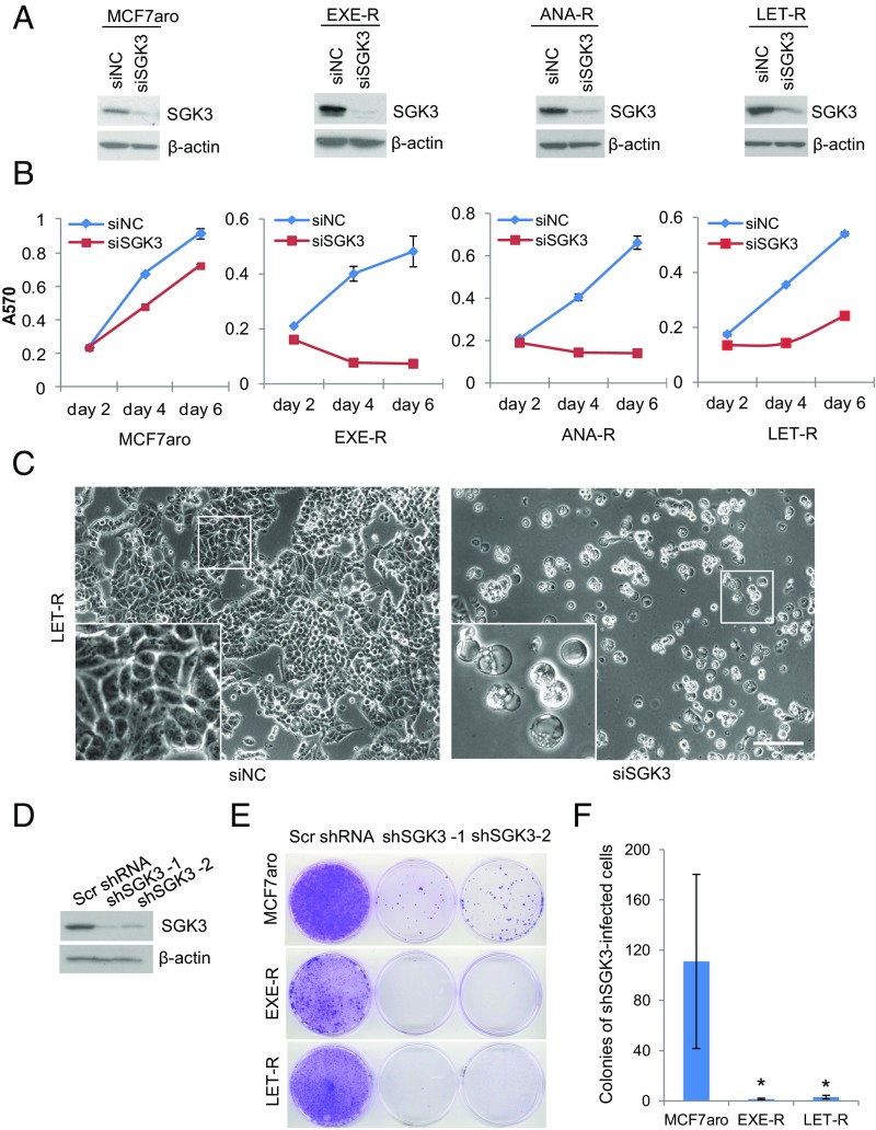
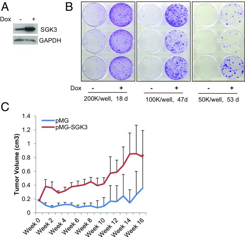
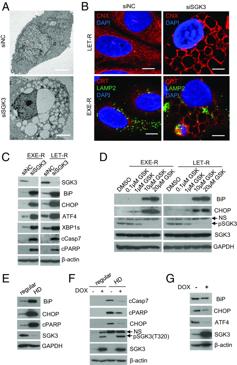
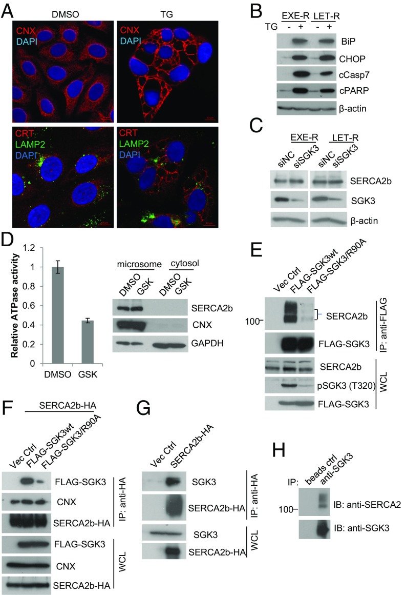
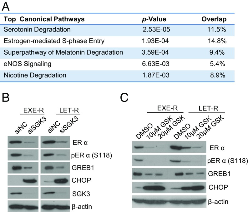
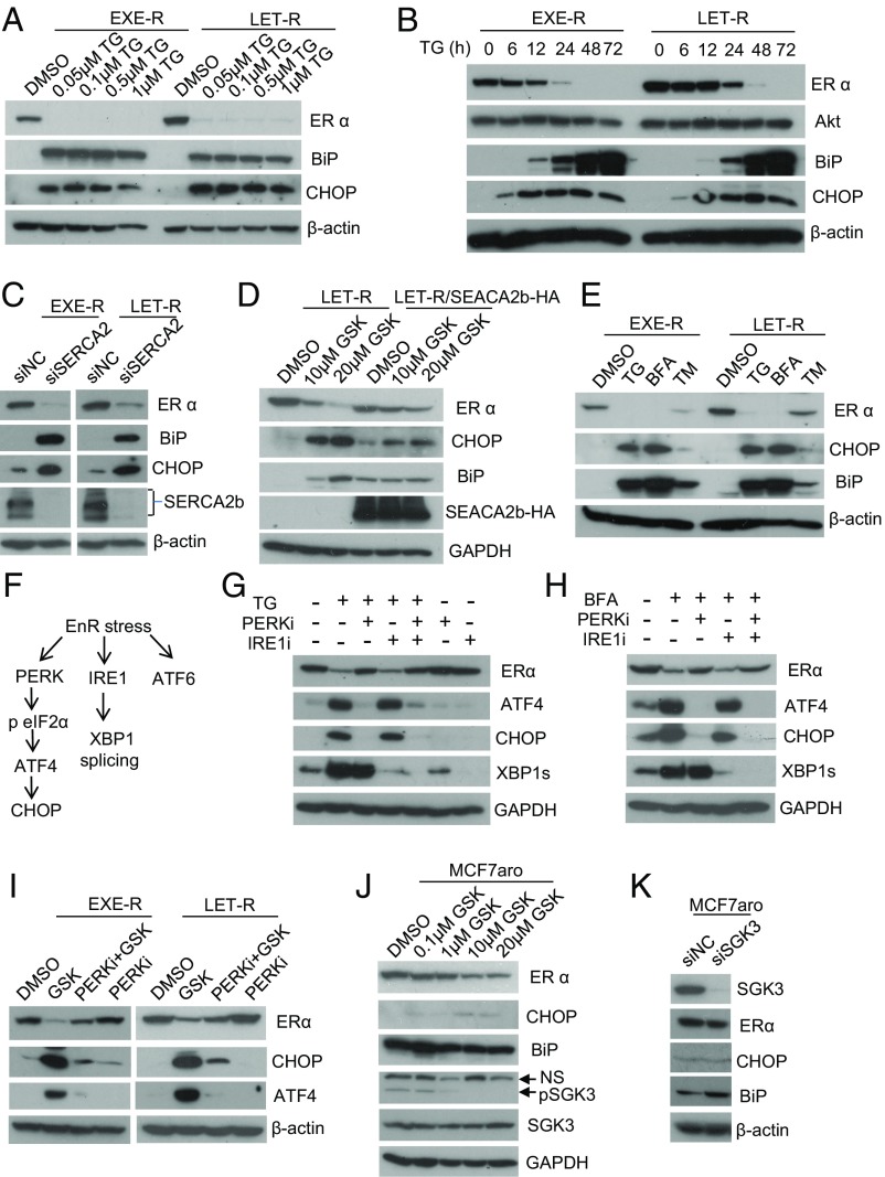
Many estrogen receptor alpha (ERα)-positive breast cancers initially respond to aromatase inhibitors (AIs), but eventually acquire resistance. Here, we report that serum- and glucocorticoid-inducible kinase 3 (SGK3), a kinase transcriptionally regulated by ERα in breast cancer, sustains ERα signaling and drives acquired AI resistance. SGK3 is up-regulated and essential for endoplasmic reticulum (EnR) homeostasis through preserving sarcoplasmic/EnR calcium ATPase 2b (SERCA2b) function in AI-resistant cells. We have further found that EnR stress response down-regulates ERα expression through the protein kinase RNA-like EnR kinase (PERK) arm, and SGK3 retains ERα expression and signaling by preventing excessive EnR stress. Our study reveals regulation of ERα expression mediated by the EnR stress response and the feed-forward regulation between SGK3 and ERα in breast cancer. Given SGK3 inhibition reduces AI-resistant cell survival by eliciting excessive EnR stress and also depletes ERα expression/function, we propose SGK3 inhibition as a potential effective treatment of acquired AI-resistant breast cancer.

Keywords: SERCA2; SGK3; aromatase inhibitor; endoplasmic reticulum stress; estrogen receptor.


Conflict of interest statement

The authors declare no conflict of interest.

Figures

Fig. 1.
Fig. 1.
SGK3 expression is up-regulated in ERα+ breast cancer cells during development of acquired AI resistance. (A) Relative SGK3 mRNA levels in different cell lines determined by microarray. MCF7aro-1nM T, MCF7aro cells were cultured in hormone-depleted medium and treated with 1 nM T for 48 h; TAM-R, tamoxifen-resistant MCF7aro cells, generated by long-term culture of MCF7aro cells in hormone-depleted medium added with T plus tamoxifen (>6 mo); LTEDaro, long-term estrogen-deprived MCF7aro cells, which were generated by long-term culture of MCF7aro cells in hormone-depleted medium (>6 mo), and estrogen was not required for proliferation of the resulting cells. (B) Western blotting analysis of MCF7aro cells cultured in different conditions and LET-R cells. HD, cultured in hormone-depleted medium for 48 h; T only, cultured in hormone-depleted medium and treated with 1 nM T for 48 h; regular, cultured in the normal growth medium [MEM medium with 10% (vol/vol) FBS]. LET-R cells were cultured in hormone-depleted medium and treated with 1 nM T plus 200 nM letrozole. (C) Western blotting analysis of MCF7aro cells, AI-resistant cells, and LTEDaro cells. All the cells were cultured in their normal growth media, as described in Materials and Methods. (D) Western blotting analysis of levels of SGK3 and ERα in EXE-R and LET-R cells after being treated with 100 nM ICI 182,780, 5 μM enzalutamide, or their combination for 48 h.
Fig. 2.
Fig. 2.
SGK3 is critical for AI-resistant cell proliferation and survival. (A) Western blotting analysis of SGK3 levels in AI-sensitive and AI-resistant cells transfected with siRNA negative control or SGK3 siRNA for 72 h. (B) The effect of SGK3 knockdown on cell proliferation/survival in MCF7aro, EXE-R, ANA-R, and LET-R cells measured by MTT. (C) Light microscopy of LET-R cells transfected with siRNA negative control or SGK3 siRNA for 4 d. (Magnification: 100×.) (Scale bar: 50 μm.) (D) Western blotting analysis of SGK3 levels in MCF7aro cells transduced with scramble shRNA or two distinct SGK3 shRNA viral vectors. (E) Effect of knockdown of SGK3 by shRNA on colony formation. MCF7aro, EXE-R, and LET-R cells were cultured in 6-cm dishes and transduced with scrambled shRNA or two distinct SGK3 shRNA lentiviral vectors, respectively. Two days after transduction, cells were treated with 5 μg/mL puromycin to eliminate untransducted cells. Once the scrambled shRNA-infected cells reached confluency, both the scrambled shRNA-transduced cells and the SGK3 shRNA-transduced cell counterpart from the same cell line were fixed and stained with crystal violet simultaneously. (F) Quantization of colonies of the SGK3 shRNA-transduced cells. Both the scrambled shRNA-transduced cells and the SGK3 shRNA-transduced cell counterpart from the same cell line were fixed and stained with crystal violet simultaneously when the former reached confluency. Colonies of the SGK3 shRNA-transduced cells were counted and analyzed. *P < 0.05, by Student t test.
Fig. 3.
Fig. 3.
SGK3 promotes AI resistance. (A) Western blotting analysis of SGK3 levels in MCF7aro/TO/SGK3 cells cultured with or without DOX. (B) Colony formation of MCF7aro/TO/SGK3 cells grown in hormone-depleted medium with T plus letrozole in the presence and absence of DOX. MCF7aro/TO/SGK3 cells seeded in three six-well plates at 200,000 cells per well, 100,000 cells per well, and 50,000 cells per well, respectively, were cultured in hormone-depleted medium with T plus letrozole and treated with or without 250 ng/mL DOX. After the indicated time, the cells were fixed and stained with crystal violet. (C) The growth curve of breast cancer xenografts derived from MC7aro/pMG or MCF7aro/pMG-SGK3 cells in ovariectomized mice inserted with T tablets and treated with anastrozole. The tumor size was measured every week.
Fig. 4.
Fig. 4.
SGK3 maintains EnR homeostasis. (A) Transmission electron microscopy of LET-R cells transfected with siRNA negative control or SGK3 siRNA for 72 h. (Magnification: 1,100×.) (Scale bar: 4 μm.) (B) Immunofluorescence microscopy of AI-resistant cells transfected with SGK3 siRNA or siRNA negative control. EXER-R and LET-R cells were transfected with siRNA negative control or SGK3 siRNA for 72 h and then fixed and immunostained with anti-calnexin (CNX) or anti-calreticulin (CRT) plus anti-LAMP2. After immunostaining, the cells were mounted in DAPI solution and imaged under a confocal microscope. DAPI stained nuclei blue. CNX and CRT were shown in red, and LAMP2 was shown in green. (Scale bar: 5 μm.) LAMP2 is a membrane protein of lysosome and late endosome, and thus serves as a marker of lysosome and later endosome. (C) Western blotting analysis of EXE-R and LET-R cells after being transfected with siRNA negative control or SGK3 siRNA for 72 h. (D) Western blotting analysis of EXE-R and LET-R cells after being treated with the increasing concentrations of GSK650394 for 48 h. (E) Western blotting analysis of MCF7aro cells cultured in regular growth medium or in hormone-depleted medium for 3 d. (F) Western blotting analysis of MCF7aro/TO/SGK3 cells grown in different conditions. MCF7aro/TO/SGK3 cells were cultured in normal growth medium or hormone-depleted medium and treated with or without 250 ng/mL DOX for 3 d. (G) Western blotting analysis of MCF7aro/TO/SGK3 cells grown in the medium with T plus letrozole in the presence or absence of 250 ng/mL DOX for 14 d.
Fig. 5.
Fig. 5.
SGK3 maintains EnR homeostasis through interaction with SERCA2b. (A) Confocal microscopy of EXE-R cells treated with 0.5 μM thapsigargin (TG) for 48 h. Cells were immunostained with anti-CNX or anti-CRT plus anti-LAMP2 and mounted in DAPI solution. DAPI stained nuclei blue. CNX and CRT were shown in red, and LAMP2 was shown in green. (Scale bar: 10 μm.) (B) Western blotting analysis of EXE-R cells and LET-R cells after being treated with 0.5 μM TG for 72 h. (C) Western blotting analysis of EXE-R and LET-R cells after being transfected with SGK3 siRNA or siRNA negative control for 72 h. (D) Effect of SGK3 inhibition on SERCA2b ATPase activity. EXE-R cells were treated with DMSO or 20 μM GSK650394 overnight and harvested for microsome isolation. Five micrograms microsomes were used for analysis of SERCA2b ATPase activity in vitro (Left). The same amount of proteins from microsomal and cytosolic fractions was loaded for Western blotting analysis (Right). (E) Immunoprecipitation analysis of the interaction between SGK3 and SERCA2b. 293T cells were transfected with empty vector or vectors expressing either FLAG-wtSGK3 or FLAG-SGK3/R90A. An equivalent amount of cell lysate was immunoprecipitated with anti-FLAG resin, and the precipitated proteins were detected by Western blotting, using the relevant antibodies. (F) 293T cells were cotransfected with SERCA2b-HA expression vector and empty vectors or vector expressing FLAG-SGK3 wt or FLAG-SGK3/R90A. The cell lysates were immunoprecipitated with anti-HA resin, and precipitated proteins were detected by Western blotting. (G) MCF7aro cells were transfected with empty vector or SERCA2b-HA expression vector for 30 h. Cell lysates were immunoprecipitated with anti-HA resin, and precipitated proteins were detected by Western blotting analysis. (H) Immunoprecipitation analysis of the association between endogenous SGK3 and SERCA2b. The same amount of EXE-R cell lysates were incubated with beads alone or beads coated with anti-SGK3, and the precipitated proteins were detected by Western blotting using anti-SERCA2 or anti-SGK3.
Fig. 6.
Fig. 6.
SGK3 retains ERα expression and signaling in AI-resistant cells. (A) The five most significantly down-regulated canonical pathways in siSGK3-transfected LET-R cells vs. siNC-transfected LET-R cells analyzed by IPA. LER-R cells were transfected with siRNA negative control or SGK3 siRNA for 72 h and then subjected to RNA-seq analysis. Differentially expressed genes from RNA-seq data (>1.5-fold change in siSGK3 cells vs. siNC cells; P < 0.05) were analyzed using IPA. (B) Western blotting analysis of EXE-R and LET-R cells after being transfected with siRNA negative control or SGK3 siRNA for 72 h. (C) Western blotting analysis of EXE-R and LET-R cells after being treated with 10- or 20 μM GSK650394 for 72 h.
Fig. 7.
Fig. 7.
SGK3 retains ERα expression through maintaining EnR homeostasis via SERCA2b. (A) Western blotting analysis of EXE-R and LET-R cells after being treated with different concentrations of TG, as indicated, for 48 h. (B) Western blotting analysis of EXE-R and LET-R cells after being treated with 0.1 μM TG for different time, as indicated. (C) Western blotting analysis of EXE-R and LET-R cells after being transfected with siRNA negative control or SERCA2b siRNA for 72 h. (D) Western blotting analysis of LET-R and LET-R/SERCA2b-HA cells after being treated with different concentrations of GSK650394, as indicated, for 48 h. (E) Western blotting analysis of EXE-R and LET-R cells after being treated with 1 μM TG, 5 μg/mL brefeldin A (BFA), and 4 μg/mL tunicamycin (TM) for 48 h. (F) Schematic diagram of the three arms of EnR stress response or UPR. (G) Western blotting analysis of MCF7aro cells treated with 0.5 μM TG alone or plus 1 μM GSK2656157 (PERK inhibitor) or 25 μM 4μ8C (IRE1 inhibitor) or two in combination for 24 h. (H) Western blotting analysis of MCF7aro cells treated with 2.5 μg/mL BFA alone or plus 1 μM GSK2656157 (PERK inhibitor) or 25 μM 4μ8C (IRE1 inhibitor) or two in combination for 24 h. (I) Western blotting analysis of EXE-R cells and LET-R cells after being treated with 20 μM GSK650394, 2 μM PERK inhibitor (GSK2656157), alone or in combination for 24 h. (J) Western blotting analysis of MCF7aro cells treated with different concentrations of GSK650394, as indicated, for 48 h. (K) Western blotting analysis of MCF7aro cells transfected with siRNA negative control or SGK3 siRNA for 72 h.

Similar articles

Cited by

References

    1. Ma CX, Reinert T, Chmielewska I, Ellis MJ. Mechanisms of aromatase inhibitor resistance. Nat Rev Cancer. 2015;15(5):261–275. - PubMed
    1. Chen S, et al. What do we know about the mechanisms of aromatase inhibitor resistance? J Steroid Biochem Mol Biol. 2006;102(1-5):232–240. - PMC - PubMed
    1. Robertson JF, et al. Endocrine treatment options for advanced breast cancer--the role of fulvestrant. Eur J Cancer. 2005;41(3):346–356. - PubMed
    1. Ali S, Coombes RC. Endocrine-responsive breast cancer and strategies for combating resistance. Nat Rev Cancer. 2002;2(2):101–112. - PubMed
    1. Krebs J, Agellon LB, Michalak M. Ca(2+) homeostasis and endoplasmic reticulum (ER) stress: An integrated view of calcium signaling. Biochem Biophys Res Commun. 2015;460(1):114–121. - PubMed

Publication types

MeSH terms

Substances