. 2017 Mar 17;292(11):4743-4752.
doi: 10.1074/jbc.M116.757666. Epub 2017 Feb 7.

Central Regulatory Role for SIN1 in Interferon γ (IFNγ) Signaling and Generation of Biological Responses

Affiliations

Central Regulatory Role for SIN1 in Interferon γ (IFNγ) Signaling and Generation of Biological Responses

Barbara Kroczynska et al. J Biol Chem. .

Abstract

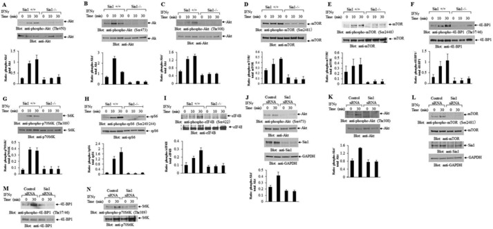
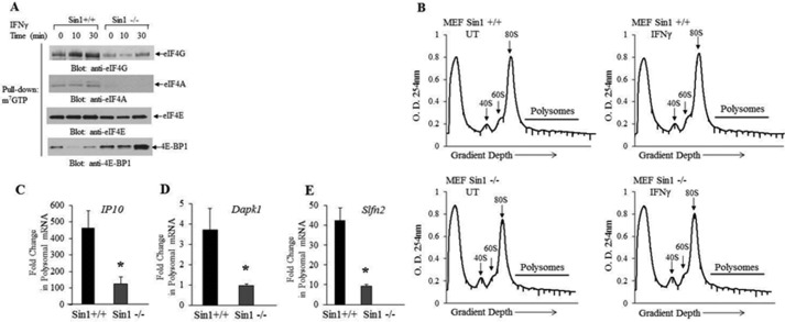
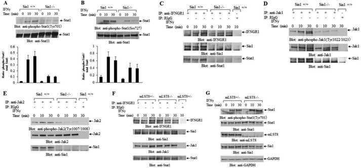
The precise signaling mechanisms by which type II IFN receptors control expression of unique genes to induce biological responses remain to be established. We provide evidence that Sin1, a known element of the mammalian target of rapamycin complex 2 (mTORC2), is required for IFNγ-induced phosphorylation and activation of AKT and that such activation mediates downstream regulation of mTORC1 and its effectors. These events play important roles in the assembly of the eukaryotic translation initiation factor 4F (eIF4F) and mRNA translation of IFN-stimulated genes. Interestingly, IFNγ-induced tyrosine phosphorylation of STAT1 is reduced in cells with targeted disruption of Sin1, leading to decreased transcription of several IFNγ-inducible genes in an mTORC2-independent manner. Additionally, our studies establish that Sin1 is essential for generation of type II IFN-dependent antiviral effects and antiproliferative responses in normal and malignant hematopoiesis. Together, our findings establish an important role for Sin1 in both transcription and translation of IFN-stimulated genes and type II IFN-mediated biological responses, involving both mTORC2-dependent and -independent functions.

Keywords: RNA editing; antiviral agent; innate immunity; interferon; signal transduction; translation control.


Conflict of interest statement

The authors declare that they have no conflicts of interest with the contents of this article

Figures

FIGURE 1.
FIGURE 1.
IFNγ-dependent engagement of mTOR effectors is Sin1-dependent. A–I, serum-starved Sin1+/+ and Sin1−/− MEFs were treated with mouse IFNγ (5 × 10³ IU/ml) for the indicated times. A–I (top panels), cell lysates were prepared, and equal amounts of protein were resolved by SDS-PAGE and then subjected to immunoblotting with the indicated specific anti-phosphoantibodies. The blots in the respective top panels were stripped and probed with anti-AKT (A–C), anti-mTOR (D and E), anti-4E-BP1 (F), anti-p70S6K (G), anti-rpS6 (H), and anti-eIF4B (I) antibodies. J–N, serum-starved U937 cells transfected with control siRNA or Sin1 siRNA were treated with human IFNγ (5 × 10³ IU/ml) for the indicated times. Cell lysates were prepared, and proteins were resolved by SDS-PAGE and then processed for immunoblotting with anti- phospho-Ser-473 AKT, AKT, Sin1, and GAPDH antibodies (J), anti-phospho-Thr-308 AKT and anti-AKT antibodies (K), antibodies against phospho-Ser-2481 mTOR, mTOR, Sin1, or GAPDH (L), anti-phospho-Thr-37/46 4E-BP1 and anti-4E-BP1 antibodies (M), and anti-phospho-Thr-389 p70S6K and anti-p70S6K antibodies (N). A–K (bottom panels), bands from three independent experiments (including the blots shown) were quantified by densitometry. Data are expressed as ratios of phosphoprotein over respective total protein values, and bar graphs represent means ± S.E. (error bars) of three independent experiments for each experimental condition.
FIGURE 2.
FIGURE 2.
Requirement of Sin1 for type II IFN-induced ISG protein expression. A and B, serum-starved Sin1+/+ and Sin1−/− MEFs were treated with mouse IFNγ (1.5 × 10³ IU/ml) as indicated. Cell lysates were prepared, and proteins were resolved by SDS-PAGE and immunoblotted with anti-IP10 (A) or anti-DAPK1 (B). C and D, serum-starved U937 cells transfected with control siRNA or Sin1 siRNA were treated with human IFNγ (1.5 × 10³ IU/ml) for the indicated times. Cell lysates were prepared, and proteins were resolved by SDS-PAGE and then processed for immunoblotting with anti-IP10 (C) and anti-DAPK1 and anti-SLFN5 (D) antibodies. A–D, anti-GAPDH antibody was used for loading control.
FIGURE 3.
FIGURE 3.
Essential role for Sin1 in mRNA translation of type II ISGs. A, serum-starved Sin1+/+ and Sin1−/− MEFs were either left untreated or treated with mouse IFNγ (5 × 10³ IU/ml) for the indicated times. Cell lysates were bound to the cap analog m7GTP biotin-labeled and streptavidin beads (39). After extensive washing, bound proteins were resolved by SDS-PAGE and immunoblotted with antibodies against eIF4G, eIF4A, eIF4E, and 4E-BP1. The cell lysates used for this experiment are from the same experiment shown in Fig. 1F. B, Sin1+/+ and Sin1−/− MEFs were either left untreated or treated with mouse IFNγ (1.5 × 10³ IU/ml) in DMEM containing 0.5% FBS for 24 h. Cell lysates were layered on 5–50% sucrose gradients and subjected to density gradient centrifugation, and fractions were collected by continuous monitoring of optical density (O.D.) at 254 nm. Optical density at 254 nm is shown as a function of gradient depth and the 40S, 60S, and 80S peaks, and polysomal fractions are indicated. C–E, polysomal fractions were pooled, and RNA was isolated. Subsequently, quantitative real-time PCR was carried out to determine Ip10 (C) Dapk1 (D), and Slfn2 (E) mRNA expression in the polysomal fractions, using Gapdh for normalization. Data are expressed as -fold change over control untreated cells, and bar graphs represent means ± S.E. (error bars) of three independent experiments, including the ones shown in B. Statistical analyses were performed using Student's t test (*, p < 0.05).
FIGURE 4.
FIGURE 4.
Essential role of Sin1 in transcription of Type II ISGs. Serum-starved Sin1+/+ or Sin1−/− MEFs were either left untreated or were treated with mouse IFNγ (2.5 × 10³ IU/ml) for 6 h, and total RNA was isolated as described under “Experimental Procedures.” mRNA expression of Dapk1 (A), Ip10 (B), and Slfn2 (C) was evaluated by quantitative real-time PCR, and Gapdh was used for normalization. Data are expressed as -fold change over control untreated cells, and bar graphs represent means ± S.E. (error bars) of three independent experiments. Statistical analyses were performed using Student's t test (*, p < 0.05).
FIGURE 5.
FIGURE 5.
IFNγ-induced tyrosine phosphorylation of STAT1 is Sin1-dependent. A and B, serum-starved Sin1+/+ and Sin1−/− MEFs were treated with mouse IFNγ (5 × 10³ IU/ml) for the indicated times. Cell lysates were prepared, and equal amounts of protein were resolved by SDS-PAGE and then subjected to immunoblotting analyses with the indicated antibodies. A and B (bottom panels), bands from three independent experiments (including the blots shown) were quantified by densitometry. Data are expressed as ratios of phospho-Stat1 over total Stat1, and bar graphs represent means ± S.E. (error bars) of three independent experiments for each experimental condition. C–E, serum-starved Sin1+/+ and Sin1−/− MEFs were treated with mouse IFNγ (5 × 10³ IU/ml) for the indicated times. C, equal amounts of protein were processed for immunoprecipitation (IP) with anti-IFNGR1 antibody or control IgG, as indicated. The immunoprecipitated proteins were resolved by SDS-PAGE and then subjected to immunoblot analyses with anti-IFNGR1, anti-Sin1, and anti-Stat1 antibodies. D and E, equal amounts of protein were processed for immunoprecipitation with anti-Jak1 (D) or anti-Jak2 (E) and control IgG, as indicated. The immunoprecipitated proteins were resolved by SDS-PAGE and then subjected to immunoblot analyses with anti-phospho-Tyr-1022/1023 Jak1, Jak1, and Sin1 antibodies (D) or with anti-phospho-Tyr-1007/1008 Jak2, Jak2, and Sin1 antibodies (E). F, serum-starved mLST8+/− and mLST8−/− MEFs were treated with mouse IFNγ (5 × 10³ IU/ml) for the indicated times. Equal amounts of protein were processed for immunoprecipitation with anti-IFNGR1 antibody or control IgG as indicated. The immunoprecipitated proteins were resolved by SDS-PAGE and then subjected to immunoblot analyses with anti-IFNGR1, Sin1, Jak1, and Stat1 antibodies. G, the same cell lysates used in H were resolved by SDS-PAGE and then subjected to immunoblot analyses with anti-phospho-Tyr-701 Stat1, Stat1, mLST8, Sin1, and GAPDH antibodies.
FIGURE 6.
FIGURE 6.
IFNγ-induced phosphorylation of Sin1 at Thr-86 is AKT-dependent but not required for STAT1 phosphorylation. A–C, serum-starved Sin1+/+ MEFs were treated with rapamycin (20 nm) or MK-2206 (10 μm) for 30 min, followed by co-treatment with mouse IFNγ (5 × 10³ IU/ml) for the indicated times. Cell lysates were prepared, and equal amounts of protein were resolved by SDS-PAGE and then subjected to immunoblotting with the indicated specific anti-phosphoantibodies. These blots were stripped and probed with the respective anti-total antibodies.
FIGURE 7.
FIGURE 7.
Requirement of Sin1 for generation of IFNγ biological effects. A, U937 cells were transfected with either control siRNA or siRNA specifically targeting Sin1, as indicated. The cells were subsequently plated in methylcellulose, in the absence or presence of human IFNγ, and leukemic CFU-L colony formation was assessed. Data are expressed as percentage of colony formation of untreated control siRNA-transfected cells, and bar graphs represent means ± S.E. (error bars) of three independent experiments. Statistical significance was calculated using one-way ANOVA followed by Tukey's test: *, p < 0.05. B, normal human CD34+ bone marrow-derived cells were transfected with control siRNA or Sin1 siRNA and incubated in methylcellulose in the presence or absence of human IFNγ, as indicated. CFU-GM and BFU-E progenitor colonies were scored after 14 days in culture. Data are expressed as percentage of colony formation of untreated control siRNA-transfected cells, and bar graphs represent means ± S.E. of three independent experiments. Statistical significance was calculated using one-way ANOVA followed by Tukey's test: *, p < 0.05. C, Sin1+/+ and Sin1−/− MEFs were seeded in wells of a 96-well plate in DMEM. 24 h later, the cells were either left untreated or treated with the indicated doses of mouse IFNγ for 16 h and subsequently challenged with EMCV (0.02 MOI). Virus-induced CPE were quantified 24 h later, as described under “Experimental Procedures.” Data are expressed as percentage protection from EMCV CPE and are representative of three independent experiments. Values are means ± S.E. of quadruplicate assays.

Similar articles

Cited by

References

    1. Stark G. R., Kerr I. M., Williams B. R., Silverman R. H., and Schreiber R. D. (1998) How cells respond to interferons. Annu. Rev. Biochem. 67, 227–264 - PubMed
    1. Borden E. C., Sen G. C., Uze G., Silverman R. H., Ransohoff R. M., Foster G. R., and Stark G. R. (2007) Interferons at age 50: past, current and future impact on biomedicine. Nat. Rev. Drug Discov. 6, 975–990 - PMC - PubMed
    1. Donnelly R. P., and Kotenko S. V. (2010) Interferon-λ: a new addition to an old family. J. Interferon Cytokine Res. 30, 555–564 - PMC - PubMed
    1. González-Navajas J. M., Lee J., David M., and Raz E. (2012) Immunomodulatory functions of type I interferons. Nat. Rev. Immunol. 12, 125–135 - PMC - PubMed
    1. Hall J. C., and Rosen A. (2010) Type I interferons: crucial participants in disease amplification in autoimmunity. Nat. Rev. Rheumatol. 6, 40–49 - PMC - PubMed

Publication types

Substances