. 2017 Feb 7;8(1):e02267-16.
doi: 10.1128/mBio.02267-16.

ATP-Dependent Persister Formation in Escherichia coli

Affiliations

ATP-Dependent Persister Formation in Escherichia coli

Yue Shan et al. mBio. .

Abstract

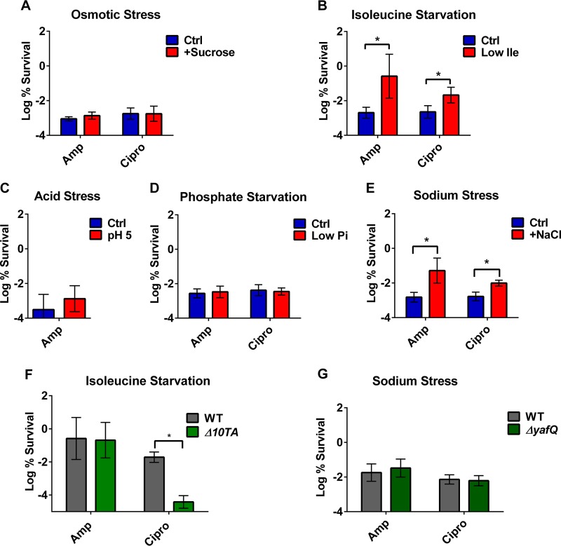
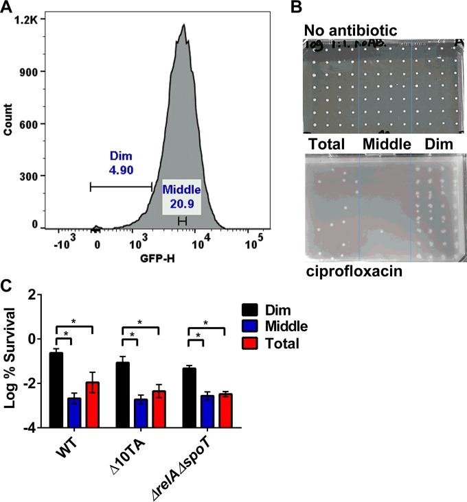
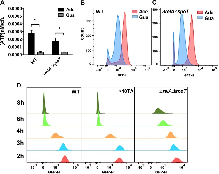
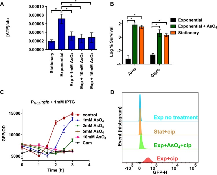
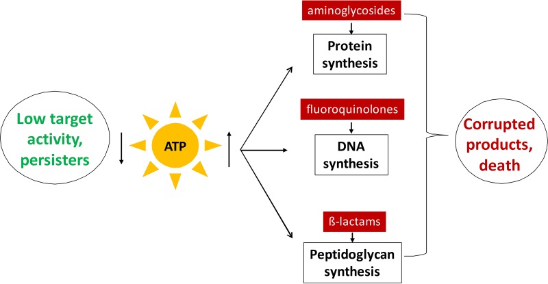
Persisters are dormant variants that form a subpopulation of cells tolerant to antibiotics. Persisters are largely responsible for the recalcitrance of chronic infections to therapy. In Escherichia coli, one widely accepted model of persister formation holds that stochastic accumulation of ppGpp causes activation of the Lon protease that degrades antitoxins; active toxins then inhibit translation, resulting in dormant, drug-tolerant persisters. We found that various stresses induce toxin-antitoxin (TA) expression but that induction of TAs does not necessarily increase persisters. The 16S rRNA promoter rrnB P1 was proposed to be a persister reporter and an indicator of toxin activation regulated by ppGpp. Using fluorescence-activated cell sorting (FACS), we confirmed the enrichment for persisters in the fraction of rrnB P1-gfp dim cells; however, this is independent of toxin-antitoxins. rrnB P1 is coregulated by ppGpp and ATP. We show that rrnB P1 can report persisters in a relA/spoT deletion background, suggesting that rrnB P1 is a persister marker responding to ATP. Consistent with this finding, decreasing the level of ATP by arsenate treatment causes drug tolerance. Lowering ATP slows translation and prevents the formation of DNA double-strand breaks upon fluoroquinolone treatment. We conclude that variation in ATP levels leads to persister formation by decreasing the activity of antibiotic targets.

Importance: Persisters are a subpopulation of antibiotic-tolerant cells responsible for the recalcitrance of chronic infections. Our current understanding of persister formation is primarily based on studies of E. coli The activation of toxin-antitoxin systems by ppGpp has become a widely accepted model for persister formation. In this study, we found that stress-induced activation of mRNA interferase-type toxins does not necessarily cause persister formation. We also found that the persister marker rrnB P1 reports persister cells because it detects a drop in cellular ATP levels. Consistent with this, lowering the ATP level decreases antibiotic target activity and, thus, leads to persister formation. We conclude that stochastic variation in ATP is the main mechanism of persister formation. A decrease in ATP provides a satisfactory explanation for the drug tolerance of persisters, since bactericidal antibiotics act by corrupting energy-dependent targets.


Figures

FIG 1
FIG 1
Upregulation of TA modules plays a limited role in persister formation. (A to E) MG1655 (wild type [WT]) was grown under stress as described in Materials and Methods to approximately the same starting density as the control. Cultures were then challenged with ciprofloxacin (0.5 μg/ml) or ampicillin (100 μg/ml) for 4 h (A, C, D, E) or 5 h (B). (F, G) Strains with deletions of toxins showing increased expression under sodium stress or isoleucine starvation were tested for persister formation. MG1655 (WT) and isogenic mutant strains were grown to the same cell density under the indicated stress and then challenged with ampicillin (100 μg/ml) or ciprofloxacin (0.5 μg/ml) for 5 h (F) or 4 h (G). Results are expressed as percent survival by comparison to untreated culture prior to the addition of antibiotic. Data are the average results from at least two independent experiments performed with three biological replicates (n ≥ 6). An asterisk indicates a significant difference (P < 0.05) by two-tailed Student’s t test. Error bars represent standard deviations.
FIG 2
FIG 2
rrnB P1 promoter activity correlates with persisters independently of mRNA interferases and ppGpp. (A) Exponentially growing cells of MG1655-ASV carrying an rrnB P1::gfpunstable transcription fusion were exposed to 1-µg/ml ciprofloxacin for 4 h. The antibiotic-treated cells were then analyzed by FACS. The dim (5%), middle (20%), and total (100%) fractions of the population were isolated by cell sorting. Cells from the dim, middle, and total fractions of the population were sorted onto agar plates, and the persisters were quantified by CFU. (B) A representative plate image after sorting. (Top) One cell was sorted onto each spot from an exponentially growing culture without antibiotic challenge. (Bottom) One thousand cells were sorted onto each spot from a ciprofloxacin-challenged culture. (C) MG1655-ASV (WT), isogenic Δ10TA, and ΔrelA ΔspoT cultures were exposed to 1-µg/ml ciprofloxacin for 4 h and underwent FACS analysis and cell sorting. The percent survival for each fraction was determined by comparing the CFU count with the total number of sorted cells. Data are the average results from at least two independent experiments performed with three biological replicates (n ≥ 6). Error bars represent standard deviations. An asterisk indicates a significant difference (P < 0.05) by two-tailed Student’s t test.
FIG 3
FIG 3
rrnB P1 senses ATP level independently of ppGpp and is repressed upon entrance into stationary phase. (A) Overnight cultures of ASV ΔpurE (WT) and an isogenic ASV ΔpurE ΔrelA ΔspoTrelA ΔspoT) mutant were diluted 1:100 into MOPS minimal medium supplemented with 0.2% glucose, 0.2% Casamino acid, and 10-μg/ml thiamine and with either adenine or guanine (0.2 mM), as indicated. Cells were grown for 2 h for the WT and 3 h for the ΔrelA ΔspoT mutant before measuring ATP level and GFP fluorescence. Data are the average results from two independent experiments performed with three biological replicates (n = 6). Error bars represent standard deviations. An asterisk indicates a significant difference (P < 0.05) by two-tailed Student’s t test. (B, C) GFP fluorescence was analyzed by FACS to determine the transcription level of rrnB P1. (D) Stationary-phase E. coli MG1655-ASV (WT) and isogenic Δ10TA and ΔrelA ΔspoT cultures were diluted 1:100 into LB medium. At each time point, GFP fluorescence was analyzed by FACS to determine the transcription activity of rrnB P1.
FIG 4
FIG 4
Drop in intracellular ATP level leads to increased persister formation through lowering target activity. (A) ATP levels were measured in stationary and exponentially growing MG1655 (WT). Cells were treated with arsenate for 30 min where indicated. (B) MG1655 culture was grown in LB medium to either exponential phase or stationary phase and then treated with 10 mM arsenate (+AsO4) for 30 min where indicated and challenged with ampicillin or ciprofloxacin for 4 h. Results are expressed as percent survival by comparison to untreated culture prior to the addition of antibiotic. Data are the average results from at least two independent experiments performed with three biological replicates (n ≥ 6). Error bars represent standard errors. An asterisk indicates a significant difference (P < 0.05) by two-tailed Student’s t test. (C) E. coli MG1655 strain harboring a plasmid-borne PlacZ::gfp fusion was grown to exponential phase. Cultures were pretreated with different concentrations of arsenate or chloramphenicol (Cam) for 30 min. Isopropyl-β-d-thiogalactopyranoside (IPTG) (1 mM) was then added where indicated to induce expression of GFP from the lacZ promoter (time zero). GFP fluorescence (excitation at 485 nm and emission at 528 nm) was measured every 30 min. Data points are the average results from the experiment performed in triplicate (n = 3). Error bars represent standard deviations. (D) TUNEL assay for DNA fragmentation measurement. E. coli MG1655 strain was grown to either exponential phase or stationary phase, and cultures were pretreated with arsenate for 30 min where indicated. Cells were then treated with 1-μg/ml ciprofloxacin for 3 h. No ciprofloxacin was added to the no-treatment control. Cells were then used for TUNEL assay analysis. Free 3′-end DNA was labeled with FITC-dUTP, and FITC intensity was measured by FACS.
FIG 5
FIG 5
Proposed model for persister formation. Antibiotics kill by corrupting active cellular processes that require ATP. A decrease in ATP diminishes the target activity and leads to antibiotic tolerance.

Comment in

Similar articles

Cited by

References

    1. Conlon BP. 2014. Staphylococcus aureus chronic and relapsing infections: evidence of a role for persister cells: an investigation of persister cells, their formation and their role in S. aureus disease. Bioessays 36:991–996. doi:10.1002/bies.201400080. - DOI - PubMed
    1. Vuong C, Voyich JM, Fischer ER, Braughton KR, Whitney AR, DeLeo FR, Otto M. 2004. Polysaccharide intercellular adhesin (PIA) protects Staphylococcus epidermidis against major components of the human innate immune system. Cell Microbiol 6:269–275. doi:10.1046/j.1462-5822.2004.00367.x. - DOI - PubMed
    1. Leid JG, Willson CJ, Shirtliff ME, Hassett DJ, Parsek MR, Jeffers AK. 2005. The exopolysaccharide alginate protects Pseudomonas aeruginosa biofilm bacteria from IFN-gamma-mediated macrophage killing. J Immunol 175:7512–7518. doi:10.4049/jimmunol.175.11.7512. - DOI - PubMed
    1. Lewis K. 2010. Persister cells. Annu Rev Microbiol 64:357–372. doi:10.1146/annurev.micro.112408.134306. - DOI - PubMed
    1. Keren I, Wu Y, Inocencio J, Mulcahy LR, Lewis K. 2013. Killing by bactericidal antibiotics does not depend on reactive oxygen species. Science 339:1213–1216. doi:10.1126/science.1232688. - DOI - PubMed

MeSH terms

Substances