. 2016 Jun 25;2(6):796-810.
doi: 10.1016/j.jcmgh.2016.06.003. eCollection 2016 Nov.

Hypoxia Positively Regulates the Expression of pH-Sensing G-Protein-Coupled Receptor OGR1 (GPR68)

Affiliations

Hypoxia Positively Regulates the Expression of pH-Sensing G-Protein-Coupled Receptor OGR1 (GPR68)

Cheryl de Vallière et al. Cell Mol Gastroenterol Hepatol. .

Abstract

Background & aims: A novel family of proton-sensing G-protein-coupled receptors, including ovarian cancer G-protein-coupled receptor 1 (OGR1) (GPR68) has been identified to play a role in pH homeostasis. Hypoxia is known to change tissue pH as a result of anaerobic glucose metabolism through the stabilization of hypoxia-inducible factor-1α. We investigated how hypoxia regulates the expression of OGR1 in the intestinal mucosa and associated cells.

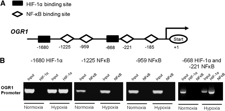
Methods: OGR1 expression in murine tumors, human colonic tissue, and myeloid cells was determined by quantitative reverse-transcription polymerase chain reaction. The influence of hypoxia on OGR1 expression was studied in monocytes/macrophages and intestinal mucosa of inflammatory bowel disease (IBD) patients. Changes in OGR1 expression in MonoMac6 (MM6) cells under hypoxia were determined upon stimulation with tumor necrosis factor (TNF), in the presence or absence of nuclear factor-κB (NF-κB) inhibitors. To study the molecular mechanisms involved, chromatin immunoprecipitation analysis of the OGR1 promoter was performed.

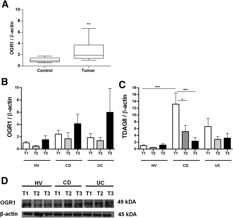
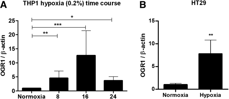
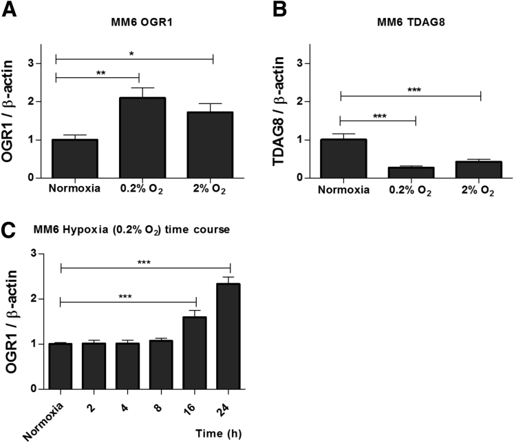
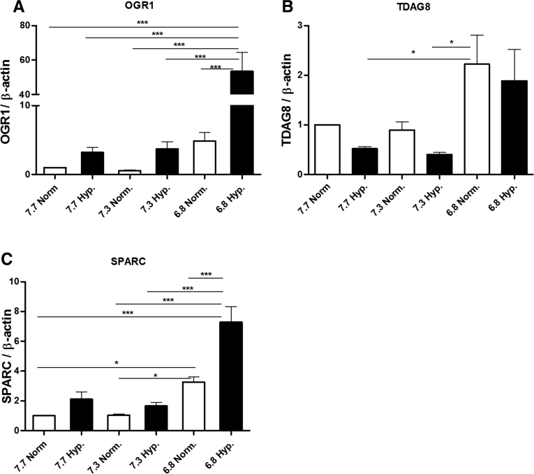
Results: OGR1 expression was significantly higher in tumor tissue compared with normal murine colon tissue. Hypoxia positively regulated the expression of OGR1 in MM6 cells, mouse peritoneal macrophages, primary human intestinal macrophages, and colonic tissue from IBD patients. In MM6 cells, hypoxia-enhanced TNF-induced OGR1 expression was reversed by inhibition of NF-κB. In addition to the effect of TNF and hypoxia, OGR1 expression was increased further at low pH. Chromatin immunoprecipitation analysis showed that HIF-1α, but not NF-κB, binds to the promoter of OGR1 under hypoxia.

Conclusions: The enhancement of TNF- and hypoxia-induced OGR1 expression under low pH points to a positive feed-forward regulation of OGR1 activity in acidic conditions, and supports a role for OGR1 in the pathogenesis of IBD.

Keywords: AICAR, 5-aminoimidazole-4-carboxamide-1-β-4-ribofuranoside; CD, Crohn's disease; ChIP, chromatin immunoprecipitation; FCS, fetal calf serum; GPR, G-protein–coupled receptor; GRP65; HIF, hypoxia-inducible factor; HV, healthy volunteer; IBD, inflammatory bowel disease; IEC, intestinal epithelial cell; IFN, interferon; IL, interleukin; Inflammation; Inflammatory Bowel Disease; MM6, MonoMac 6; NF-κB, nuclear factor-κB; OGR1, ovarian cancer G-protein–coupled receptor 1 (GPR68); Ovarian Cancer G-Protein–Coupled Receptor; RT-qPCR, quantitative reverse-transcription polymerase chain reaction; SPARC, secreted protein acidic and rich in cysteine; TDAG8; TDAG8, T-cell death-associated gene 8 (GPR65); TNF, tumor necrosis factor; Th, T-helper; UC, ulcerative colitis; WT, wild type; mRNA, messenger RNA.


Figures

Figure 1
Figure 1
OGR1 expression increases in tumor and under hypoxia. (A) OGR1 expression increases in murine tumors. To induce colon carcinomas, mice were injected intraperitoneally with a single dose of azoxymethane (7.4 mg/kg), followed by 3 cycles of 3% dextran sodium sulfate in drinking water for 1 week and normal drinking water for 2 weeks. Mice were killed 10 days after the last cycle for colonic tumor collection. OGR1 mRNA expression levels were significantly higher (P = .0220) in murine colonic tumors compared with the normal colonic mucosa. Groups: control (n = 7), tumor group (n = 7). (B and C) Hypoxia induces a tendency to increase OGR1 mRNA expression and significantly decreases TDAG8 mRNA expression in the colon of IBD patients. HVs (n = 10), CD patients (n = 11), and UC patients (n = 9) were subjected to hypoxic conditions in a hypobaric chamber resembling an altitude of 4000 m for 3 hours. Distal colon biopsy specimens were taken the day before entering the hypobaric chamber (T1), immediately after hypoxia (T2), and 1 week after the first biopsy (T3). Total RNA was isolated, reverse-transcribed, and hypoxia-induced changes in gene expression were analyzed using RT-qPCR. (B) Although not significant, OGR1 mRNA expression after hypoxia showed an increasing tendency at T3 in CD and UC patients when compared with HVs. (C) Conversely, mRNA levels of TDAG8 were reduced significantly at T1 and T3 in CD patients subjected to hypoxia. Expression changes were calculated relative to samples taken at T1 after normalizing with human β-actin endogenous control. (D) Immunoblotting analysis of OGR1 in colon biopsy specimens from HVs, CD patients, and UC patients as described earlier. β-actin was used as loading control. Results represent means ± SEM. Statistical analysis was performed using 1-way analysis of variance followed by the Tukey test (*P < .05, **P < .01, ***P < .001).
Figure 2
Figure 2
Hypoxia induces the mRNA expression of OGR1 in cultured human IECs and monocytes. (A and B) HT29 and THP1 cells were incubated under hypoxic conditions (0.2% O2) for 8, 16, and 24 hours. Total RNA was isolated, reverse-transcribed, and hypoxia-induced changes in gene expression were analyzed using RT-qPCR. Hypoxia significantly induced the mRNA expression of OGR1. Results represent means ± SEM of 2 independent experiments. Statistical analysis was performed using the Student t test (n = 5; *P < .05, **P < .01, ***P < .001).
Figure 3
Figure 3
Expression of pH-sensing receptors OGR1 and TDAG8 changed under hypoxic conditions. MonoMac 6 cells were exposed to quite severe hypoxia (0.2% O2) or modest hypoxia (2% O2) for 18 hours. (A) OGR1 expression increased 2-fold and 1.7-fold at 0.2% and 2% O2, respectively. (B) Conversely, TDAG8 mRNA expression decreased 0.7- and 0.6-fold at 0.2% and 2% O2, respectively. (C) OGR1 mRNA expression remained unchanged from 0 to 8 hours, but increased 1.6- and 2.3-fold after 16 and 24 hours, respectively. Each figure is representative of at least 3 independent experiments. Results are expressed as means ± SD. *P < .05, **P < .01, and ***P < .001 using 1-way analysis of variance followed by the Tukey test.
Figure 4
Figure 4
Expression of pH-sensing receptors OGR1 and TDAG8 under hypoxic conditions was influenced further by pH in MM6 cells. MonoMac 6 cells were exposed to hypoxia (0.2% O2) for 24 hours at pH 7.7, pH 7.3, and pH 6.8. Under hypoxic conditions: (A) OGR1 mRNA expression increased at acidic pH compared with expression at pH 7.7 or pH 7.3, (B) TDAG8 mRNA expression decreased in all conditions under hypoxia (ns), and (C) SPARC mRNA expression was increased significantly under hypoxia at pH 6.8. Results represent means ± SEM of 5 independent experiments. Asterisks denote significant differences from the respective control: *P < .05, and ***P < .001.
Figure 5
Figure 5
Expression of pH-sensing receptors OGR1 and TDAG8 under hypoxic conditions was influenced by pH in peritoneal macrophages from WT mice but not from Ogr1-/-mice. Mature quiescent peritoneal macrophages from Ogr1-/- and WT mice were isolated and cultured at different pH values (7.7, 7.3, and 6.8) under normoxia or hypoxia (0.2% O2) for 24 hours. mRNA isolation was performed and the expression of several genes was measured by RT-qPCR. (A) OGR1 mRNA expression was significantly higher in hypoxia at acidic pH. (B) TDAG8 mRNA expression was significantly higher in hypoxia at acidic pH values in WT cells. (C and D) The mRNA expression of proinflammatory cytokines, IL6, and TNF was significantly higher in hypoxia at pH 6.8, with an approximately 2-fold increase in WT cells. (E) At acidic pH under hypoxia expression of SPARC was not increased. Results are expressed as means ± SEM (n = 3). ***P < .001 using 1-way analysis of variance followed by the Tukey test.
Figure 6
Figure 6
Expression of pH-sensing receptors OGR1 and TDAG8 under hypoxic conditions was influenced further by pH in human intestinal macrophages. Intestinal macrophages from healthy mucosa of carcinoma patients were isolated and cultured at different pH values (7.7, 7.3, and 6.8) under normoxia or hypoxia (0.2% O2) for 24 hours. mRNA isolation was performed and the expression of several genes was measured by RT-qPCR. (A) OGR1 mRNA expression was significantly higher in hypoxia at acidic pH. (B) TDAG8 mRNA expression was reduced in hypoxia in all pH levels tested. (C–E) The mRNA expression of proinflammatory cytokines, IL6, IL8, and TNF was significantly higher in hypoxia at pH 6.8. (F) No differences in the mRNA expression of SPARC. Results are expressed as means ± SEM (n = 5). *P < .05, **P < .01, and ***P < .001 using 1-way analysis of variance followed by the Tukey test.
Figure 7
Figure 7
Hypoxia enhances TNF induction of OGR1 expression and was reversed by NF-κB inhibitors in MM6 cells. TNF treatment under hypoxia (0.2% O2). (A and B) After 6 hours, OGR1 mRNA expression was enhanced, but after 24 hours there was a synergistic effect by hypoxia on TNF induction. The inhibitory effect of the NF-κB inhibitor SC-514 (25 μmol/L) was less effective under hypoxia. (C) AICAR (0.05 mmol/L) showed less inhibitory effects under hypoxia. Each figure is representative of 3 independent experiments. Results are expressed as means ± SD. *P < .05, **P < .01, and ***P < .001 using 1-way analysis of variance followed by the Tukey test.
Figure 8
Figure 8
Hypoxia induces the recruitment of HIF-1α, but not NF-κB, to the OGR1 promoter. (A) Putative binding sites for HIF-1a and NF-κB were found in the OGR1 promoter using Genomatix software tools (Munich, Germany). (B) THP1 cells were incubated under hypoxic conditions (0.2% O2) for 24 hours. ChIP analysis was performed using antibodies against HIF-1α and NF-kB, for immunoprecipitation. RT-qPCR was performed using the TaqMan system with specific primers for the OGR1 promoter-binding sites of the nuclear factors HIF-1α and NF-κB. Aliquots taken before immunoprecipitation were used as input control. PCR products were run on 2% agarose gel. The results are representative of 2 independent experiments. HIF-1α, but not NF-κB, was recruited to the promoter of OGR1 in THP1 cells subjected to hypoxia for 24 hours.

References

    1. Semenza G.L. HIF-1 and mechanisms of hypoxia sensing. Curr Opin Cell Biol. 2001;13:167–171. - PubMed
    1. Semenza G.L. Oxygen sensing, homeostasis, and disease. N Engl J Med. 2011;365:537–547. - PubMed
    1. Semenza G.L. Oxygen sensing, hypoxia-inducible factors, and disease pathophysiology. Annu Rev Pathol. 2014;9:47–71. - PubMed
    1. Eltzschig H.K., Bratton D.L., Colgan S.P. Targeting hypoxia signalling for the treatment of ischaemic and inflammatory diseases. Nat Rev Drug Discov. 2014;13:852–869. - PMC - PubMed
    1. Palazon A., Goldrath A.W., Nizet V. HIF transcription factors, inflammation, and immunity. Immunity. 2014;41:518–528. - PMC - PubMed