Microglial Function during Glucose Deprivation: Inflammatory and Neuropsychiatric Implications
- PMID: 28176274
- PMCID: PMC5820372
- DOI: 10.1007/s12035-017-0422-9
Microglial Function during Glucose Deprivation: Inflammatory and Neuropsychiatric Implications
Abstract
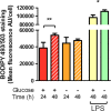
Inflammation is increasingly recognized as a contributor to the pathophysiology of neuropsychiatric disorders, including depression, anxiety disorders and autism, though the factors leading to contextually inappropriate or sustained inflammation in pathological conditions are yet to be elucidated. Microglia, as the key mediators of inflammation in the CNS, serve as likely candidates in initiating pathological inflammation and as an ideal point of therapeutic intervention. Glucose deprivation, as a component of the pathophysiology of ischemia or occurring transiently in diabetes, may serve to modify microglial function contributing to inflammatory injury. To this end, primary microglia were cultured from postnatal rat brain and subject to glucose deprivation in vitro. Microglia were characterized for their proliferation, phagocytic function and secretion of inflammatory factors, and tested for their capacity to respond to a potent inflammatory stimulus. In the absence of glucose, microglia remained capable of proliferation, phagocytosis and inflammatory activation and showed increased release of inflammatory factors after presentation of an inflammatory stimulus. Glucose-deprived microglia demonstrated increased phagocytic activity and decreased accumulation of lipids in lipid droplets over a 48-h timecourse, suggesting they may use scavenged lipids as a key alternate energy source during metabolic stress. In the present manuscript, we present novel findings that glucose deprivation may sensitize microglial release of inflammatory mediators and prime microglial functions for both survival and inflammatory roles, which may contribute to psychiatric comorbidities of ischemia, diabetes and/or metabolic disorder.
Keywords: Depression; Diabetes; Hypoglycemia; Inflammation; Ischemia; Microglia.
Conflict of interest statement
Conflict of Interest
The authors declare that they have no competing interests.
Figures


Similar articles
-
Rosuvastatin enhances anti-inflammatory and inhibits pro-inflammatory functions in cultured microglial cells.Neuroscience. 2016 Feb 9;314:47-63. doi: 10.1016/j.neuroscience.2015.11.053. Epub 2015 Nov 26. Neuroscience. 2016. PMID: 26633263
-
β-Hydroxybutyrate Improves the Redox Status, Cytokine Production and Phagocytic Potency of Glucose-Deprived HMC3 Human Microglia-like Cells.J Neuroimmune Pharmacol. 2024 Jul 23;19(1):35. doi: 10.1007/s11481-024-10139-5. J Neuroimmune Pharmacol. 2024. PMID: 39042253
-
Features of bilirubin-induced reactive microglia: from phagocytosis to inflammation.Neurobiol Dis. 2010 Dec;40(3):663-75. doi: 10.1016/j.nbd.2010.08.010. Epub 2010 Aug 19. Neurobiol Dis. 2010. PMID: 20727973
-
Anti-inflammatory activities of resveratrol in the brain: role of resveratrol in microglial activation.Eur J Pharmacol. 2010 Jun 25;636(1-3):1-7. doi: 10.1016/j.ejphar.2010.03.043. Epub 2010 Mar 31. Eur J Pharmacol. 2010. PMID: 20361959 Review.
-
Microglial phagocytosis and regulatory mechanisms: Key players in the pathophysiology of depression.Neuropharmacology. 2025 Jun 15;271:110383. doi: 10.1016/j.neuropharm.2025.110383. Epub 2025 Feb 22. Neuropharmacology. 2025. PMID: 39993469 Review.
Cited by
-
Metabolic Reprogramming in Gliocyte Post-cerebral Ischemia/ Reperfusion: From Pathophysiology to Therapeutic Potential.Curr Neuropharmacol. 2024;22(10):1672-1696. doi: 10.2174/1570159X22666240131121032. Curr Neuropharmacol. 2024. PMID: 38362904 Free PMC article. Review.
-
Lipid droplet accumulation in microglia and their potential roles.Lipids Health Dis. 2025 Jun 14;24(1):215. doi: 10.1186/s12944-025-02633-3. Lipids Health Dis. 2025. PMID: 40514678 Free PMC article. Review.
-
Consequences of a Metabolic Glucose-Depletion on the Survival and the Metabolism of Cultured Rat Astrocytes.Neurochem Res. 2019 Oct;44(10):2288-2300. doi: 10.1007/s11064-019-02752-1. Epub 2019 Feb 20. Neurochem Res. 2019. PMID: 30788754
-
Cytokine Signalling at the Microglial Penta-Partite Synapse.Int J Mol Sci. 2021 Dec 7;22(24):13186. doi: 10.3390/ijms222413186. Int J Mol Sci. 2021. PMID: 34947983 Free PMC article. Review.
-
Phosphorylation of Microglial IRF5 and IRF4 by IRAK4 Regulates Inflammatory Responses to Ischemia.Cells. 2021 Jan 30;10(2):276. doi: 10.3390/cells10020276. Cells. 2021. PMID: 33573200 Free PMC article.
References
Publication types
MeSH terms
LinkOut - more resources
Full Text Sources
Other Literature Sources
Medical

