. 2017 Feb 8:8:14329.
doi: 10.1038/ncomms14329.

RIP1 autophosphorylation is promoted by mitochondrial ROS and is essential for RIP3 recruitment into necrosome

Affiliations

RIP1 autophosphorylation is promoted by mitochondrial ROS and is essential for RIP3 recruitment into necrosome

Yingying Zhang et al. Nat Commun. .

Abstract

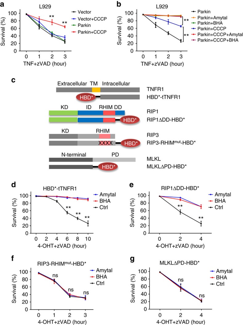
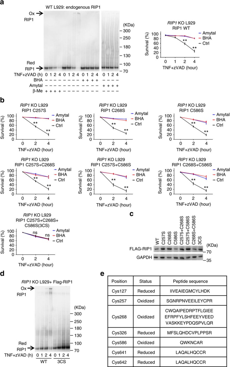
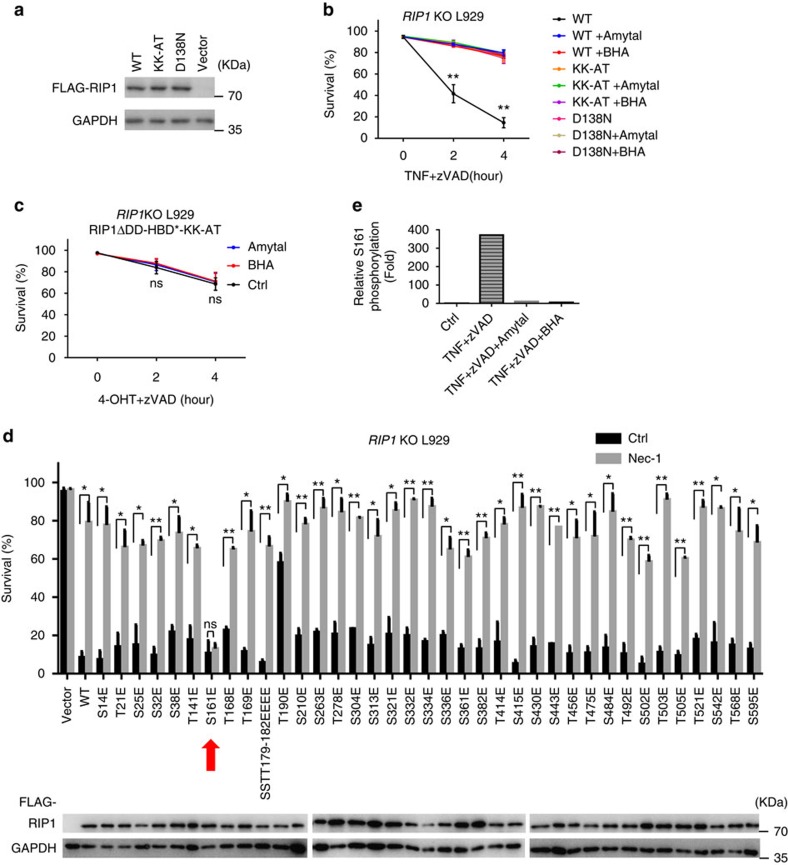
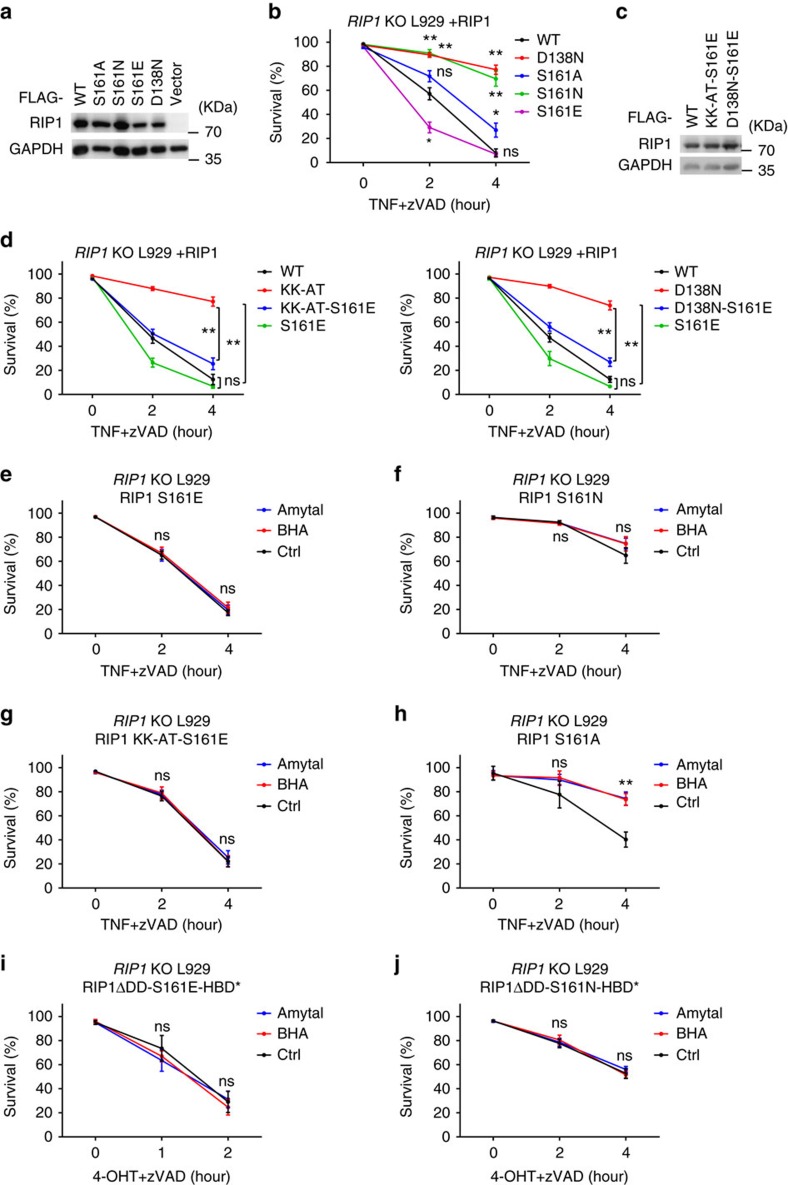
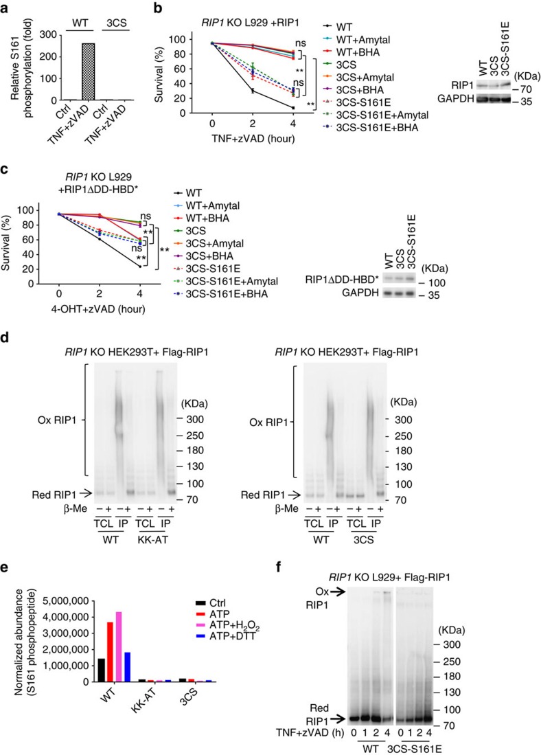
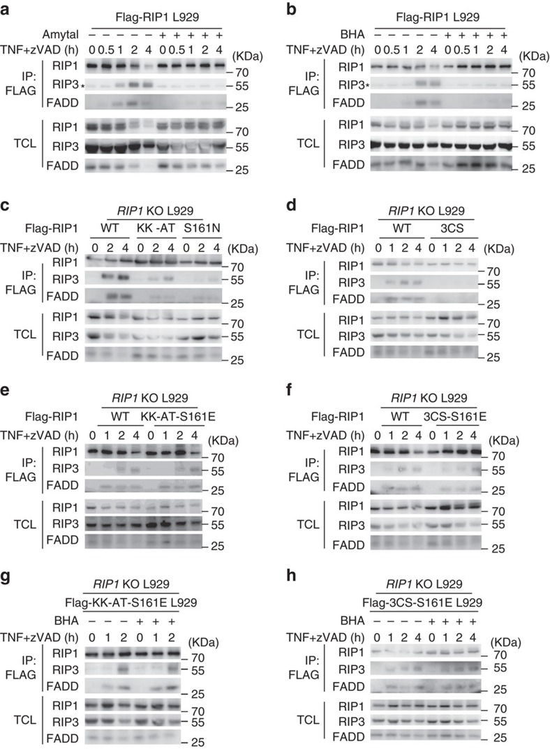
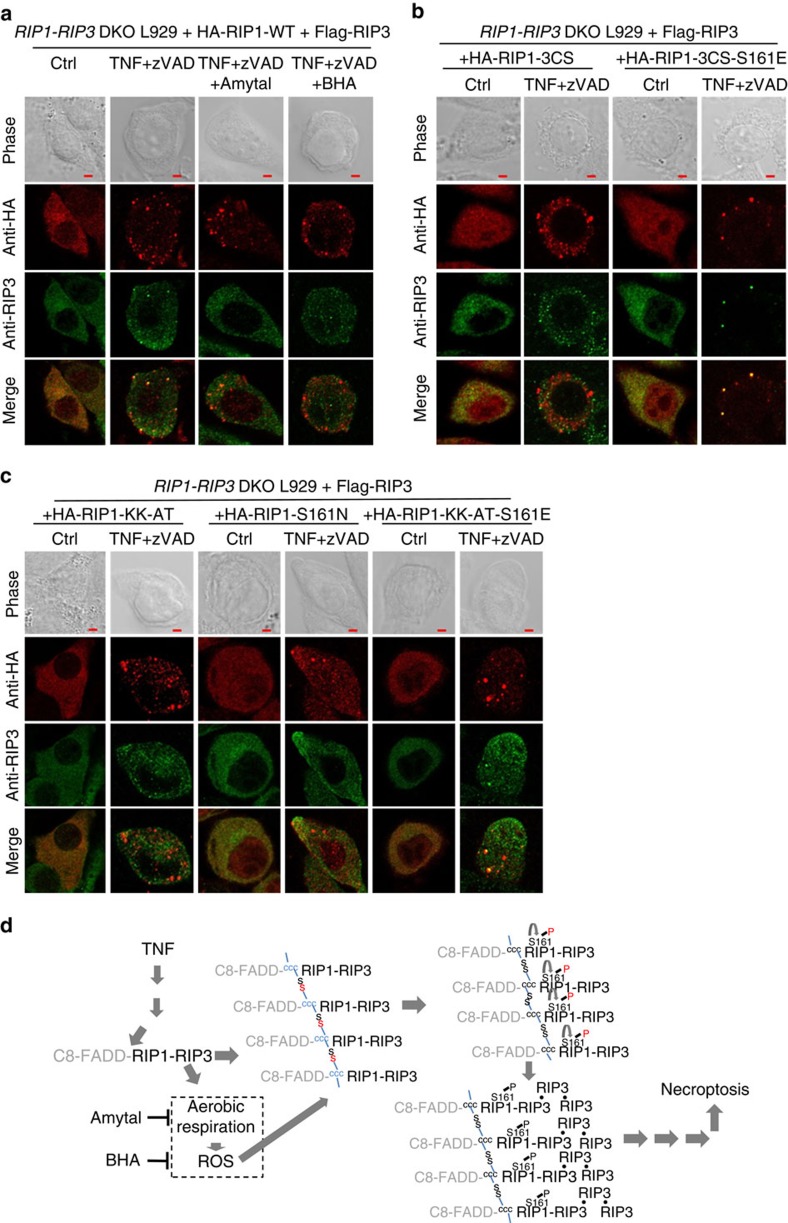
Necroptosis is a type of programmed cell death with great significance in many pathological processes. Tumour necrosis factor-α(TNF), a proinflammatory cytokine, is a prototypic trigger of necroptosis. It is known that mitochondrial reactive oxygen species (ROS) promote necroptosis, and that kinase activity of receptor interacting protein 1 (RIP1) is required for TNF-induced necroptosis. However, how ROS function and what RIP1 phosphorylates to promote necroptosis are largely unknown. Here we show that three crucial cysteines in RIP1 are required for sensing ROS, and ROS subsequently activates RIP1 autophosphorylation on serine residue 161 (S161). The major function of RIP1 kinase activity in TNF-induced necroptosis is to autophosphorylate S161. This specific phosphorylation then enables RIP1 to recruit RIP3 and form a functional necrosome, a central controller of necroptosis. Since ROS induction is known to require necrosomal RIP3, ROS therefore function in a positive feedback circuit that ensures effective induction of necroptosis.


Conflict of interest statement

The authors declare no competing financial interests.

Figures

Figure 1
Figure 1. Mitochondrial ROS target a site(s) upstream of RIP3 and downstream of RIP1 oligomerization in TNF-induced necroptosis of L929 cells.
(a) Wildtype (WT) L929 cells transfected with Flag-Parkin expression vector or empty vector were treated with CCCP (10 μM) for 48 h. These cells, together with corresponding CCCP non-treated control cells were treated with murine TNF (mTNF) +zVAD for different periods of time as indicated. Survival rate was determined by PI exclusion using flow cytometry. The final concentrations of mTNF and zVAD used in this paper were always 10 ng/ml and 20 μM, respectively, unless specially noted. (b) L929 cells expressing Flag-Parkin were treated with CCCP for 48 h. These cells, together with CCCP non-treated control, were incubated with mTNF +zVAD for different periods of time. BHA or amytal was added 1 h before TNF stimulation. Viabilities were measured with PI exclusion. The concentrations of BHA and amytal used in this paper were always 100 μM and 1 mM, respectively. (c) Schematic representation of HBD* fused tTNFR1, RIP1ΔDD, RIP3-RHIMmut and MLKLΔPD. HBD* represents the mutated version with G521R, and RHIMmut stands for RIP3 QIG449–451AAA mutation in RHIM domain. TM, KD, ID, DD are short for transmembrane, kinase domain, intermediate domain and death domain, respectively. (dg) Lentivirus encoding Flag-tagged HBD*-tTNFR1, RIP1ΔDD-HBD*, RIP3-RHIMmut-HBD* or MLKLΔPD-HBD* was packaged in HEK293T cells, and was used to infect corresponding gene KO L929 cells. These reconstituted L929 cells were then treated with 4-OHT (1 μM) +zVAD for the indicated period of time with or without BHA/amytal. Viabilities were measured with PI exclusion. Data in (a,b) and (d,g) represented the mean±s.e.m. of three independent experiments. **P<0.01; ns: no significant difference. See also Supplementary Fig. 1.
Figure 2
Figure 2. C257, C268 and C586 residues in RIP1 are oxidized in response to TNF-induced ROS in L929 cells.
(a) L929 cells were treated with mTNF+zVAD for indicated periods of time with or without BHA/amytal. Western blotting of RIP1 with anti-RIP1 antibody was performed under reducing (with β-Mercaptoethanol (β-Me)) and non-reducing (without β-Me) conditions (left panel). Cell viabilities were measured with PI exclusion (right panel). Ox: oxidized form; Red: reduced form. (b) The three cysteines, C257, C268 and C586 in RIP1 were mutated to serines individually or in combination as annotated. The Flag-tagged mutants were then expressed in RIP1 KO L929 cells by lentivirus vector for 24 h. Viabilities were measured by PI exclusion at different time points after treatment of mTNF+zVAD with/without BHA or amytal. (c) The expression levels of reconstituted WT and mutant RIP1 in RIP1 KO cells in (b) were measured by western blotting with anti-RIP1 antibody. (d) RIP1 KO cells reconstituted with Flag-RIP1 or Flag-RIP1 3CS as in (b) were treated with mTNF+zVAD for indicated periods of time. Western blotting of RIP1 was performed under non-reducing condition. (e) Oxidized RIP1 complex was isolated from non-reducing SDS-PAGE and was analysed by MS. The cysteine residues, their redox statuses and the sequences of the tryptic peptides containing those cysteines were summarized. For details see Fig. S2c. Data in (a,b) represented the mean±s.e.m. of three independent experiments. **P<0.01; ns: no significant difference. Data shown in (a,c,d) are representatives of two to three independent experiments. See also Supplementary Fig. 2.
Figure 3
Figure 3. Identification of S161 as an autophosphorylation site of RIP1 in response to ROS activation.
(a) RIP1 KO L929 cells were infected with lentivirus encoding Flag-tagged WT, KK-AT or D138N RIP1, respectively, for 24 h, and then subjected to western blotting for examination of expression level of these proteins. Anti-RIP1 and anti-GAPDH antibodies were used. (b) RIP1 KO L929 cells carrying WT, KK-AT or D138N RIP1 were treated with mTNF+zVAD for time as indicated with/without the presence of BHA/amytal. Viabilities were measured by PI exclusion. (c) RIP1 KO L929 cells reconstituted with Flag-tagged RIP1ΔDD-HBD*-KK-AT were treated with 4-OHT+zVAD for indicated periods of time with or without BHA/amytal. Viabilities were measured by PI exclusion. (d) RIP1 KO L929 cells were reconstituted with RIP1 carrying different serine/threonine mutations and stimulated with mTNF+zVAD for 4 h. Necrostatin-1 (Nec-1) was used at 30 μM to pretreat the cells for 1 h and then kept in the media till viabilities were measured by PI exclusion. RIP1 expression levels were determined by western blotting with anti-RIP1 antibody. (e) Flag-tagged RIP1 reconstituted RIP1 KO L929 cells were treated with mTNF+zVAD for 2 h with or without BHA/amytal followed by Flag immunoprecipitation and targeted MS analysis. Phosphopeptides containing S161 phosphorylation were detected and the MS2 intensities of the S161 phosphopeptide in each sample were extracted and the relative folds were calculated and shown. Data in (bd) represented the mean±s.e.m. of three and two independent experiments, respectively. *P<0.05; **P<0.01; ns: no significant difference. See also Supplementary Fig. 3.
Figure 4
Figure 4. ROS promote RIP1 autophosphorylation on S161 and this phosphorylation is essential for RIP1 to effectively transduce necroptotic signal.
(a,c) RIP1 KO L929 cells were infected with lentivirus encoding Flag-tagged RIP1 WT, D138N, S161A, S161N, S161E, KK-AT-S161E and D138N-S161E, respectively, for 24 h. Expression level of RIP1 was measured by western blotting with anti-RIP1 antibody. (b,d) Viabilities of each reconstituted cell line were measured by PI exclusion after mTNF+zVAD treatment for different periods of time. (ej) RIP1 KO L929 cells were infected with lentivirus encoding Flag-tagged RIP1 S161E, S161N, KK-AT-S161E, S161A, RIP1ΔDD-S161E-HBD* or RIP1ΔDD-S161N-HBD*, respectively, for 24 h. Viabilities of each cell line were determined at different time points under stimulation of mTNF/4-OHT+zVAD with or without BHA/amytal. Data in (b,dj) represented the mean±s.e.m. of three independent experiments. *P<0.05; **P<0.01; ns: no significant difference. See also Supplementary Fig. 4.
Figure 5
Figure 5. 3CS mutation blocks S161 phosphorylation and S161E mutation can bypass the defect of 3CS mutation in necroptosis.
(a) Flag-tagged RIP1 WT or 3CS reconstituted RIP1 KO L929 cells were treated with mTNF+zVAD for 2 h followed by Flag immunoprecipitation and targeted MS analysis. Phosphopeptides containing phosphorylated S161 were detected and the MS2 intensities of the S161 phosphopeptide in each sample were extracted and the relative folds were calculated and shown. (b) RIP1 KO L929 cells were infected with lentivirus encoding Flag-tagged WT, 3CS or 3CS-S161E RIP1 for 24 h. Viabilities were determined at different time points after mTNF+zVAD treatment with or without BHA/amytal. The RIP1 expression were determined by western blotting with anti-RIP1 antibody. (c) The same as in (b) except that the cells were infected with lentivirus encoding Flag-tagged RIP1ΔDD-HBD*, RIP1ΔDD-HBD*-3CS or RIP1ΔDD -HBD*-3CS-S161E for 24 h. Viabilities were determined at different time points after 4-OHT+zVAD treatment with or without BHA/amytal. (d) Flag-tagged WT, KK-AT or 3CS RIP1 was expressed in RIP1 KO HEK293T cells and purified by M2 beads. These proteins were analysed by western blotting with anti-RIP1 antibody under reducing and non-reducing conditions. (e) Proteins described in (d) were pre-treated with nothing, 10 μM H2O2, or 1 mM DTT for 10 min at 20 °C, and then in vitro kinase reactions of these proteins were performed by adding ATP to final concentration of 10 μM and incubated at 30 °C for 30 min. The reaction was stopped by adding TCA to 20%. The TCA precipitates were subjected to targeted MS analysis of S161 phosphorylation. The normalized abundances of S161 phosphopeptide were calculated by normalizing MS2 intensities of the S161 phosphopeptide with that of RIP1 tryptic peptide DLKPENILVDRDFHIK. (f) RIP1 KO L929 cells reconstituted with Flag-RIP1 or Flag-RIP1 3CS-S161E were treated with mTNF+zVAD for indicated periods of time. Western blotting with anti-RIP1 antibody was performed under non-reducing condition. Data in (b,c) represented the mean±s.e.m. of two independent experiments. **P<0.01; ns: no significant difference. Data shown in (d,f) are representatives of two to three independent experiments. See also Supplementary Fig. 5.
Figure 6
Figure 6. TNF-induced ROS enhance necrosome formation in a RIP1 S161 phosphorylation-dependent manner.
(a,b) Flag-RIP1 knock-in L929 cells were treated with mTNF+zVAD for the indicated time periods with/without BHA/amytal. Cell lysates were subjected to immunoprecipitation with mouse anti-Flag M2 beads and then western blotting with anti-RIP1, anti-RIP3 and anti-FADD antibodies as indicated. TCL: total cell lysate; *: non-specific band. (cf) RIP1 KO L929 cells were infected with lentivirus encoding RIP1 WT, KK-AT, S161N, 3CS, KK-AT-S161E or 3CS-S161E for 24 h, and treated and analysed as in (a). (g,h) RIP1 KO L929 cells expressing RIP1 KK-AT-S161E or 3CS-S161E were treated with mTNF+zVAD for different periods of time with or without the presence of BHA. Cells were then analysed as in (a). Data shown in (ah) are representatives of two to three independent experiments. See also Supplementary Figs 6,7.
Figure 7
Figure 7. ROS as well as S161 phosphorylation of RIP1 facilitate RIP1-RIP3 co-localization in the cells undergoing TNF-induced necroptosis.
(a) RIP1 and RIP3 DKO L929 cells reconstituted with HA-RIP1-WT and Flag-RIP3 were treated with mTNF+zVAD with or without the presence of BHA/amytal. Two hours after TNF stimulation, cells were fixed and immunostained for HA and RIP3 simultaneously and then subjected to confocal microscopy. (b,c) RIP1 and RIP3 DKO L929 cells reconstituted with Flag-RIP3 were then reconstituted with HA-RIP1 3CS, 3CS-S161E, KK-AT, S161N, KK-AT-S161E. The cells were treated with or without mTNF+zVAD for 2 h, and then fixed and immunostained for HA and RIP3 simultaneously. (d) A proposed model for how ROS lead to RIP1 autophosphorylation, which enhances TNF-induced necroptosis. Scale bar: 3 μm. The images are representatives of pictures taken from at least 10 fields. See also Supplementary Figs 6,7.

References

    1. Christofferson D. E., Li Y. & Yuan J. Control of life-or-death decisions by RIP1 kinase. Annu. Rev. Physiol. 76, 129–150 (2014). - PubMed
    1. Han J., Zhong C. Q. & Zhang D. W. Programmed necrosis: backup to and competitor with apoptosis in the immune system. Nat. Immunol. 12, 1143–1149 (2011). - PubMed
    1. Sun L. & Wang X. A new kind of cell suicide: mechanisms and functions of programmed necrosis. Trends Biochem. Sci. 39, 587–593 (2014). - PubMed
    1. Weinlich R. & Green D. R. The two faces of receptor interacting protein kinase-1. Mol. Cell 56, 469–480 (2014). - PMC - PubMed
    1. Chan F. K., Luz N. F. & Moriwaki K. Programmed necrosis in the cross talk of cell death and inflammation. Annu. Rev. Immunol. 33, 79–106 (2014). - PMC - PubMed

Publication types