. 2017 Sep;42(10):2096-2104.
doi: 10.1038/npp.2017.30. Epub 2017 Feb 8.

Disrupted Glutamatergic Transmission in Prefrontal Cortex Contributes to Behavioral Abnormality in an Animal Model of ADHD

Affiliations

Disrupted Glutamatergic Transmission in Prefrontal Cortex Contributes to Behavioral Abnormality in an Animal Model of ADHD

Jia Cheng et al. Neuropsychopharmacology. 2017 Sep.

Abstract

Spontaneously hypertensive rats (SHR) are the most widely used animal model for the study of attention deficit hyperactivity disorder (ADHD). Here we sought to reveal the neuronal circuits and molecular basis of ADHD and its potential treatment using SHR. Combined electrophysiological, biochemical, pharmacological, chemicogenetic, and behavioral approaches were utilized. We found that AMPAR-mediated synaptic transmission in pyramidal neurons of prefrontal cortex (PFC) was diminished in SHR, which was correlated with the decreased surface expression of AMPAR subunits. Administration of methylphenidate (a psychostimulant drug used to treat ADHD), which blocks dopamine transporters and norepinephrine transporters, ameliorated the behavioral deficits of adolescent SHR and restored AMPAR-mediated synaptic function. Activation of PFC pyramidal neurons with a CaMKII-driven Gq-coupled designer receptor exclusively activated by designer drug also led to the elevation of AMPAR function and the normalization of ADHD-like behaviors in SHR. These results suggest that the disrupted function of AMPARs in PFC may underlie the behavioral deficits in adolescent SHR and enhancing PFC activity could be a treatment strategy for ADHD.


Figures

Figure 1
Figure 1
AMPAR-excitatory postsynaptic current (EPSC) and AMPAR surface expression are diminished in prefrontal cortex (PFC) pyramidal neurons of SHR. (a and b) Left: Input-output curves of AMPAR-EPSCs (a) and NMDAR-EPSCs (b) evoked by a series of stimulation intensities in PFC pyramidal neurons from control (Wistar–Kyoto, WKY) rats and spontaneously hypertensive rats (SHR). *p<0.05, **p<0.01, analysis of variance (ANOVA). Right: representative EPSC traces. (c and d) Immunoblots and quantification analysis of surface (c) and total (d) protein levels of AMPAR subunits and NMDAR subunits in PFC slices from control and SHR. **p<0.01, t-test.
Figure 2
Figure 2
Methylphenidate (MPH) treatment rescues behavioral deficits in spontaneously hypertensive rat (SHR). (a) Bar graphs (mean±SEM) showing the discrimination ratio of the temporal order recognition memory (TORM) task in SHR or control (Sprague–Dawley, SD) rats treated with saline or MPH (0.5 mg/kg, i.p., 1 h before testing). **p<0.01, analysis of variance (ANOVA). (b) Bar graphs showing the number of midline crossing in locomotion test in SHR or control rats treated with saline or MPH. **p<0.01, ANOVA. (c) Dot plots showing the number of midline crossing in SHR before and after MPH treatment. **p<0.01, paired t-test. (d) Bar graphs showing the time in the center and the number of center entries in the open-field test of SHR or control rats treated with saline or MPH. *p<0.05, ANOVA.
Figure 3
Figure 3
Methylphenidate (MPH) treatment enhances AMPAR-excitatory postsynaptic current (EPSC) in prefrontal cortex (PFC) pyramidal neurons of spontaneously hypertensive rat (SHR). (a) Left: Input–output curves of AMPAR-EPSC evoked by a series of stimulation intensities in PFC pyramidal neurons from SHR or control (Sprague–Dawley, SD) rats treated with saline or MPH (0.5 mg/kg). *p<0.05, **p<0.01, analysis of variance (ANOVA), compared with SHR+MPH. Right: representative EPSC traces. (b) Left: Bar graphs (mean±SEM) showing the miniature EPSC (mEPSC) amplitude or frequency in PFC pyramidal neurons from SHR or control rats treated with saline or MPH. *p<0.05, ANOVA. Right: representative mEPSC traces.
Figure 4
Figure 4
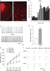
Chemicogenetic activation of prefrontal cortex (PFC) pyramidal neurons in spontaneously hypertensive rat (SHR) enhances AMPAR-excitatory postsynaptic current (EPSC). (a) Low- and high-magnification images showing hM3Dq adeno-associated virus (AAV) delivery to PFC pyramidal neurons. (b) Time course showing the frequencies of spontaneous AP (sAP) before and after bath application of clozapine N-oxide (CNO) (5 μM) in a PFC pyramidal neuron from a rat with the injection of hM3Dq AAV to medial PFC. (c) Representative sAP traces before and after CNO application. (d) Bar graphs (mean±SEM) showing sAP frequencies in the absence (−) or presence of CNO in PFC neurons from hM3Dq AAV-infected rats. ***p<0.001, t-test. (e) AMPAR-EPSC I/O in PFC pyramidal neurons (hM3Dq AAV-infected) from SHR or control (Sprague–Dawley, SD) rats injected with saline or CNO. *p<0.05, analysis of variance. f, Representative AMPAR-EPSC traces.
Figure 5
Figure 5
Chemicogenetic activation of prefrontal cortex (PFC) pyramidal neurons in spontaneously hypertensive rat (SHR) rescues behavioral deficits. (a) Bar graphs (mean±SEM) showing the discrimination ratio of the temporal order recognition memory (TORM) task in SHR or control (Sprague–Dawley, SD) rats (hM3Dq adeno-associated virus (AAV)-infected) injected with saline or clozapine N-oxide (CNO). *p<0.05, analysis of variance (ANOVA). (b) Bar graphs showing the number of midline crossing in locomotion test in SHR or control rats (hM3Dq AAV-infected) injected with saline or CNO. *p<0.05, ANOVA. (c) Bar graphs showing the time in the center and the number of center entries in the open-field test of SHR or control rats (hM3Dq AAV-infected) injected with saline or CNO. *p<0.05, ANOVA.

References

    1. Adhikari A, Topiwala MA, Gordon JA (2009). Synchronized activity between the ventral hippocampus and the medial prefrontal cortex during anxiety. Neuron 65: 257–269. - PMC - PubMed
    1. Alexander GM, Rogan SC, Abbas AI, Armbruster BN, Pei Y, Allen JA et al (2009). Remote control of neuronal activity in transgenic mice expressing evolved G protein-coupled receptors. Neuron 63: 27–39. - PMC - PubMed
    1. Arnsten AF (2009). Toward a new understanding of attention-deficit hyperactivity disorder pathophysiology: an important role for prefrontal cortex dysfunction. CNS Drugs 23: 33–41. - PubMed
    1. Arnsten AF, Rubia K (2012). Neurobiological circuits regulating attention, cognitive control, motivation, and emotion: disruptions in neurodevelopmental psychiatric disorders. J Am Acad Child Adolesc Psychiatry 51: 356–367. - PubMed
    1. Barker GR, Bird F, Alexander V, Warburton EC (2007). Recognition memory for objects, place, and temporal order: a disconnection analysis of the role of the medial prefrontal cortex and perirhinal cortex. J Neurosci 27: 2948–2957. - PMC - PubMed

MeSH terms