. 2017 Feb 8:7:41871.
doi: 10.1038/srep41871.

Unusual life cycle and impact on microfibril assembly of ADAMTS17, a secreted metalloprotease mutated in genetic eye disease

Affiliations

Unusual life cycle and impact on microfibril assembly of ADAMTS17, a secreted metalloprotease mutated in genetic eye disease

Dirk Hubmacher et al. Sci Rep. .

Abstract

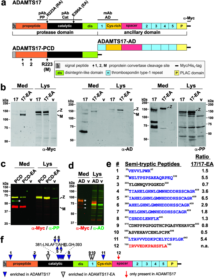
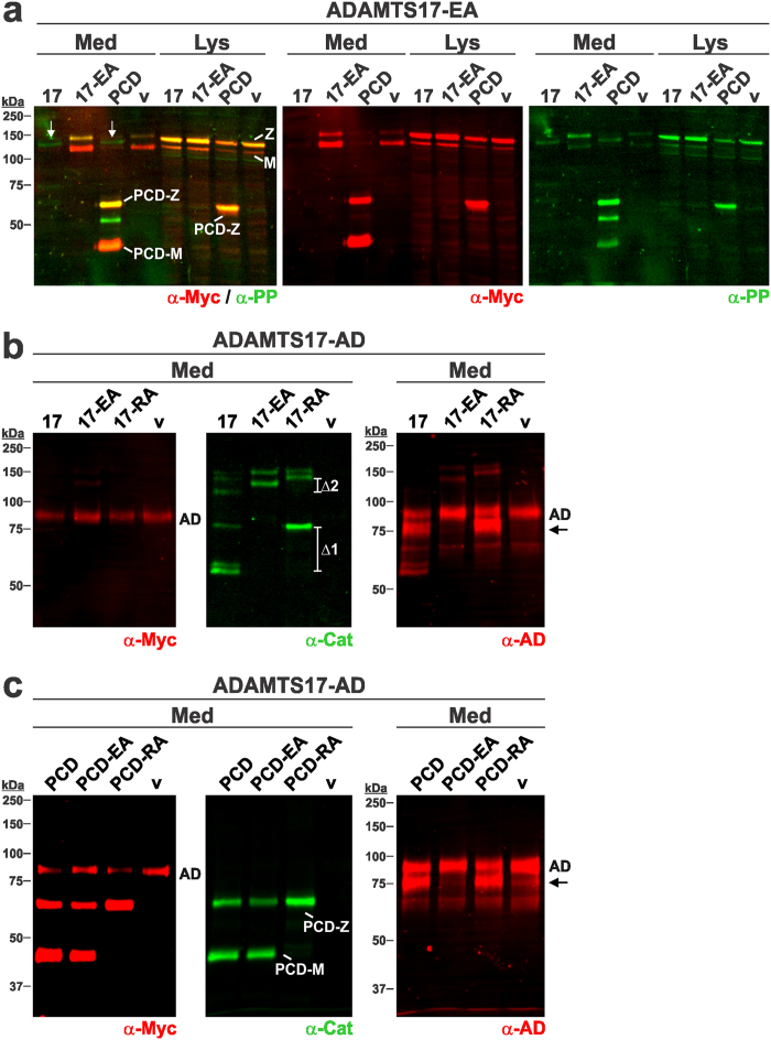
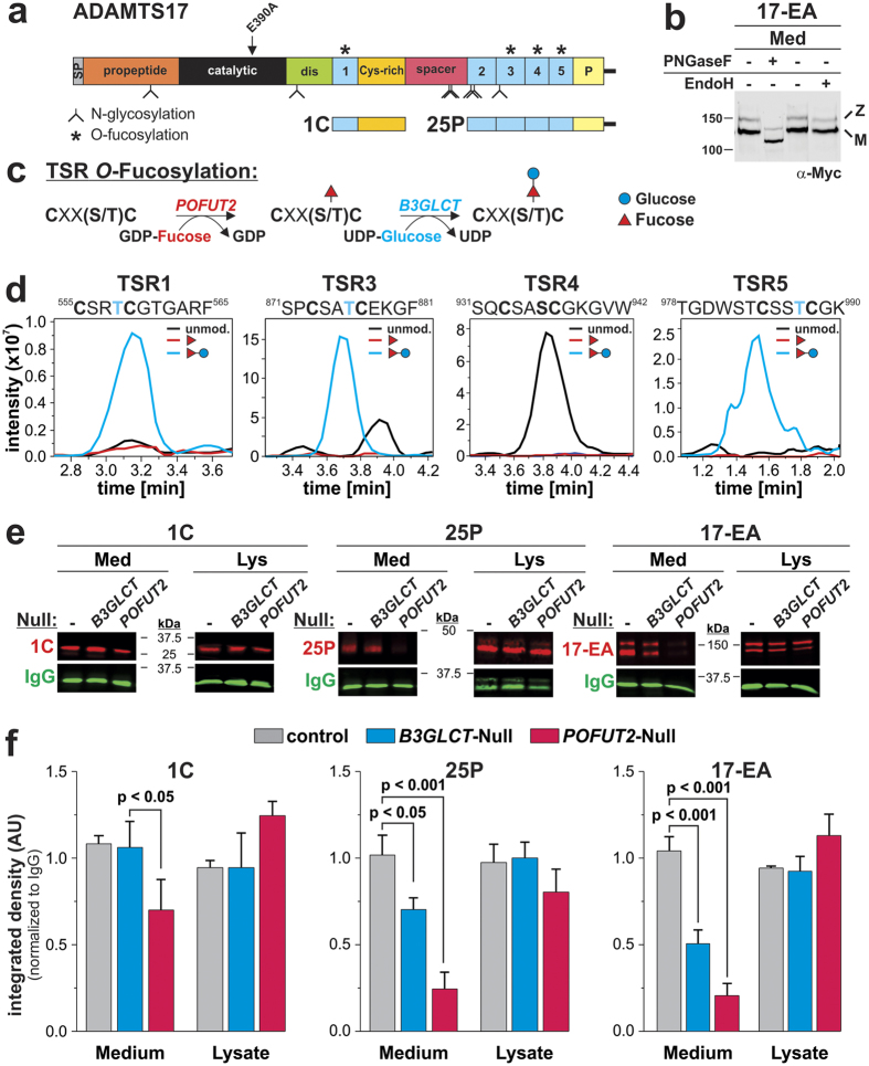
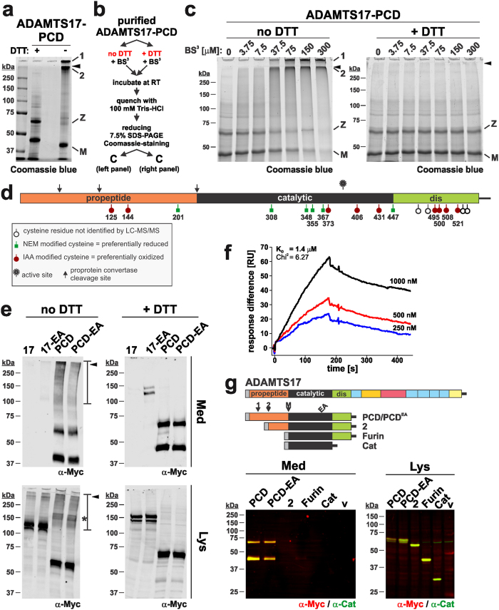
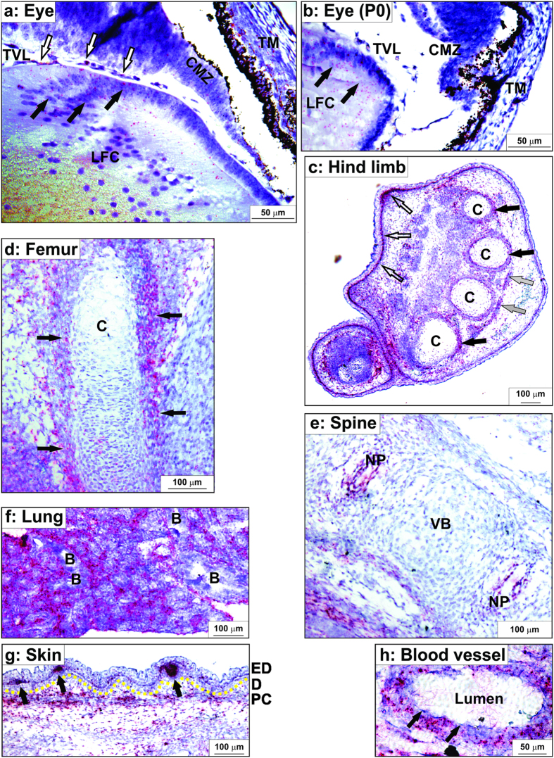
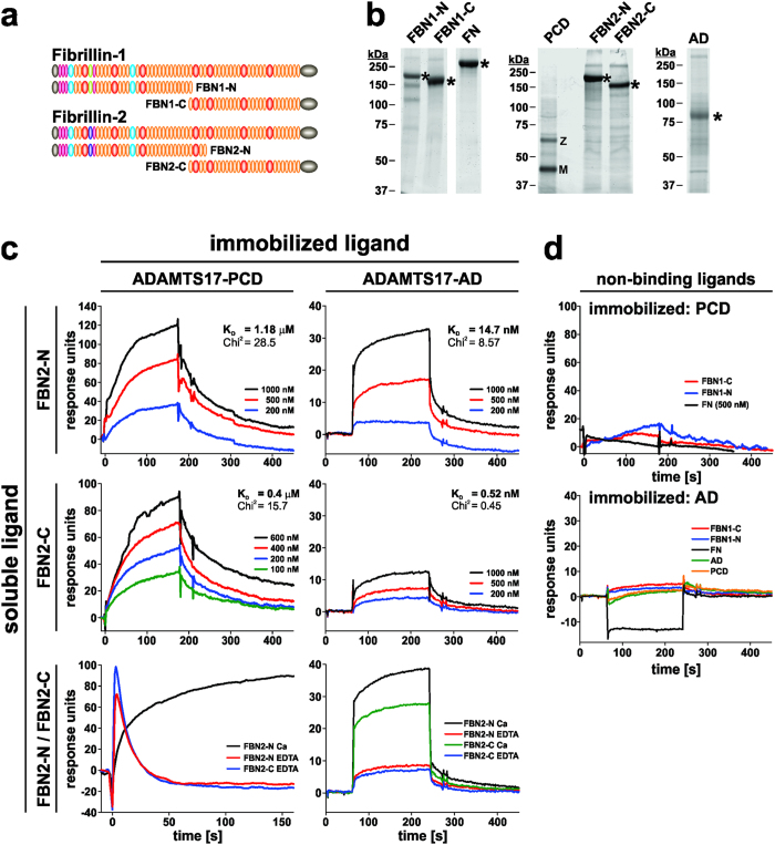
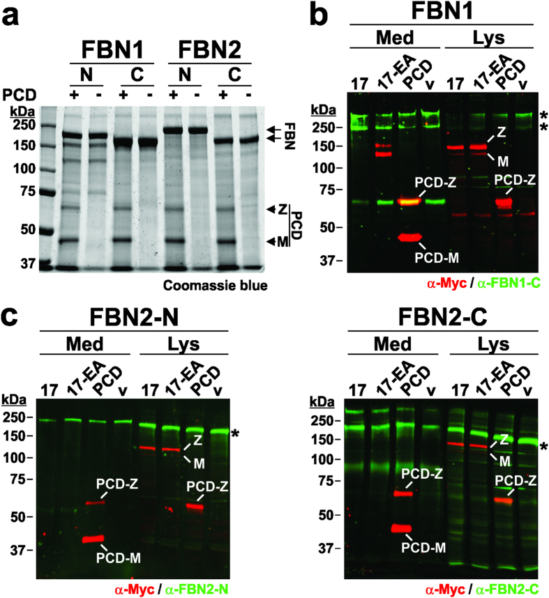
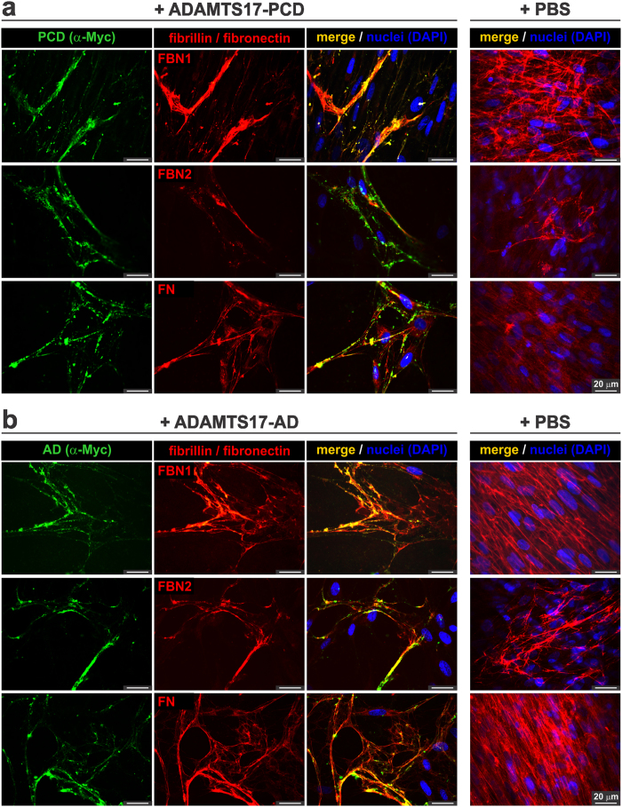
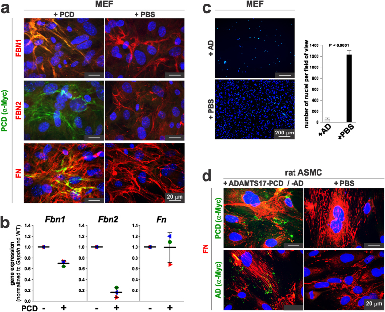
Secreted metalloproteases have diverse roles in the formation, remodeling, and the destruction of extracellular matrix. Recessive mutations in the secreted metalloprotease ADAMTS17 cause ectopia lentis and short stature in humans with Weill-Marchesani-like syndrome and primary open angle glaucoma and ectopia lentis in dogs. Little is known about this protease or its connection to fibrillin microfibrils, whose major component, fibrillin-1, is genetically associated with ectopia lentis and alterations in height. Fibrillin microfibrils form the ocular zonule and are present in the drainage apparatus of the eye. We show that recombinant ADAMTS17 has unique characteristics and an unusual life cycle. It undergoes rapid autocatalytic processing in trans after its secretion from cells. Secretion of ADAMTS17 requires O-fucosylation and its autocatalytic activity does not depend on propeptide processing by furin. ADAMTS17 binds recombinant fibrillin-2 but not fibrillin-1 and does not cleave either. It colocalizes to fibrillin-1 containing microfibrils in cultured fibroblasts and suppresses fibrillin-2 (FBN2) incorporation in microfibrils, in part by transcriptional downregulation of Fbn2 mRNA expression. RNA in situ hybridization detected Adamts17 expression in specific structures in the eye, skeleton and other organs, where it may regulate the fibrillin isoform composition of microfibrils.


Conflict of interest statement

The authors declare no competing financial interests.

Figures

Figure 1
Figure 1. ADAMTS17 undergoes autocatalytic processing.
(a) Domain organization of ADAMTS17 and the constructs ADAMTS17-PCD and ADAMTS17-AD. The location of the ADAMTS17 antibody epitopes (black line) and the sites of site-directed mutagenesis (furin-site: R223A, active site: E390A) are indicated above the ADAMTS17 domains. The predicted furin/PACE cleavage sites are shown below the ADAMTS17-PCD construct. Cleavage at site R223 results in mature (“M”) ADAMTS17. (b) Western blot analysis of conditioned medium (Med) and cell lysate (Lys) from full-length ADAMTS17 (17), the active site mutant ADAMTS17EA (17-EA), and empty vector (v) expressing HEK293F cells. Western blots were probed with the indicated antibodies and detected with enhanced chemiluminescence. (c) Western blot analysis of conditioned medium (Med) and cell lysate (Lys) from ADAMTS17-PCD (PCD), ADAMTS17-PCDEA (PCD-EA), and empty vector (v) expressing HEK293F cells. The asterisk indicates a band which is reactive with anti-propeptide antibody (anti-PP, green), but not anti-myc (red), and which is absent in ADAMTS17-PCDEA. (d) Western blot analysis of conditioned medium (Med) and cell lysate (Lys) from ADAMTS17-AD (AD) and empty vector (v) expressing HEK293F cells. The asterisk indicates intact ADAMTS17-AD (yellow), reactive with anti-myc (red) and anti-ancillary domain antibody (anti-AD, green). Arrowheads indicate species reactive with only anti-myc, but not anti-AD (red). (e) Semi-tryptic peptides resulting from ADAMTS17 autoproteolysis and identified by LC-MS/MS (blue: enriched in ADAMTS17, black: enriched in ADAMTS17EA, red: present only in wild-type ADAMTS17) (f) Location of semi-tryptic peptides in ADAMTS17. Numbers and color coding correspond to the list in e. mAb, monoclonal antibody; M, mature enzyme; pAb, polyclonal antibody; v, vector; Z, zymogen.
Figure 2
Figure 2. ADAMTS17 undergoes autocatalysis in trans that is independent of furin processing.
(a) Western blot analysis of conditioned medium (Med) and cell lysate (Lys) of HEK293F cells co-transfected with ADAMTS17EA (17-EA) and ADAMTS17 (17), ADAMTS17EA (17-EA), ADAMTS17-PCD (PCD), or empty vector (v) indicate trans-cleavage of ADAMTS17EA by ADAMTS17 and ADAMTS17-PCD. White arrows indicate lanes with cleavage products, detected with the anti-propeptide antibody (anti-PP, green). Anti-myc (red) detects zymogen and mature forms of ADAMTS17 in the cell lysates and zymogen and mature form of ADAMTS17-PCD in the cell lysate and conditioned medium. (b) Western blot analysis of conditioned medium (Med) from HEK293F cells, stably expressing ADAMTS17-AD (AD) and transiently expressing ADAMTS17 (17), ADAMTS17EA (17-EA), ADAMTS17RA (17-RA) or empty vector (v). Note reduced levels of ADAMTS17-AD in the presence of ADAMTS17 or ADAMTS17RA (left-hand panel, red) and mass shift of furin-resistant species compared to wild type (brackets indicated by Δ1 and Δ2) (middle panel, green). The proteolytic product detected with anti-AD is indicated with an arrow (right-hand panel, red) and only appears upon transfection of ADAMTS17-AD expressing cells with ADAMTS17 or ADAMTS17RA. (c) Western blot analysis of conditioned medium (Med) from HEK293F cells, stably expressing ADAMTS17-AD (AD) and transiently expressing ADAMTS17-PCD (PCD), ADAMTS17-PCDEA (PCD-EA), ADAMTS17-PCDRA (PCD-RA) or empty vector (v). The mature form of ADAMTS17-PCD (PCD-M) was not generated in ADAMTS17-PCDRA, indicating lack of furin processing (left-hand panel, red and middle panel, green). Furin processing did not abolish proteolytic activity as observed by reduction in the intensity of anti-myc reactive ADAMTS17-AD (left-hand panel, top band) or the appearance of an additional band reactive with the anti-AD antibody (arrow, right panel, red). Note that the α-Myc antibody (red) detects both ADAMTS17-PCD and ADAMTS17-AD. M, mature enzyme; Z, zymogen.
Figure 3
Figure 3. ADAMTS17 is N-glycosylated and O-fucosylated and requires O-fucosylation for secretion.
(a) Predicted N-glycosylation (branched stems) and O-fucosylation sites (asterisks) in ADAMTS17 and constructs used for LC-MS/MS and the ADAMTS17 secretion analysis. (b) Western blot analysis of ADAMTS17EA-containing medium (Med) with or without treatment with peptide-N-glycosidase F (PNGaseF) or endoglycosidase H (EndoH). M = mature enzyme; Z = zymogen. For the full-length western blot see Supplemental Fig. 5a. (c) Schematic of TSR O-fucosylation depicting the enzymes and substrates involved. (d) Extracted ion chromatograms of the ions corresponding to unmodified (black), O-fucose (red), and O-fucose-glucose (blue) glycoforms of peptides from ADAMTS17 TSRs as identified by nano-LC-MS/MS. The O-fucosylation consensus sequence of the respective TSR and the modified residue (blue) are indicated above each chromatogram. (e) Western blot analysis of medium (Med) and lysate (Lys) from HEK293T cells with inactivated B3GLCT or POFUT2 (null) and transiently transfected with ADAMTS17-1C (1C), ADAMTS17-25P (25P) and ADAMTS17EA (red). Co-transfected IgG (green) was used as the secretion control for quantification since it does not contain TSRs. For the full-length western blots see Supplemental Fig. 5b,c. (f) Quantification of the integrated densities of the respective bands normalized to IgG (n = 3). Statistical significance was calculated using a two-sided Student t-test and compared to the band intensity measured after secretion from wild-type cells.
Figure 4
Figure 4. ADAMTS17-PCD self-associates via disulfide bonds involving the propeptide.
(a) Coomassie blue stained 7.5% SDS-PAGE of purified ADAMTS17-PCD (20 μg/lane) separated under reducing (+ DTT) and non-reducing (− DTT) conditions. The black arrowhead depicts the border between the stacking and separating gel; 1 and 2 indicate large molecular weight complexes of ADAMTS17-PCD, which in the absence of DTT, did not enter the stacking or separating gel, respectively. The “+ DTT” and “− DTT” lanes are separated by an empty lane. M, mature enzyme; Z, zymogen. (b) Flow chart outlining the design of cross-linking experiments in c. (c) Coomassie blue-stained reducing SDS-PAGE of purified ADAMTS17-PCD (10 μg/lane) incubated with increasing concentrations of BS3 cross-linker in the presence (+ DTT) or absence of reducing agent (no DTT) (Annotations as in a). (d) Schematic of ADAMTS17-PCD showing cysteine residues and indicating their oxidized or reduced status determined by LC-MS/MS. (e) Western-blot analysis of conditioned medium (Med) and cell lysate (Lys) from cells expressing ADAMTS17 (17), ADAMTS17EA (17-EA), ADAMTS17-PCD (PCD), and ADAMTS17-PCDEA (PCD-EA) under reducing (+ DTT) and non-reducing conditions (no DTT). Areas of the gel in the high MW range (>100 kDa) reacting with anti-myc in the absence of DTT in medium and lysate are outlined with a bar. The asterisk indicates prominent anti-myc reactive ADAMTS17-PCD band in the lysate under non-reducing conditions. (f) Surface plasmon resonance indicates interaction of ADAMTS17-PCD to surface immobilized ADAMTS17-PCD in a dose dependent manner. The dissociation constant (KD) was calculated assuming 1:1 binding. (g) Schematic of ADAMTS17-PCD truncation mutants (top panel). Western blot analysis of conditioned medium and cell lysate from HEK293 cells transiently transfected with ADAMTS17-PCD truncation mutants indicates anti-myc and anti-catalytic domain antibody reactive bands (yellow) of the predicted molecular weight in cell lysates for each mutant (bottom, right-hand panel). However, the entire propeptide is required for secretion (bottom, left-hand panel).
Figure 5
Figure 5. Adamts17 in situ hybridization in 16.5 day-old mouse embryos or neonates showing expression in tissues relevant to Weill-Marchesani syndrome.
The RNAScope method results in red signal over cells containing Adamts17 mRNA. Sections were counterstained with hematoxylin (blue). (a,b) At embryonic day (E) 16.5 (a), Adamts17 was expressed in equatorial lens fiber cells (LFC, black arrows), capillaries of the tunica vasculosa lentis (TVL, white arrows), the non-pigmented epithelium of the ciliary margin zone (CMZ), and the trabecular meshwork cells (TM) of the eye. In neonates (b) Adamts17 continued to be expressed in LFC, but expression was reduced in the other ocular tissues. (ce) Adamts17 mRNA expression in the skeleton. In the hind limb autopod (c), Adamts17 was detected in the perichondrium around cartilage (C, black arrows), in tendon (grey arrows), and in the dermis and hair follicles (white arrows). Adamts17 was expressed in the perichondrium of the femur (d) (arrows) and in the nucleus pulposus (NP) of the intervertebral disks but not in the vertebral bodies (VB) (e). (f) Strong Adamts17 mRNA expression was found in the lung parenchyma, but not the bronchial epithelium (B). (g) Adamts17 mRNA expression in skin was localized to the hair follicles and the panniculus carnosus (PC). A yellow line marks the dermal (D) – epidermal (ED) junction. Arrows indicate developing hair follicles. (h) In blood vessels, Adamts17 was expressed in the smooth muscle cells of the blood vessel wall, but not endothelial cells (arrows).
Figure 6
Figure 6. ADAMTS17-PCD binds to fibrillin-2 but not fibrillin-1 or fibronectin.
(a) Domain structure of recombinant fibrillin-1 and fibrillin-2 peptides used to analyze interactions with ADAMTS17-PCD (PCD) or ADAMTS17-AD (AD). (b) Coomassie blue stained SDS-PAGE showing the integrity and purity of the recombinant proteins used in the interaction studies. The asterisks indicate the full-length constructs. M, mature enzyme; Z, zymogen. (c) Surface plasmon resonance shows binding of FBN2-N and FBN2-C to ADAMTS17-PCD (left-hand panels) or ADAMTS17-AD (right-hand panels). Binding was dose-dependent and Ca2+-dependent. The dissociation constant (KD) was calculated assuming 1:1 stoichiometry. (d) ADAMTS17-PCD (top panel) or ADAMTS17-AD (bottom panel) did not bind to FBN1-N, FBN1-C, or cellular fibronectin (FN) analytes and ADAMTS17-AD did not self-interact or bind to ADAMTS17-PCD.
Figure 7
Figure 7. ADAMTS17-PCD does not cleave fibrillin-1 or fibrillin-2.
(a) Coomassie-blue stained SDS-PAGE of fibrillin-1 (FBN1-N, FBN1-C) and fibrillin-2 (FBN2-N, FBN2-C) peptides (5 μg/lane) (see Fig. 6a) incubated with (+) or without (−) recombinant ADAMTS17-PCD (5 μg/lane) shows no evidence for proteolysis of fibrillin-1 or fibrillin-2 in vitro. M = mature enzyme; Z = zymogen. (b) Western blot analysis of conditioned medium (Med) and cell lysate (Lys) from HEK293F cells stably expressing fibrillin-1 and transiently expressing ADAMTS17 (17), ADAMTS17EA (17-EA), ADAMTS17-PCD (PCD) or empty vector (v). Western blots were probed with antibodies against fibrillin-1 (anti-FBN1-C, green) and the recombinant ADAMTS17 peptides (anti-myc, red). (c) Western blot analysis of conditioned medium (Med) and cell lysate (Lys) from HEK293F cells stably expressing FBN2-N or FBN2-C with ADAMTS17 (17), ADAMTS17EA (17-EA), ADAMTS17-PCD (PCD), or empty vector (v) shows no evidence of cleavage. Western blots were probed with antibodies detecting fibrillin-2 (anti-FBN2-N or anti-FBN2-C, green) and the recombinant ADAMTS17 peptides (anti-myc, red).
Figure 8
Figure 8. ADAMTS17-PCD and ADAMTS17-AD localize to fibrillin microfibrils deposited by cultured human dermal fibroblasts (HDF).
(a,b) HDF were cultured in the presence of 50 μg ADAMTS17-PCD (a) or 50 μg ADAMTS17-AD (b) for 6 days and co-stained with anti-myc (for ADAMTS17-PCD (PCD) or ADAMTS17-AD (AD)) and antibodies against fibrillin-1 (FBN1), fibrillin-2 (FBN2), or fibronectin (FN) as indicated. Since background staining with the anti-myc antibody was very low, only the merged images are shown for the control cells (right-hand panels).
Figure 9
Figure 9. ADAMTS17-PCD interferes with fibrillin-2 microfibril formation by mouse embryo fibroblasts.
(a) Mouse embryo fibroblasts (MEFs) were cultured in the presence of 50 μg ADAMTS17-PCD (PCD) or an equivalent volume of buffer (PBS) for 48 h and co-stained for ADAMTS17-PCD with anti-myc and with antibodies against fibrillin-1 (FBN1), fibrillin-2 (FBN2), or fibronectin (FN) as indicated. (b) qRT-PCR analysis of Fbn1, Fbn2, and Fn in MEFs shows reduction in Fbn2 gene expression in the presence of ADAMTS17-PCD (PCD). Note the lesser reduction in Fbn1 expression and no change in Fn gene expression. (c) MEF cell number was greatly reduced in the presence of ADAMTS17-AD. (d) Rat aortic smooth muscle cells (ASMC, A7r5) were cultured in the presence of ADAMTS17-PCD (PCD), ADAMTS17-AD (AD) or the equivalent volume of PBS for 72 h and costained using anti-myc and fibronectin (FN). Note the punctate staining of ADAMTS17-PCD and ADAMTS17-AD, which does not co-localize with fibronectin fibrils.

Similar articles

Cited by

References

    1. Apte S. S. A disintegrin-like and metalloprotease (reprolysin-type) with thrombospondin type 1 motif (ADAMTS) superfamily: functions and mechanisms. J. Biol. Chem. 284, 31493–31497 (2009). - PMC - PubMed
    1. Bekhouche M. & Colige A. The procollagen N-proteinases ADAMTS2, 3 and 14 in pathophysiology. Matrix Biol. 44–46, 46–53 (2015). - PubMed
    1. Nandadasa S., Foulcer S. & Apte S. S. The multiple, complex roles of versican and its proteolytic turnover by ADAMTS proteases during embryogenesis. Matrix Biol. 35, 34–41 (2014). - PMC - PubMed
    1. Fujikawa K., Suzuki H., McMullen B. & Chung D. Purification of human von Willebrand factor-cleaving protease and its identification as a new member of the metalloproteinase family. Blood 98, 1662–1666. (2001). - PubMed
    1. Stanton H. et al.. ADAMTS5 is the major aggrecanase in mouse cartilage in vivo and in vitro. Nature 434, 648–652 (2005). - PubMed

Publication types

LinkOut - more resources