. 2017 Feb 8:7:41838.
doi: 10.1038/srep41838.

Intestinal cancer stem cells marked by Bmi1 or Lgr5 expression contribute to tumor propagation via clonal expansion

Affiliations

Intestinal cancer stem cells marked by Bmi1 or Lgr5 expression contribute to tumor propagation via clonal expansion

Hirotsugu Yanai et al. Sci Rep. .

Abstract

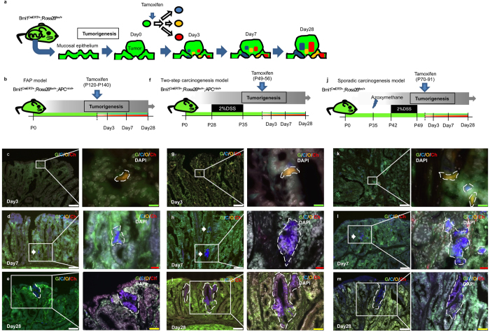
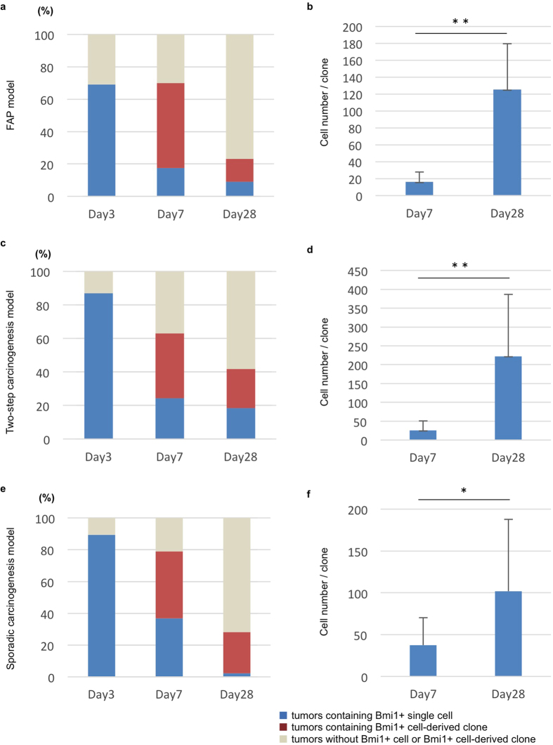
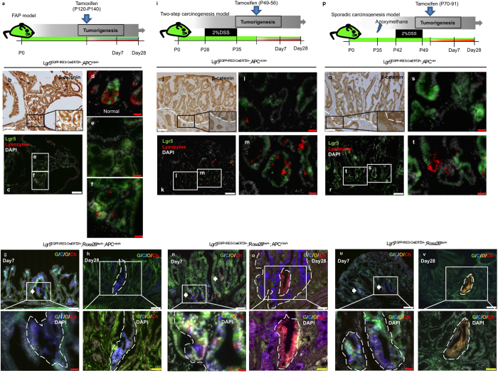
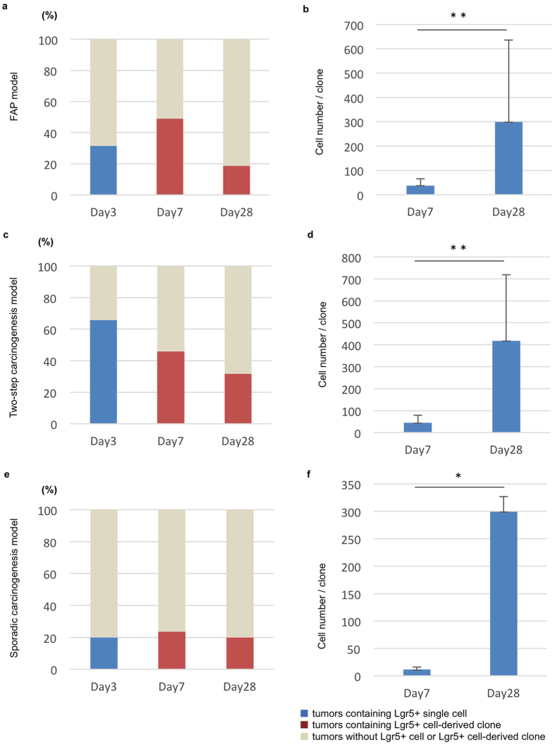
Although the existence of cancer stem cells in intestine tumors has been suggested, direct evidence has not been yet provided. Here, we showed, using the multicolor lineage-tracing method and mouse models of intestinal adenocarcinoma and adenoma that Bmi1- or Lgr5- positive tumorigenic cells clonally expanded in proliferating tumors. At tumor initiation and during tumor propagation in the colon, the descendants of Lgr5-positive cells clonally proliferated to form clusters. Clonal analysis using ubiquitous multicolor lineage tracing revealed that colon tumors derived from Lgr5-positive cells were monoclonal in origin but eventually merged with neighboring tumors, producing polyclonal tumors at the later stage. In contrast, the origin of small intestine tumors was likely polyclonal, and during cancer progression some clones were eliminated, resulting in the formation of monoclonal tumors, which could merge similar to colon tumors. These results suggest that in proliferating intestinal neoplasms, Bmi1- or Lgr5-positive cells represent a population of cancer stem cells, whereas Lgr5-positive cells also function as cells-of-origin for intestinal tumors.


Conflict of interest statement

The authors declare no competing financial interests.

Figures

Figure 1
Figure 1. Lineage-tracing using tamoxifen-inducible multicolor labeling system to track the cell-fate of Bmi1-positive tumorigenic cells in various models of intestinal tumors.
(a) Schematic protocol of the CreERT2-mediatid multicolor labeling using Bmi1CreERT/+; Rosa26rbw/+ mice. Rainbow (rbw) construct is knocked-in into Rosa26 locus and consists of three loxP variants: lox2271, loxN, and loxP, and subsequent cDNA for Green Fluorescent protein (GFP) followed by cDNAs for mCerulean, mOrange and mCherry, which are proceeded by lox2271, loxN, and loxP, respectively. This mouse was crossed with Bmi1CreERT/+ line for the purpose of multicolor lineage tracing of Bmi1-positive cells. When this mouse, all the cells of which express GFP, received tamoxifen, recombination is occurred in Bmi1-positive cells mediated by CreERT2, leading to GFP deletion and random exercise between two lox2271s, loxNs, or loxPs. As a result, mCerulean, mOrange or mCherry moves next to CAG promoter, resulting in random expression of either fluorescent protein. After tumor was confirmed to occur, the mice were injected with tamoxifen to induce multicolor labelling. (b) Schematic protocol of lineage-tracing of Bmi1-positive cells in tumor-bearing FAP model mice. FAP spontaneously occurs in APCmin/+ mice before the age of 140. P, Postnatal day. (f) Schematic protocol of lineage-tracing of Bmi1-positive cells in tumor-bearing two-step carcinogenesis model mice. When APCmin/+ mice, which develop FAP, are administrated with 2% DSS from the age of 28 to 35, tumorigenesis is accelerated to generate colon adenoma. P, Postnatal day. (j) Schematic protocol of lineage-tracing of Bmi1-positive cells in tumor-bearing sporadic carcinogenesis model mice. P, Postnatal day. (c–e,g–i,k–m) Representative traced images of Bmi1+ cell induced in developing tumors. Tumors were harvested at 3 days after induction (c,g and k), 7 days after induction (d,h and l) and 28 days after induction (e,i and m) in FAP model, two-step carcinogenesis model, and sporadic carcinogenesis model, respectively. G; GFP, C; mCerulean, O; mOrange, Ch; mCherry. Scale bars, white; 100 μm, yellow; 50 μm, red; 20 μm, green; 10 μm.
Figure 2
Figure 2. Existence and manner of clonal expansion of Bmi1+ cells in developing small intestinal tumors and colon tumors.
(a,c,e) To assess the existence of Bmi1+ cells in developing tumor and the percentage of the tumors containing Bmi1+ cell-derived clone, FAP model (a), two-step caricinogenesis model (c), and sporadic carcinogenesis model (e) were set up using Bmi1CreERT/+; Rosa26rbw/+ mice. The percentage of the developing tumors that contained Bmi1+ positive cells was calculated by tamoxifen induction of tumor-bearing mice followed by 3-day chase and observation of the cells with rainbow color; mCerulean, mOrange, and mCherry (Day3). To evaluate the percentage of the tumors containing Bmi1+ cell-derived clone, tumor-bearing mice of three models were injected with tamoxifen and clones with rainbow color were examined at 7 days or 28 days after induction (a,c,e): Day7, and Day28. At the same time, tumors that contained rainbow-colored single cell, which was suggested to express Bmi1 when tamoxifen induction but had not divided, was categorized as “tumors containing Bmi1+ single cell”. The number of mice and tumors analyzed were shown in Supplementary Table 1. (b,d,f) The number of the cells that comprised each Bmi1+ cell-derived clone were measured, and the average is shown. Cell number per clone at day28 after tamoxifen induction was compared with day7. Error bars indicate standard deviation. **p < 0.01, *p < 0.05. The number of tumors analyzed and the raw data are shown in Supplementary Table 2.
Figure 3
Figure 3. Cell-fate mapping of the Lgr5-positive cells though tumor development in various mouse models.
(a) Schematic protocol of lineage-tracing of Lgr5-positive cells in tumor-bearing FAP model mice. FAP spontaneously occurs in APCmin/+ mice before the age of 140. P, Postnatal day. (i) Schematic protocol of lineage-tracing of Lgr5-positive cells in tumor-bearing two-step carcinogenesis model mice. When APCmin/+ mice, which develop FAP, are administrated with 2% DSS from the age of 28 to 35, tumorigenesis is accelerated to generate colon adenoma. P, Postnatal day. (p) Schematic protocol of lineage-tracing of Lgr5-positive cells in tumor-bearing sporadic carcinogenesis model mice. P, Postnatal day. (b,c,e,f,j–m,q–t) To investigate the localization of Lgr5+ cells and Paneth cells in the small intestinal tumors and colon tumors, serial sections were prepared from dissected tumors in FAP model mice, two-step carcinogenesis model mice, and sporadic carcinogenesis model mice. One of them was stained for β-catenin to determine the area of tumor mass according to its nuclear localization (b,j and q) and their serial sections were served for immunostaining of lysozyme together with observation of EGFP expression in order to detect Paneth cells and Lgr5+ cells, respectively (c,k and r). The area circled with white dotted line represents tumor area, evaluated by nuclear accumulation of β-Catenin. Insets represent higher magnification images of the boxed area. Green; EGFP fluorescence, Red; Lysozyme, White; DAPI. Scale bars, white; 100 μm, yellow; 50 μm, red; 20 μm. (d) Representative images of Lgr5+ normal cells in small intestine. (f,m,t) Representative images of Lgr5+ tumorigenic cells that coincided with Paneth cells. (e,l,s) Representative images of Lgr5+ tumorigenic cells that were independent of Paneth cells. (g,h,n,o,u,v) Representative traced images of Lgr5+ cell induced in developing tumors. Tumors were harvested at 7 days after induction (g,n and u) and 28 days after induction (h,o and v) in FAP model, two-step carcinogenesis model, and sporadic carcinogenesis model, respectively. G; GFP, C; mCerulean, O; mOrange, Ch; mCherry. Scale bars, white; 100 μm, yellow; 50 μm, red; 20 μm.
Figure 4
Figure 4. Existence and manner of clonal expansion of Lgr5+ cells in developing small intestinal tumors and colon tumors.
(a,c,e) The percentage of the developing tumors that contained Lgr5+ positive cells was calculated by observing EGFP using tumor-bearing Lgr5EGFP-IRES-CreERT2/ mice of three models; FAP model (a), two-step carcinogenesis model (c), and sporadic carcinogenesis model (e). To assess the percentage of the tumors containing Lgr5+ cell-derived clone, FAP model, two-step caricinogenesis model, and sporadic carcinogenesis model were set up using Lgr5EGFP-IRES-CreERT2/+; Rosa26rbw/+ mice, followed by tamoxifen induction and chase to trace the lineage of Lgr5+ cell (a,c,e): Day7 and Day28. This analysis excluded GFP+ clones because we cannot judge whether the origin of the GFP+ clone is the cell that expressed GFP accompanied by Lgr5 or the Lgr5-negative cell, owing to ubiquitous expression of GFP in Rosarbw mice. The number of mice and tumors analyzed were shown in Supplementary Table 3. (b,d,f) The number of the cells that comprised each Lgr5+ cell-derived clone was measured, and the average is shown. Cell number per clone at day28 after tamoxifen induction was compared with day7. Error bars indicate standard deviation. **p < 0.01, *p < 0.05. The number of tumors analyzed and the raw data are shown in Supplementary Table 2.

Similar articles

Cited by

References

    1. Sangiorgi E. & Capecchi M. R. Bmi1 is expressed in vivo in intestinal stem cells. Nature genetics 40, 915–920 (2008). - PMC - PubMed
    1. Barker N. et al.. Identification of stem cells in small intestine and colon by marker gene Lgr5. Nature 449, 1003–1007 (2007). - PubMed
    1. Barker N. et al.. Crypt stem cells as the cells-of-origin of intestinal cancer. Nature 457, 608–611 (2008). - PubMed
    1. Red-Horse K., Ueno H., Weissman I. L. & Krasnow M. A. Coronary arteries form by developmental reprogramming of venous cells. Nature 464, 549–553 (2010). - PMC - PubMed
    1. Tanaka T. et al.. Identification of stem cells that maintain and regenerate lingual keratinized epithelial cells. Nature cell biology (2013). - PubMed

Publication types

MeSH terms

LinkOut - more resources