

Spatial variation and inconsistency between estimates of onset of muscle activation from EMG and ultrasound
- PMID: 28176821
- PMCID: PMC5296741
- DOI: 10.1038/srep42011
Spatial variation and inconsistency between estimates of onset of muscle activation from EMG and ultrasound
Abstract
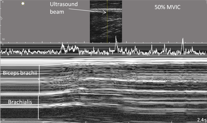
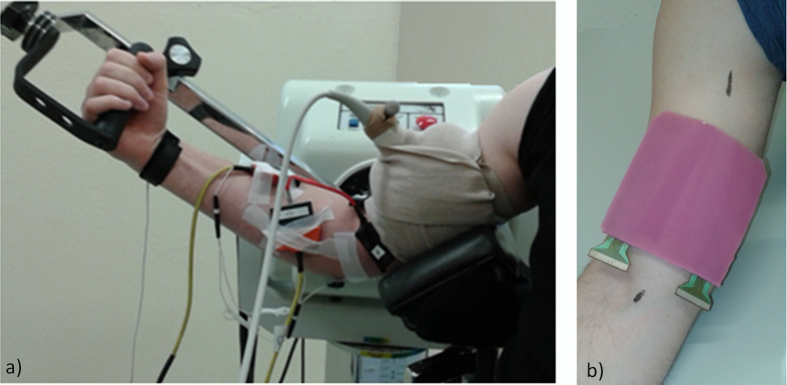
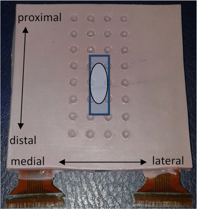
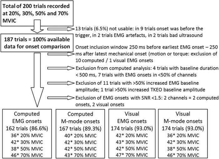
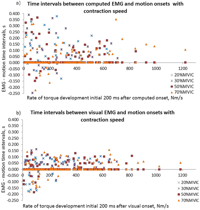
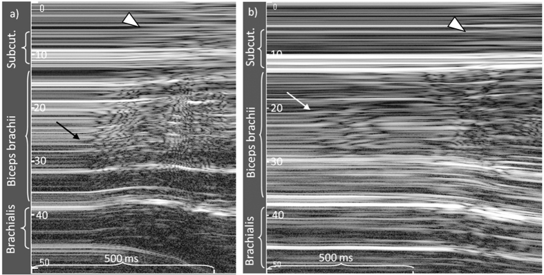
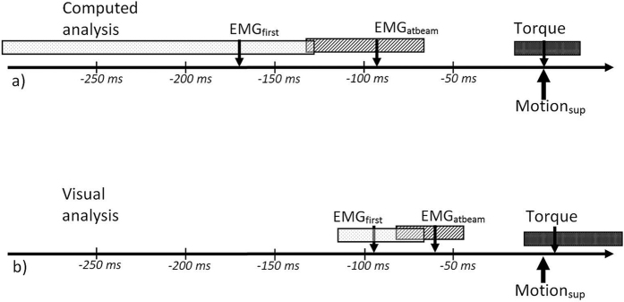
Delayed onset of muscle activation can be a descriptor of impaired motor control. Activation onset can be estimated from electromyography (EMG)-registered muscle excitation and from ultrasound-registered muscle motion, which enables non-invasive measurements in deep muscles. However, in voluntary activation, EMG- and ultrasound-detected activation onsets may not correspond. To evaluate this, ten healthy men performed isometric elbow flexion at 20% to 70% of their maximal force. Utilising a multi-channel electrode transparent to ultrasound, EMG and M(otion)-mode ultrasound were recorded simultaneously over the biceps brachii muscle. The time intervals between automated and visually estimated activation onsets were correlated with the regional variation of EMG and muscle motion onset, contraction level and speed. Automated and visual onsets indicated variable time intervals between EMG- and motion onset, median (interquartile range) 96 (121) ms and 48 (72) ms, respectively. In 17% (computed analysis) or 23% (visual analysis) of trials, motion onset was detected before local EMG onset. Multi-channel EMG and M-mode ultrasound revealed regional differences in activation onset, which decreased with higher contraction speed (Spearman ρ ≥ 0.45, P < 0.001). In voluntary activation the heterogeneous motor unit recruitment together with immediate motion transmission may explain the high variation of the time intervals between local EMG- and ultrasound-detected activation onset.
Conflict of interest statement
The authors declare no competing financial interests.
Figures
Similar articles
-
Differences in muscle contraction onset as determined by ultrasound and electromyography.Muscle Nerve. 2019 Apr;59(4):494-500. doi: 10.1002/mus.26395. Epub 2019 Jan 6. Muscle Nerve. 2019. PMID: 30536792
-
M-mode ultrasound used to detect the onset of deep muscle activity.J Electromyogr Kinesiol. 2015 Apr;25(2):224-31. doi: 10.1016/j.jelekin.2014.12.006. Epub 2015 Jan 12. J Electromyogr Kinesiol. 2015. PMID: 25636500
-
Muscle activity onset in the lumbar multifidus muscle recorded simultaneously by ultrasound imaging and intramuscular electromyography.Clin Biomech (Bristol). 2006 Nov;21(9):905-13. doi: 10.1016/j.clinbiomech.2006.05.003. Epub 2006 Jul 5. Clin Biomech (Bristol). 2006. PMID: 16822599 Clinical Trial.
-
Advances in EMG measurement techniques, analysis procedures, and the impact of muscle mechanics on future requirements for the methodology.J Biomech. 2023 Jul;156:111687. doi: 10.1016/j.jbiomech.2023.111687. Epub 2023 Jun 15. J Biomech. 2023. PMID: 37339541 Review.
-
Multi-Sensing Techniques with Ultrasound for Musculoskeletal Assessment: A Review.Sensors (Basel). 2022 Nov 27;22(23):9232. doi: 10.3390/s22239232. Sensors (Basel). 2022. PMID: 36501933 Free PMC article. Review.
Cited by
-
Influence of the Inter-Trial Interval, Movement Observation, and Hand Dominance on the Previous Trial Effect.Front Hum Neurosci. 2021 Oct 28;15:761514. doi: 10.3389/fnhum.2021.761514. eCollection 2021. Front Hum Neurosci. 2021. PMID: 34776910 Free PMC article.
-
A Study of the Central Motor Drives Interactions Between the Eyes, and an Index Finger, and a Little Finger.Brain Sci. 2025 Apr 20;15(4):422. doi: 10.3390/brainsci15040422. Brain Sci. 2025. PMID: 40309893 Free PMC article.
-
Skeletal Muscle Extracellular Matrix - What Do We Know About Its Composition, Regulation, and Physiological Roles? A Narrative Review.Front Physiol. 2020 Mar 19;11:253. doi: 10.3389/fphys.2020.00253. eCollection 2020. Front Physiol. 2020. PMID: 32265741 Free PMC article. Review.
-
Analyzing muscle thickness changes in lateral abdominal muscles while exercising using virtual reality.J Phys Ther Sci. 2024 Jul;36(7):372-377. doi: 10.1589/jpts.36.372. Epub 2024 Jul 1. J Phys Ther Sci. 2024. PMID: 38952461 Free PMC article.
-
Physical and electrophysiological motor unit characteristics are revealed with simultaneous high-density electromyography and ultrafast ultrasound imaging.Sci Rep. 2022 May 25;12(1):8855. doi: 10.1038/s41598-022-12999-4. Sci Rep. 2022. PMID: 35614312 Free PMC article.
References
-
- Falla D., Jull G. & Hodges P. W. Feedforward activity of the cervical flexor muscles during voluntary arm movements is delayed in chronic neck pain. Experimental Brain Research, 43–48 (2004). - PubMed
-
- Hess S. et al.. Timing of rotator cuff activation during shoulder external rotation in throwers with and without symptoms of pain. Journal of Orthopaedic and Sports Physical Therapy 35, 812–820 (2005). - PubMed
-
- Stensdotter A. K., Grip H., Hodges P. W. & Häger-Ross C. Quadriceps activity and movement reactions in response to unpredictable sagittal support-surface translations in women with patellofemoral pain. Journal of Electromyography and Kinesiology 18, 298–307 (2008). - PubMed
-
- Gubler D. et al.. Ultrasound tissue Doppler imaging reveals no delay in abdominal muscle feed-forward activity during rapid arm movements in patients with chronic low back pain. Spine 35, 1506–1513 (2010). - PubMed
-
- Vasseljen O., Unsgaard-Tondel M., Westad C. & Mork P. J. Effect of core stability exercises on feedforward activation of the deep abdominal muscles in chronic low back pain: a randomized controlled trial. Spine 37, 1101–1108 (2012). - PubMed
Publication types
MeSH terms
LinkOut - more resources
Full Text Sources
Other Literature Sources
