. 2017 Feb 8:7:42019.
doi: 10.1038/srep42019.

Structure of an octameric form of the minichromosome maintenance protein from the archaeon Pyrococcus abyssi

Affiliations

Structure of an octameric form of the minichromosome maintenance protein from the archaeon Pyrococcus abyssi

Giuseppe Cannone et al. Sci Rep. .

Abstract

Cell division is a complex process that requires precise duplication of genetic material. Duplication is concerted by replisomes. The Minichromosome Maintenance (MCM) replicative helicase is a crucial component of replisomes. Eukaryotic and archaeal MCM proteins are highly conserved. In fact, archaeal MCMs are powerful tools for elucidating essential features of MCM function. However, while eukaryotic MCM2-7 is a heterocomplex made of different polypeptide chains, the MCM complexes of many Archaea form homohexamers from a single gene product. Moreover, some archaeal MCMs are polymorphic, and both hexameric and heptameric architectures have been reported for the same polypeptide. Here, we present the structure of the archaeal MCM helicase from Pyrococcus abyssi in its single octameric ring assembly. To our knowledge, this is the first report of a full-length octameric MCM helicase.


Conflict of interest statement

The authors declare no competing financial interests.

Figures

Figure 1
Figure 1. Cloning, purification and functional assay of PabMCM.
(A) Reconstruction of the full-length PabMCM devoid of inteins. (a) PCR-amplified N and C-terminus. (b) PCR-amplified fragment Ser499-Leu525. (c) PCR-amplified of the joined N-term/Ser499-Leu525. (d) PCR-amplified of the joined N-term/Ser499-Leu525. (e) PCR-amplification of the PabMCM full-length. (f) PCR performed in order to check that the fragment was successfully joined into the MCM sequence.(B) Size-exclusion chromatography. Gel-filtration was performed in 20 mM HEPES pH 7.4, 150 mM NaCl at 0.3 ml/min flow-rate. (a) Size-exclusion chromatography trace of PabMCM. The black arrow indicates the molecular weight of gel-filtration standards (BioRad). (b) SDS-PAGE analysis of eluted fractions. (C) DNA binding assay. The gel shows the PabMCM complex can bind dsDNA. A 59-mer dsDNA was used in this experiment. Size was chosen in order to avoid multi-MCMs loading on the same dsDNA and thus force a 1:1 stoichiometry. L, DNA ladder. D, dsDNA only used has control. M, PabMCM only used as control to avoid artefacts due to ethidium bromide staining. Lines 1–5 all contain 24 pmol (calculated on the monomeric MCM) of PabMCM with increasing amount of dsDNA (10, 20, 30, 40, 50 pmol). Lines 6–10 all contain 10 pmol of dsDNA with increasing amount of PabMCM (24, 48, 72, 96, 120 pmol). The band-shift experiment was performed as shown in Fletcher et al.. (D) Helicase activity of PabMCM. Reactions contained helicase buffer with either no MCM (lanes 1 and 6, positive control or lanes 2 and 7, negative control) or increasing amounts of PabMCM (150, 300, 600 nM) lanes (3–5) and SsoMCM (50, 100, 150, 200, 300 nM) lanes (8–12).
Figure 2
Figure 2. Electron microscopy and 3D refinement of the full–length PabMCM.
(A) Characteristic negatively stained electron micrograph of PabMCM. Micrography recorded at 50,000x nominal magnification in low–dose mode (20–25 e2). White circles are used to show single particles. (B) Refined 3D model of the full-length PabMCM single octamer. Volumes were rendered with Chimera. (C) Class averages and re-projections for the refined 3D reconstruction. (D) Fourier shell correlation of the refined PabMCM model.
Figure 3
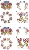
Figure 3. Model fitting of the full–length PabMCM 3D EM structure.
(A,B,C,D) Fitting of the atomic coordinates of the C–terminally truncated SsoMCM (PDB 3F9V) and C–terminal domain of SsoMCM (PDB 2M45). (E,F,G,H) Fitting of the atomic coordinates of full-length MkaMCM (PDB 3F8T). In blue is shown the fitting of the NMR structure of SsoMCM. In red, the AAA + module while in orange the N-terminal domain of both SsoMCM and MkaMCM. In light blue, EXT β-hairpin; in yellow, H2I β-hairpin; in green, PS1 β–hairpin; in dark blue, NT β–hairpin (only in panel D).
Figure 4
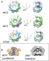
Figure 4. Structural superposition.
(A) One octameric PfuMCM AAA ring (PDB 4R7Z), the N-terminal tier of the hexameric PfuSsoMCM chimera (PDB 4R7Y) and the human Dmc1 recombinase (PDB 1V5W) superposed on PabMCM highlights the similarity with the symmetry and AAA ring subunits orientation of the crystallographic structure for PfuMCM. (B) Comparison of the central channel of the hexameric SsoMCM (47RY) and the octameric PabMCM (EMD-3487).

References

    1. Jenkinson E. R. & Chong J. P. Initiation of archaeal DNA replication. Biochem Soc Trans 31, 669–673, doi: 10.1042/ (2003). - PubMed
    1. Pan M., Santangelo T. J., Li Z., Reeve J. N. & Kelman Z. Thermococcus kodakarensis encodes three MCM homologs but only one is essential. Nucleic Acids Res 39, 9671–9680, doi: 10.1093/nar/gkr624 (2011). - DOI - PMC - PubMed
    1. Walters A. D. & Chong J. P. An archaeal order with multiple minichromosome maintenance genes. Microbiology 156, 1405–1414, doi: 10.1099/mic.0.036707-0 (2010). - DOI - PubMed
    1. Sakakibara N., Kelman L. M. & Kelman Z. Unwinding the structure and function of the archaeal MCM helicase. Mol Microbiol 72, 286–296 (2009). - PubMed
    1. Bell S. D. & Botchan M. R. The minichromosome maintenance replicative helicase. Cold Spring Harb Perspect Biol 5, a012807, doi: 10.1101/cshperspect.a012807 (2013). - DOI - PMC - PubMed

Publication types

MeSH terms

Substances

LinkOut - more resources