

Needle track seeding after radiofrequency ablation for hepatocellular carcinoma: prevalence, impact, and management challenge
- PMID: 28176952
- PMCID: PMC5268370
- DOI: 10.2147/JHC.S106558
Needle track seeding after radiofrequency ablation for hepatocellular carcinoma: prevalence, impact, and management challenge
Abstract
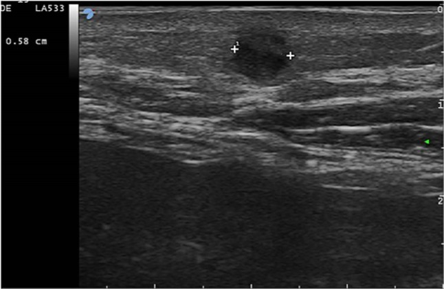
Neoplastic seeding may arise after radiofrequency ablation (RFA) for hepatocellular carcinoma (HCC). A low risk of seeding after RFA (0-1.1%) has been reported, which may rise up to 2.5% if ablation followed diagnostic biopsy. Needle track seeding presents with one or multiple rounded nodules along the needle track located within the peritoneum, along the abdominal muscles, which were penetrated by the needle, pleural surface, or in the subcutaneous and cutaneous tissues. The most widely used method for the assessment of seeding nodules is ultrasound (US), which usually displays hypoechoic nodules with intralesional vascularization. Fine needle aspiration biopsy of the nodule suspicious for malignant implant is mandatory to confirm the diagnosis and plan therapy. Wide surgical excision is the treatment of choice for neoplastic seeding. Thanks to early diagnosis and prompt treatment, development of needle track seeding is not likely to affect the long-term survival of patients.
Keywords: HCC; ablation; biopsy; liver; seeding.
Conflict of interest statement
The author reports no conflicts of interest in this work.
Figures

References
-
- Ferlay J, Soerjomataram I, Dikshit R, et al. Cancer incidence and mortality worldwide: sources, methods and major patterns in GLOBOCAN 2012 [serial online] Int J Cancer. 2015;136:E359–E386. - PubMed
-
- Ballestri S, Romagnoli D, Nascimbeni F, Francica G, Lonardo A. Role of ultrasound in the diagnosis and treatment of nonalcoholic fatty liver disease and its complications. Expert Rev Gastroenterol Hepatol. 2015;9:603–627. - PubMed
Publication types
LinkOut - more resources
Full Text Sources
Other Literature Sources
