. 2017 Feb 21;50(2):331-340.
doi: 10.1021/acs.accounts.6b00514. Epub 2017 Feb 8.

Bacterioferritin: Structure, Dynamics, and Protein-Protein Interactions at Play in Iron Storage and Mobilization

Affiliations

Bacterioferritin: Structure, Dynamics, and Protein-Protein Interactions at Play in Iron Storage and Mobilization

Mario Rivera. Acc Chem Res. .

Abstract

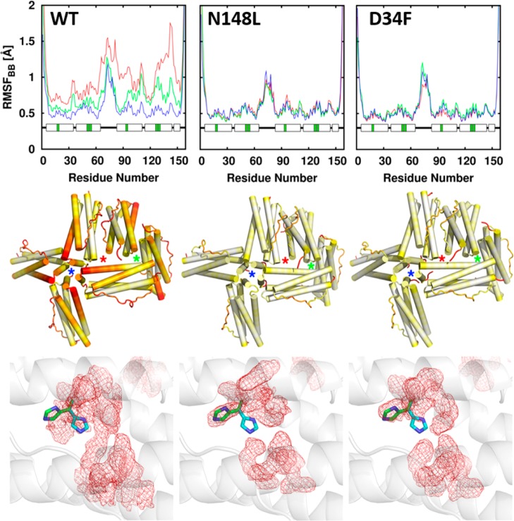
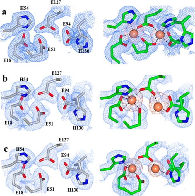
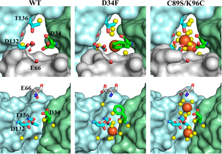
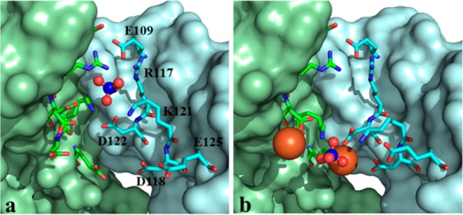
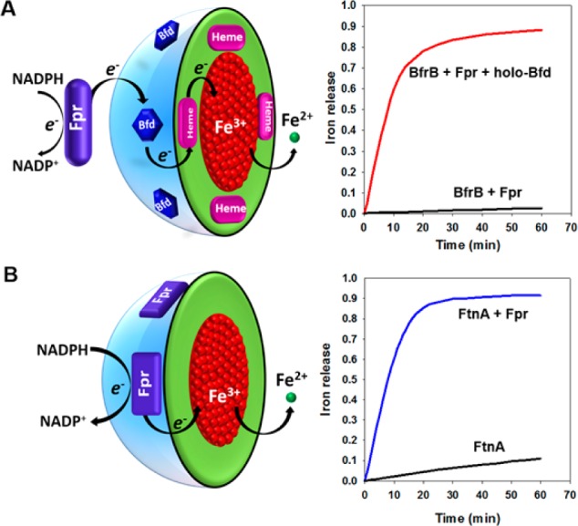
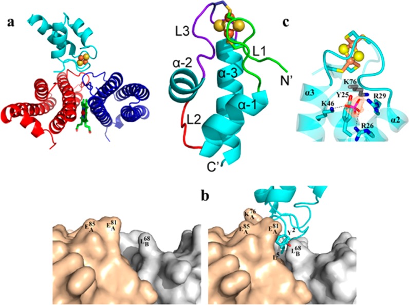
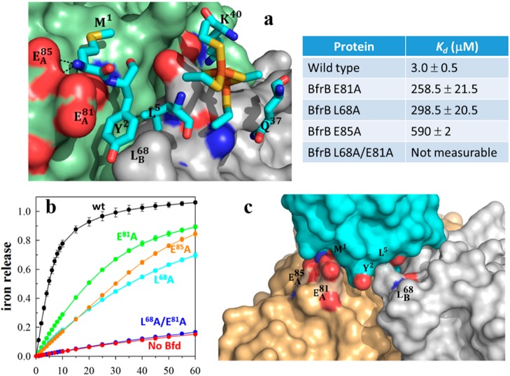
Despite its essentiality to life, iron presents significant challenges to cells: the exceedingly low solubility of Fe3+ limits its bioavailability, and the reactivity of Fe2+ toward H2O2 is a source of the toxic hydroxyl radical (HO). Consequently, cellular levels of free iron are highly regulated to ensure sufficiency while preventing iron-induced toxicity. Relatively little is known about the fate of iron in the bacterial cytosol or how cells balance the need for relatively high cytosolic iron concentrations with the potential toxicity of the nutrient. Iron storage proteins are integral to iron metabolism, and bacteria utilize two types of ferritin-like molecules to store iron, bacterial ferritin (Ftn) and bacterioferritin (Bfr). Ftn and Bfr compartmentalize iron at concentrations far above the solubility of Fe3+ and protect the reducing cell environment from unwanted Fe3+/Fe2+ redox cycling. This Account focuses on our laboratory's efforts to study iron storage proteins in the model bacterium Pseudomonas aeruginosa, an opportunistic pathogen. Prior to our studies, it was thought that P. aeruginosa cells relied on a single Bfr assembled from two distinct subunits coded by the bfrA and bfrB genes. It is now known that, like in most bacteria, two iron storage proteins coexist in P. aeruginosa cells, a bacterial Ftn (FtnA), coded by the ftnA (formerly bfrA) gene and a bacterioferritin (BfrB), coded by the bfrB gene. Studies with BfrB showed that Fe2+ oxidation occurs at ferroxidase centers (FCs), followed by gated translocation of Fe3+ to the interior cavity, a process that is, surprisingly, distinct from that observed with the extensively studied Bfr from Escherichia coli, where the FCs are stable and function only as a catalytic site for O2 reduction. Investigations with BfrB showed that the oxidation of Fe2+ at FCs and the internalization of Fe3+ depend on long-range cooperative motions, extending from 4-fold pores, via B-pores, into FCs. It remains to be seen whether similar studies with E. coli Bfr will reveal distinct cooperative motions contributing to the stability of its FCs. Mobilization of Fe3+ stored in BfrB requires interaction with a ferredoxin (Bfd), which transfers electrons to reduce Fe3+ in the internal cavity of BfrB for subsequent release of Fe2+. The structure of the BfrB/Bfd complex furnished the only known structure of a ferritin molecule in complex with a physiological protein partner. The BfrB/Bfd complex is stabilized by hot-spot residues in both proteins, which interweave into a highly complementary hot region. The hot-spot residues are conserved in the sequences of Bfr and Bfd proteins from a number of bacteria, indicating that the BfrB/Bfd interaction is of widespread significance in bacterial iron metabolism. The BfrB/Bfd structure also furnished the only known structure of a Bfd, which revealed a novel helix-turn-helix fold different from the β-strand and α-helix fold of plant and vertebrate [2Fe-2S]-ferredoxins. Bfds seem to be unique to bacteria; consequently, although mobilization of iron from eukaryotic ferritins may also be facilitated by protein-protein interactions, the nature of the protein that delivers electrons to the ferric core of eukaryotic ferritins remains unknown.


Conflict of interest statement

The author declares no competing financial interest.

Figures

Figure 1
Figure 1
BfrB is a nearly spherical molecule assembled from 24 identical subunits and 12 hemes. (A) Each subunit harbors a FC, and each heme is at the interface of 2 subunits; iron in the FCs is shown as orange spheres. The interior cavity is in contact with the exterior via 4-fold pores (B) (K+ present in each of the 4-fold pores is shown as a purple sphere), 3-fold pores (C), and B-pores (D).
Figure 2
Figure 2
(a) Superposition of two subunits in BfrB (magenta) with two subunits in FtnA (cyan); the zoomed-in view of the heme binding site in BfrB shows that M48 in FtnA cannot bind heme. The FCs in BfrB (b) are different from those in FtnA (c), which are structurally related to FCs in bacterial Ftns.
Figure 3
Figure 3
FCs in as isolated BfrB are empty (top). Soaking crystals of as isolated BfrB in Fe2+ solution allows observation of iron loaded FCs and of a conformational rearrangement of the H130 side chains from “gate open” (top) to “gate closed” (bottom). Soaking Fe-soaked crystals in crystallization solution causes the FCs to empty, with concomitant rearrangement of H130 from the “gate closed” to the “gate open” conformation.
Figure 4
Figure 4
Progress curves obtained upon mixing BfrB and iron solutions delivering 30, 50, 100, 200, and 300 Fe2+/BfrB.
Figure 5
Figure 5
(top) Per-residue backbone RMSF; blue, green, and red plots correspond to increasing ionic strength, respectively. (middle) RMSF mapped onto six subunits of a 24-mer assembly encompassing 4-fold (blue stars), 3-fold (green stars), and B-pores (red stars) shows that the flexibility near 4-fold and B-pores in wt BfrB is dampened in the mutants. In the color scale, flexibility increases from white to red. (bottom) The average conformation explored by FC residues during MD simulations is shown as red mesh; the gate open (green) and gate closed (cyan) conformations of H130 are shown in sticks.
Figure 6
Figure 6
Electron density maps of FC ligands (blue mesh) and iron atoms (orange mesh) in (a) WT, (b) N148L, and (c) D34F BfrB. FCs from as isolated structures are on the left column and FCs from Fe-soaked structures on the right.
Figure 7
Figure 7
B-pores in Fe-soaked structures viewed from the protein exterior (top) and from the side with the gray subunit removed (bottom). Iron ions are in orange and water in yellow spheres.
Figure 8
Figure 8
Comparing the 3-fold pores in as isolated (a) and Fe-soaked (b) C89S/K96C BfrB suggest a possible path for the access of phosphate to the interior cavity where they encounter Fe3+. Fe ions are shown in orange and sulfate in red and blue spheres.
Figure 9
Figure 9
Transverse view of the interior cavity in Fe-soaked C89S/K96C BfrB, depicting iron ions in orange and sulfate ions in yellow and red spheres.
Figure 10
Figure 10
Mobilization of iron stored in BfrB requires Fpr and Bfd, whereas mobilization of iron stored in FtnA requires only Fpr.
Figure 11
Figure 11
Structure of the BfrB/Bfd complex showed that Bfd binds at the interface of two BfrB subunits (a) with minimal rearrangement of the BfrB surface (b) and also revealed a unique fold for the [2Fe–2S]-Bfd (c).
Figure 12
Figure 12
(a) Zoomed-in view of the BfrB/Bfd interface depicting the BfrB surface in green (subunit A) and gray (subunit B), residues anchoring Bfd as cyan sticks, and the [2Fe–2S] cluster in orange and yellow. (b) Perturbation of the cleft defined by L68 and E81 in BfrB, which abolishes binding to Bfd, also inhibits iron mobilization from BfrB. (c) Hot region of the BfrB/Bfd complex.

References

    1. Nielsen F. H. Evolutionary events culminating in specific minerals becoming essential for life. Eur. J. Nutr. 2000, 39, 62–66. 10.1007/s003940050003. - DOI - PubMed
    1. Kehrer J. P. The Haber-Weiss reaction and mechanisms of toxicity. Toxicology 2000, 149, 43–50. 10.1016/S0300-483X(00)00231-6. - DOI - PubMed
    1. Rivera M.Bacterioferritin: Structure Function and Protein-Protein Interactions. In Handbook of Porphyrin Science; Kadish K. K., Smith K. M., Guilard R., Eds., 2014; Vol. 30; pp 135–178, DOI: 10.1142/9789814407755_0041 - DOI
    1. Andrews S. C. The Ferritin-like Superfamily: Evolution of the Biological Iron Storeman from a Ruberythrin-Like Ancestor. Biochim. Biophys. Acta, Gen. Subj. 2010, 1800, 691–705. 10.1016/j.bbagen.2010.05.010. - DOI - PubMed
    1. Lundin D.; Poole A. M.; Sjoberg B. M.; Hogbom M. Use of structural phylogenetic networks for classification of the ferritin-like superfamily. J. Biol. Chem. 2012, 287, 20565–20575. 10.1074/jbc.M112.367458. - DOI - PMC - PubMed

Publication types

MeSH terms