. 2017 Feb 1;73(Pt 2):158-170.
doi: 10.1107/S2059798316020143. Epub 2017 Feb 1.

Ligand fitting with CCP4

Affiliations

Ligand fitting with CCP4

Robert A Nicholls. Acta Crystallogr D Struct Biol. .

Abstract

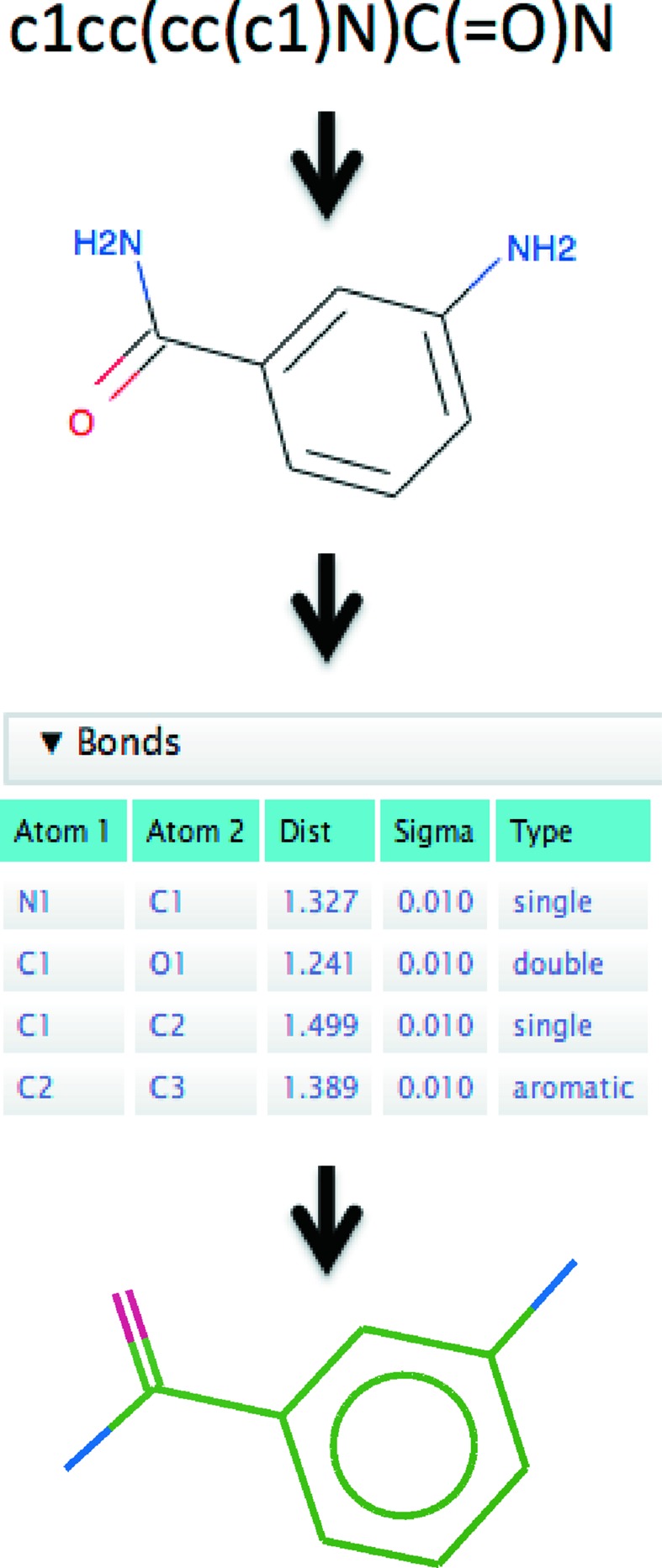
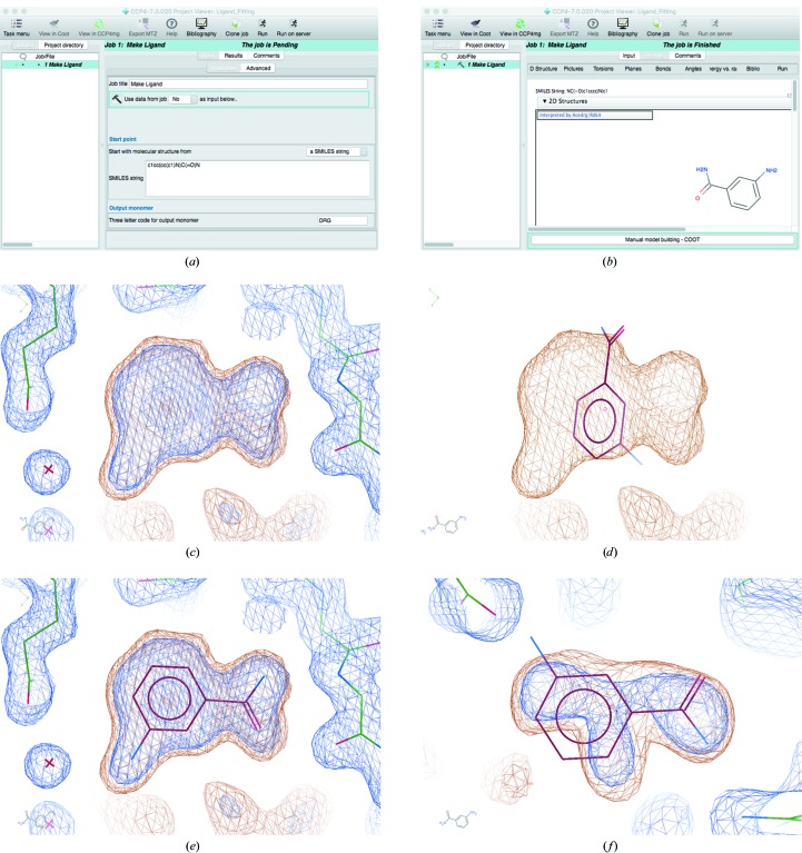
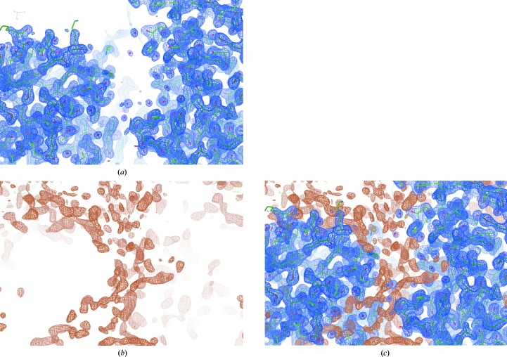
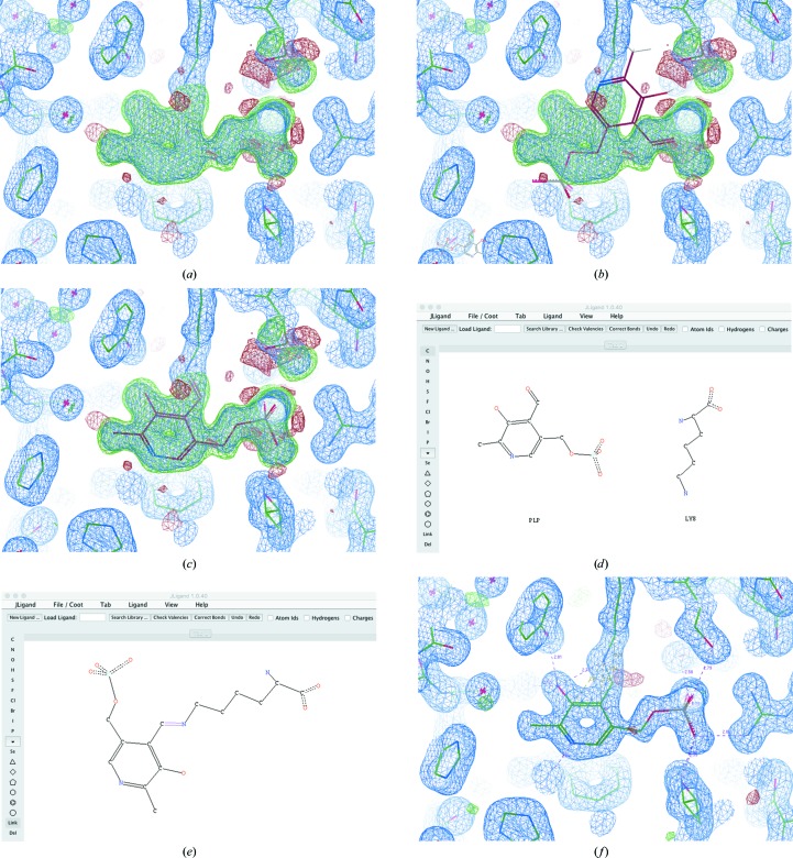
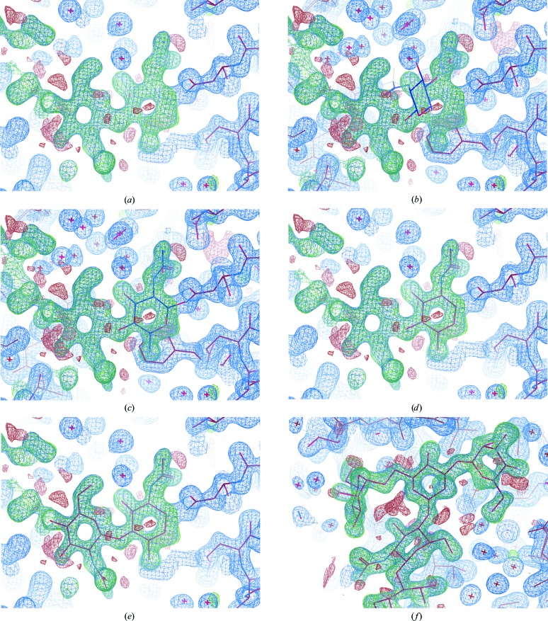
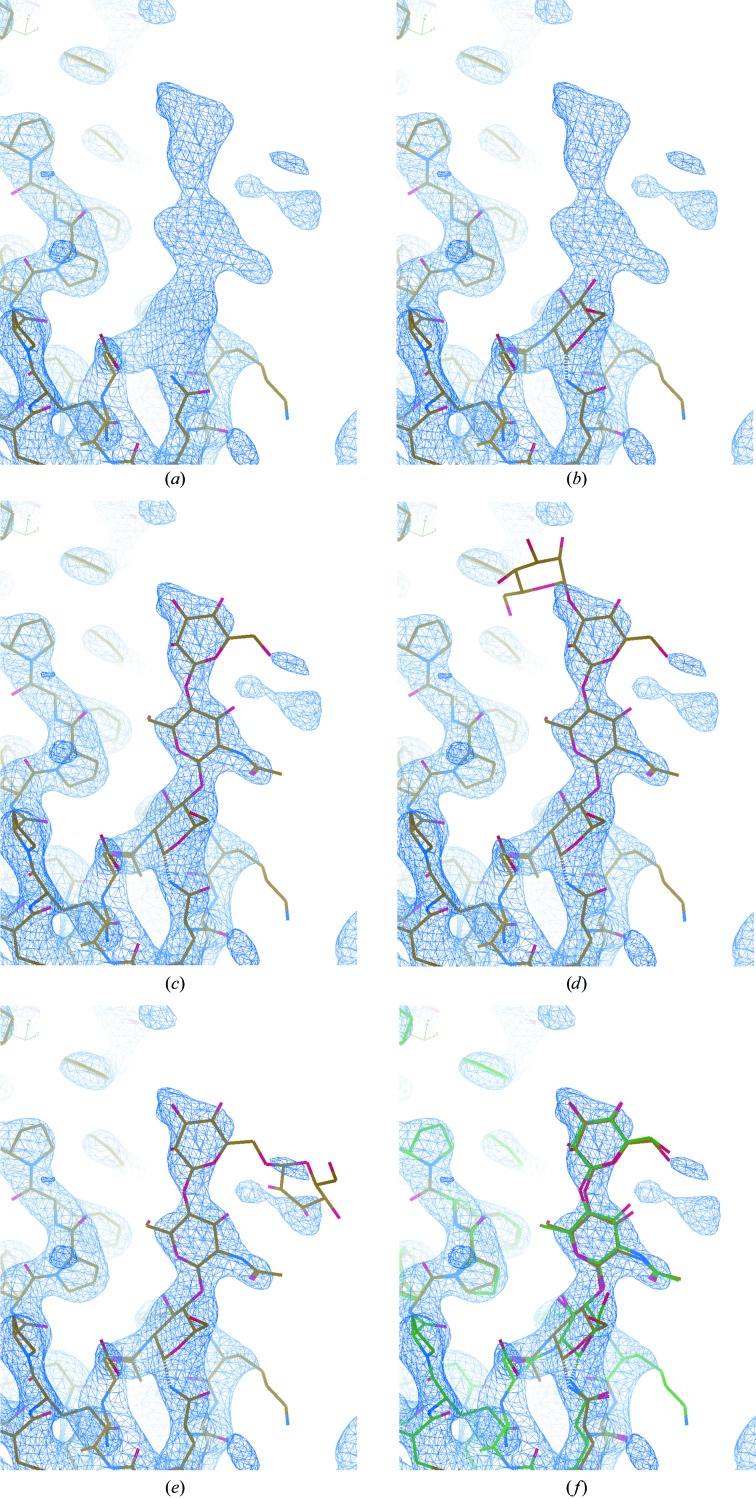
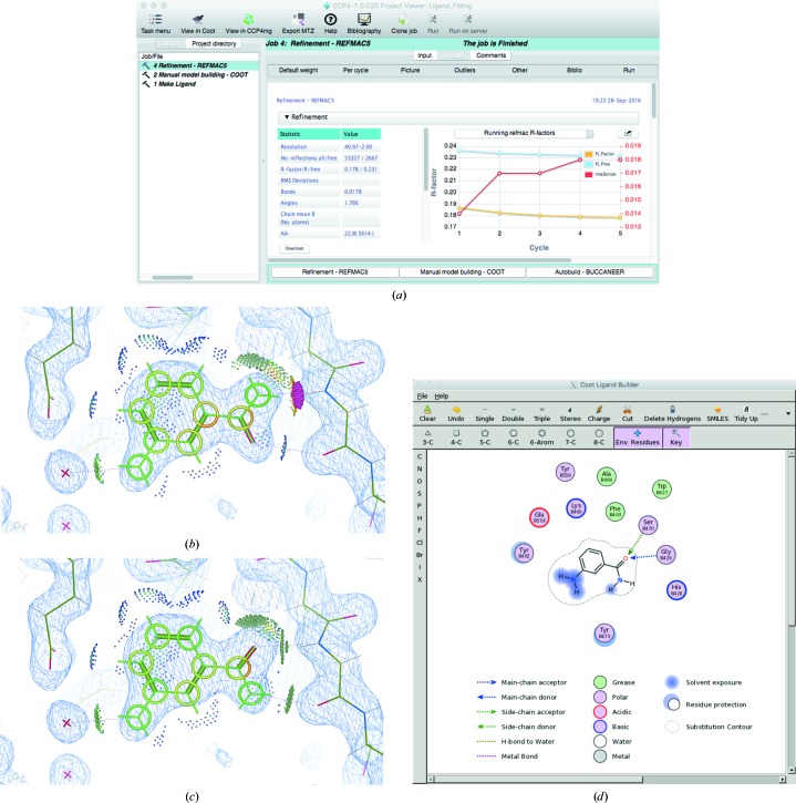
Crystal structures of protein-ligand complexes are often used to infer biology and inform structure-based drug discovery. Hence, it is important to build accurate, reliable models of ligands that give confidence in the interpretation of the respective protein-ligand complex. This paper discusses key stages in the ligand-fitting process, including ligand binding-site identification, ligand description and conformer generation, ligand fitting, refinement and subsequent validation. The CCP4 suite contains a number of software tools that facilitate this task: AceDRG for the creation of ligand descriptions and conformers, Lidia and JLigand for two-dimensional and three-dimensional ligand editing and visual analysis, Coot for density interpretation, ligand fitting, analysis and validation, and REFMAC5 for macromolecular refinement. In addition to recent advancements in automatic carbohydrate building in Coot (LO/Carb) and ligand-validation tools (FLEV), the release of the CCP4i2 GUI provides an integrated solution that streamlines the ligand-fitting workflow, seamlessly passing results from one program to the next. The ligand-fitting process is illustrated using instructive practical examples, including problematic cases such as post-translational modifications, highlighting the need for careful analysis and rigorous validation.

Keywords: CCP4; Coot; ligand fitting; model building.


Figures

Figure 1
Figure 1
The process involved in generating a ligand description and conformer generation using CCP4/AceDRG. From an input SMILES string, the first step is to create a graphical representation of the ligand using AceDRG/RDKit, which encodes basic chemical properties and interactions. This can be depicted in two dimensions and edited using Lidia. A full ligand description is generated, with reference to prior knowledge specifying details of the restraints for bond lengths, angles, torsions etc., using AceDRG. Using these restraints, RDKit generates an initial three-dimensional conformer before REFMAC5 is executed to optimize the coordinates.
Figure 2
Figure 2
Automatic ligand fitting in CCP4. The SMILES string corresponding to 3-aminobenzamide is pasted into the ‘Make Ligand’ task interface in CCP4i2 (a). Upon running the job, AceDRG is used to generate ligand restraints and RDKit is used to generate an initial conformer and a two-dimensional representation of the ligand (b). The ‘Manual Model Building’ task is then executed to open Coot. (c) displays the model (sticks) and 2mF oDF c density map (blue) corresponding to a structure solved with data extending to 2 Å resolution (PDB entry 3kcz; Karlberg et al., 2010 ▸) after manually removing the ligands. The maps are shown using Coot’s default contour levels. Automatic ligand fitting is performed, using 3-aminobenzamide as the target. The focal region corresponds to the top-ranked blob identified in the masked map (orange). The ligand coordinates are nominally positioned onto the centre of the blob (d). The ligand is then optimally oriented and rigid-body refined into the masked density (e). In this case, manual intervention would be required in order to ensure favourable hydrogen bonds are satisfied: this issue is further addressed in §6 and Fig. 7 ▸. Multiple blobs are found in the map and ligands are fitted into them; the third-highest ranked ligand is automatically fitted into a blob that actually corresponds to a glycerol molecule (f).
Figure 3
Figure 3
Creation of a mask (PDB entry 3kcz; Karlberg et al., 2010 ▸). The 2mF oDF c electron-density map corresponding to the current model is coloured blue (a). The mask, coloured orange, is created by artificially setting all modelled regions of the map to zero (b). Both maps are shown using the default contour levels in Coot. The mask is (by default) shown at a lower contour level (0.2) than the original map (0.55), emphasizing the ‘noise’ in the unmodelled regions and the fact that the masked map is set to zero in regions that have already been modelled. For reference, both masks are also shown together (c).
Figure 4
Figure 4
Conformer generation, ligand fitting and link creation, exemplified using pyridoxal 5′-phosphate (monomer code: PLP). (a) displays an unmodelled blob in the density of a structure solved using data extending to 1.6 Å resolution (PDB entry 1ajs; Rhee et al., 1997 ▸) after manually removing the ligand from the deposited model. When importing the ligand, the model will not necessarily be in the correct pose or conformation (b). Automatic conformer generation and ligand fitting results in a more reasonable pose and conformation for the coenzyme (c). Creation of a link record, describing the bond between the N atom in the side chain of Lys258 and a C atom in PLP, can be achieved by opening the Lys and PLP in JLigand (d). The O atom (O4A) in PLP can then be removed and the double-bond link created between the lysine N atom (NZ) and the C atom (C4A) in PLP (e). The model is shown following subsequent refinement by REFMAC5 (f).
Figure 5
Figure 5
Semi-automatically building an oligosaccharide using LO/Carb. (a) focuses on the glycosylation site of a structure refined using data extending to 1.6 Å resolution (PDB entry 4gos; Jeon et al., 2014 ▸), after removing the carbohydrate. The current model is shown (sticks) along with the 2mF oDF c density map (blue) and difference density map (green/red). The positive difference density corresponds to the missing carbohydrate structure, which we know comprises five sugars: two N-acetyl-d-glucosamines (NAG), one β-d-mannose (BMA) and two α-d-mannoses (MAN). The carbohydrate is N-linked to the protein residue Asn112. The carbohydrate structure is built by first placing a NAG conformer into the density next to Asn112 (b) using the ‘Get Monomer’ tool in Coot (found in the ‘File’ menu). After deleting the H atoms, Jiggle Fit (hotkey ‘J’) is used to quickly position and orient the sugar correctly, before manual adjustment and real-space refinement (hotkey ‘r’) to fit the model into the density (c). The O atom which visibly clashes with an N atom in Asn112 can then be removed from the glycosylation site (d), and the new sugar merged into the existing model (using the ‘Merge Molecules’ tool found in the ‘Calculate’ menu). LO/Carb functionality is accessible via the ‘Glyco’ menu (which is activated by selecting ‘Carbohydrate’ from the ‘Modules’ section in the ‘Extensions’ menu), through which it is possible to automatically add and fit the additional NAG (e) and finally the remaining BMA and two MAN sugars (f). Note that in this case REFMAC5 will automatically create the N-link record when the model is next refined, owing to it being a standard link present in the monomer library.
Figure 6
Figure 6
Automatically building an oligosaccharide into a low-resolution map using LO/Carb. (a) focuses on the glycosylation site of a structure refined using data extending to 3.3 Å resolution (PDB entry 4n4z; Gati et al., 2014 ▸) after removing the carbohydrate structure. The current model is shown along with the 2mF o − DF c density map, using the default contour level in Coot (0.34). Selecting ‘Add Oligomannose’ from the ‘Glyco’ menu (which is activated by selecting ‘Carbohydrate’ from the ‘Modules’ section in the ‘Extensions’ menu) results in Coot attempting to automatically build as much of the carbohydrate structure as possible. The sugar linked to the protein is built first (b), followed by additional sugars one by one, until the whole structure is built (c). Attempts to build additional sugars in chemically reasonable positions are made, but are rejected if there is insufficient density to support the model (d, e). (f) shows the final automatically built model (yellow) next to the original deposited model (green).
Figure 7
Figure 7
Refinement and validation. (a) shows the results of refinement using REFMAC5 via the CCP4i2 GUI after fitting 3-aminobenzamide into PDB entry 3kcz as demonstrated in Fig. 2 ▸. After refinement, the ligand is analysed using Coot by displaying environment distances, isolated dots and ligand distortions (b). It is evident that the ligand is in an incorrect conformation. Swapping the O and N atoms results in better stereochemistry, as is evident after re-refining the model using REFMAC5 and re-analysing the model in Coot (c). Tools such as ‘Torsion General’ are useful for such manual editing (found in the side bar to the right of the main Coot window). A two-dimensional depiction of the environment of the ligand is shown using FLEV in Coot (d).

Similar articles

Cited by

  • The missing link: covalent linkages in structural models.
    Nicholls RA, Wojdyr M, Joosten RP, Catapano L, Long F, Fischer M, Emsley P, Murshudov GN. Nicholls RA, et al. Acta Crystallogr D Struct Biol. 2021 Jun 1;77(Pt 6):727-745. doi: 10.1107/S2059798321003934. Epub 2021 May 19. Acta Crystallogr D Struct Biol. 2021. PMID: 34076588 Free PMC article.
  • Reanalysis of a μ opioid receptor crystal structure reveals a covalent adduct with BU72.
    Munro TA. Munro TA. BMC Biol. 2023 Oct 10;21(1):213. doi: 10.1186/s12915-023-01689-w. BMC Biol. 2023. PMID: 37817141 Free PMC article.
  • Optimisation of Neuraminidase Expression for Use in Drug Discovery by Using HEK293-6E Cells.
    Campbell AC, Tanner JJ, Krause KL. Campbell AC, et al. Viruses. 2021 Sep 22;13(10):1893. doi: 10.3390/v13101893. Viruses. 2021. PMID: 34696326 Free PMC article.
  • CMM-An enhanced platform for interactive validation of metal binding sites.
    Gucwa M, Lenkiewicz J, Zheng H, Cymborowski M, Cooper DR, Murzyn K, Minor W. Gucwa M, et al. Protein Sci. 2023 Jan;32(1):e4525. doi: 10.1002/pro.4525. Protein Sci. 2023. PMID: 36464767 Free PMC article.
  • The CCP4 suite: integrative software for macromolecular crystallography.
    Agirre J, Atanasova M, Bagdonas H, Ballard CB, Baslé A, Beilsten-Edmands J, Borges RJ, Brown DG, Burgos-Mármol JJ, Berrisford JM, Bond PS, Caballero I, Catapano L, Chojnowski G, Cook AG, Cowtan KD, Croll TI, Debreczeni JÉ, Devenish NE, Dodson EJ, Drevon TR, Emsley P, Evans G, Evans PR, Fando M, Foadi J, Fuentes-Montero L, Garman EF, Gerstel M, Gildea RJ, Hatti K, Hekkelman ML, Heuser P, Hoh SW, Hough MA, Jenkins HT, Jiménez E, Joosten RP, Keegan RM, Keep N, Krissinel EB, Kolenko P, Kovalevskiy O, Lamzin VS, Lawson DM, Lebedev AA, Leslie AGW, Lohkamp B, Long F, Malý M, McCoy AJ, McNicholas SJ, Medina A, Millán C, Murray JW, Murshudov GN, Nicholls RA, Noble MEM, Oeffner R, Pannu NS, Parkhurst JM, Pearce N, Pereira J, Perrakis A, Powell HR, Read RJ, Rigden DJ, Rochira W, Sammito M, Sánchez Rodríguez F, Sheldrick GM, Shelley KL, Simkovic F, Simpkin AJ, Skubak P, Sobolev E, Steiner RA, Stevenson K, Tews I, Thomas JMH, Thorn A, Valls JT, Uski V, Usón I, Vagin A, Velankar S, Vollmar M, Walden H, Waterman D, Wilson KS, Winn MD, Winter G, Wojdyr M, Yamashita K. Agirre J, et al. Acta Crystallogr D Struct Biol. 2023 Jun 1;79(Pt 6):449-461. doi: 10.1107/S2059798323003595. Epub 2023 May 30. Acta Crystallogr D Struct Biol. 2023. PMID: 37259835 Free PMC article.

References

    1. Adams, P. D. et al. (2010). Acta Cryst. D66, 213–221.
    1. Agirre, J. (2017). Acta Cryst. D73, 171–186. - PMC - PubMed
    1. Agirre, J., Davies, G., Wilson, K. & Cowtan, K. (2015). Nature Chem. Biol. 11, 303. - PubMed
    1. Agirre, J., Iglesias-Fernández, J., Rovira, C., Davies, G. J., Wilson, K. S. & Cowtan, K. D. (2015). Nature Struct. Mol. Biol. 22, 833–834. - PubMed
    1. Bax, B., Chung, C. & Edge, C. (2017). Acta Cryst. D73, 131–140. - PMC - PubMed

Publication types

MeSH terms