. 2017 Feb 1;73(Pt 2):187-194.
doi: 10.1107/S2059798316013553. Epub 2016 Sep 29.

Glycoblocks: a schematic three-dimensional representation for glycans and their interactions

Affiliations

Glycoblocks: a schematic three-dimensional representation for glycans and their interactions

Stuart McNicholas et al. Acta Crystallogr D Struct Biol. .

Abstract

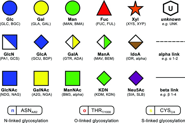
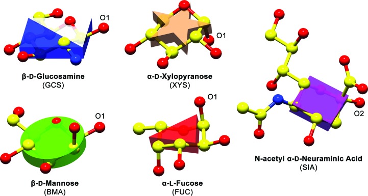
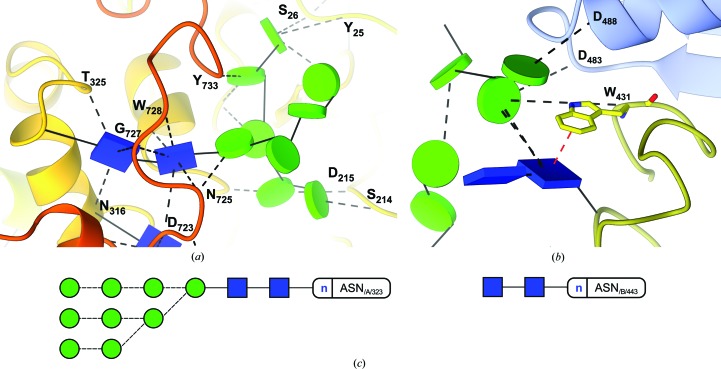
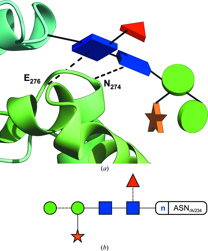
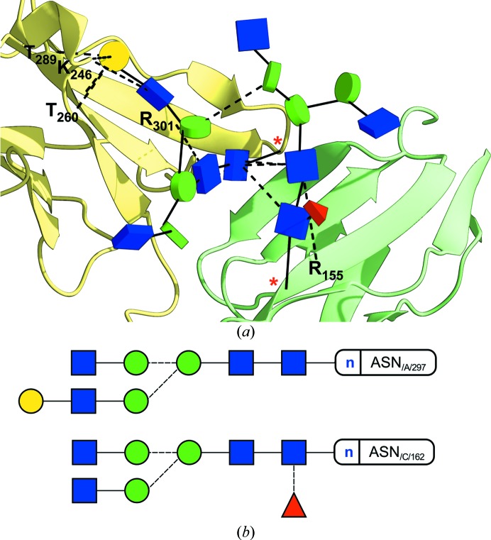
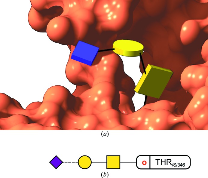
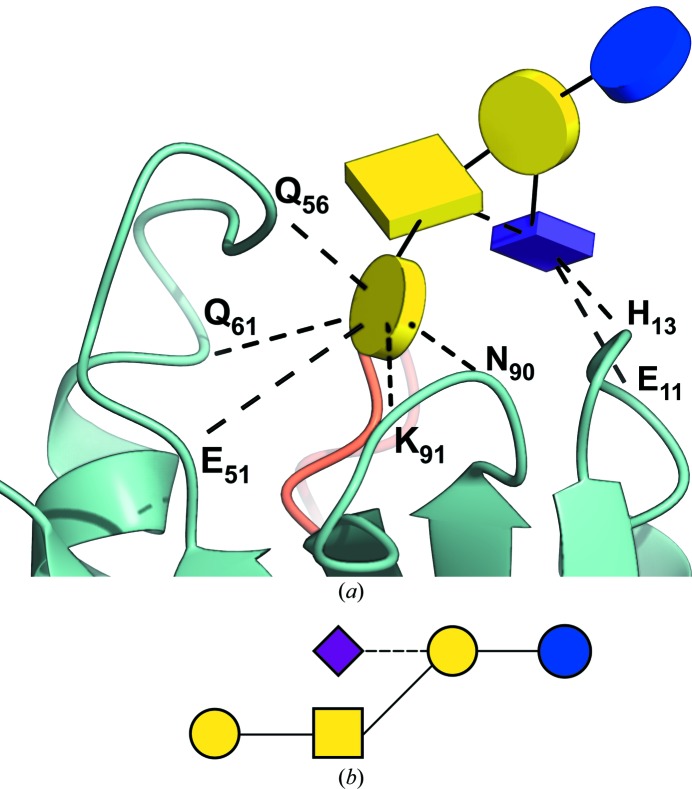
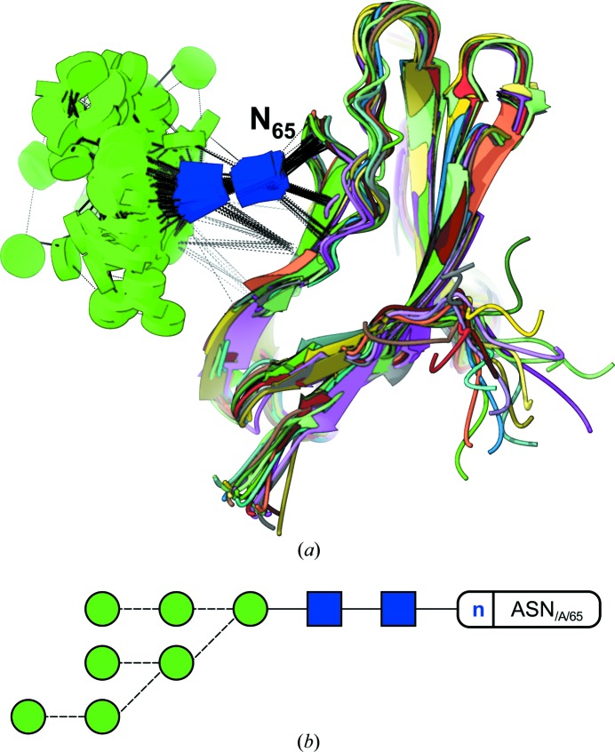
The close-range interactions provided by covalently linked glycans are essential for the correct folding of glycoproteins and also play a pivotal role in recognition processes. Being able to visualise protein-glycan and glycan-glycan contacts in a clear way is thus of great importance for the understanding of these biological processes. In structural terms, glycosylation sugars glue the protein together via hydrogen bonds, whereas non-covalently bound glycans frequently harness additional stacking interactions. Finding an unobscured molecular view of these multipartite scenarios is usually far from trivial; in addition to the need to show the interacting protein residues, glycans may contain many branched sugars, each composed of more than ten non-H atoms and offering more than three potential bonding partners. With structural glycoscience finally gaining popularity and steadily increasing the deposition rate of three-dimensional structures of glycoproteins, the need for a clear way of depicting these interactions is more pressing than ever. Here a schematic representation, named Glycoblocks, is introduced which combines a simplified bonding-network depiction (covering hydrogen bonds and stacking interactions) with the familiar two-dimensional glycan notation used by the glycobiology community, brought into three dimensions by the CCP4 molecular graphics project (CCP4mg).

Keywords: CCP4mg; Glycoblocks; Privateer; carbohydrates; glycans; interactions; molecular graphics; three-dimensional representations.


Figures

Figure 1
Figure 1
Legend to the two-dimensional representation as drawn by Privateer, and its correspondence to three-letter codes from the PDB Chemical Component Dictionary. The current version of Privateer adopts all features of the Essentials notation except the bond–angle relation, which will be available in a forthcoming update to the software. Those sugars typically found in both anomeric forms in covalently bound glycans have both three-letter codes assigned to the same shape, e.g. GLC (α-d-glucopyranose) and BGC (β-d-glucopyranose) to a blue circle. The anomeric form is mentioned explicitly for those cases where just one form is present in the PDB. As the SVG file format supports tooltips (messages that get displayed when the mouse hovers a graphical component), all information related to the linkages is displayed there in order to keep the diagrams minimal. This figure features all three-letter codes recognised by Glycoblocks up to the date of this publication.
Figure 2
Figure 2
Orientation of Glycoblocks with respect to the atomic models they represent. All monosaccharides have been oriented with the oxygen linked to the anomeric carbon (see annotations on the picture) on the right. Despite the d- and l-sugars showing 4C1 and 1C4 conformations, respectively, the orientation of the block remains representative, providing a clear hint at the stereochemistry. For clarity, object outlines and H atoms have been omitted.
Figure 3
Figure 3
Visualizing interactions with Glycoblocks. In the figure, the structure of a heavily glycosylated fungal glycosylhydrolase (PDB code 5fjj), reported by Agirre et al. (2016 ▸). (a) View of the interactions of a high-mannose tree. The glycan connected to Asn323 is perhaps the only example of a three-dimensional structure of a complete high-mannose tree in the PDB. As the protein part has been coloured in rainbow style, it can immediately be seen that the glycan establishes hydrogen bonds across multiple domains and with other glycans which, in turn, interact with other parts of the protein. (b) Visualizing stacking interactions. The first GlcNAc sugar is linked in a flipped conformation to Asn443 due to the stacking interaction with Trp431 (W431 in the picture). These interactions are depicted in red. (c) Two-dimensional representation by Privateer. Dashed lines indicate an alpha link.
Figure 4
Figure 4
(a) Glycoblocks representation of a plant N-glycan and its interactions (PDB code 5aog). The structure depicted is a cationic class III peroxidase purified from Sorghum bicolor (Nnamchi et al., 2016 ▸), which shows the typical core α1,3-fucosylated glycans covalently attached to it. The two core GlcNAc sugars establish two hydrogen bonds (dashed lines in the three-dimensional view), respectively, to one end of a neighbouring α-helix. (b) Two-dimensional representation produced by Privateer. Dashed lines indicate an α-link.
Figure 5
Figure 5
Glycan–glycan and glycan–protein contacts in an antibody–Fc γ receptor IIIa (FcγRIIIa) complex (PDB code 4a5t). (a) Glycoblocks representation. The non-fucosylated Fc fragment has been coloured in yellow, FcγRIIIa is in green. Most of the contacts that bind the two structures together occur between the glycans themselves. The missing fucose residue would have appeared at the interface between both chains, causing steric hindrance according to the authors (Mizushima et al., 2011 ▸). The glycosylation points (asparagine residues 297 and 162) have been marked with a red asterisk. (b) Two-dimensional representation produced by Privateer. Dashed lines indicate an α-link.
Figure 6
Figure 6
(a) View of an O-glycan. This shows one of the rare examples of O-glycosylation found in the PDB (code 4a5t, solved to 3.49 Å resolution), reported by Xue et al. (2012 ▸). As can be seen from the two-dimensional diagram (see b), the GalNAc–Thr linkage was originally modelled as β whilst in reality it had to be α. It is only by using all available knowledge of glycochemistry that these mistakes can be avoided, as the fit to a featureless map must always be tightly restrained to what is known in terms of link distances, angles and torsions. (b) Two-dimensional representation by Privateer. The dashed line indicates an α-link.
Figure 7
Figure 7
Visualizing ligand glycans. (a) A simplified three-dimensional view of the interactions between the GM1 pentasaccharide and the subunit B5 of the choleratoxin pentamer (PDB code 3chb), reported in Merritt et al. (1994 ▸) and re-refined in Merritt et al. (1998 ▸). Only direct hydrogen bonds are shown, as waters have been omitted from the picture. The protein part has been coloured by chain. There is an unlabelled hydrogen bond between the GalNAc and Neu5Ac monosaccharides, also drawn as a dashed line. All the depicted interactions, computed on the fly by CCP4mg, match those manually determined in the original research (Merritt et al., 1994 ▸). (b) Two-dimensional representation by Privateer. The dashed line indicates an α-link.
Figure 8
Figure 8
Simplifying NMR model representation. (a) Glycoblocks view of a partial high-mannose glycan N-linked to the adhesion domain of human CD2. This lateral view of the glycoprotein allows for an unobscured way of looking at the contacts that occur between sugars, and sugars and protein. While hydrogen bonds keep the two core GlcNAc sugars tied to the protein, the rest of the glycan shows great conformational variability. The protein part has been coloured by model. (b) Two-dimensional representation by Privateer. Dashed lines indicate an α-link.

Similar articles

Cited by

References

    1. Agirre, J., Ariza, A., Offen, W. A., Turkenburg, J. P., Roberts, S. M., McNicholas, S., Harris, P. V., McBrayer, B., Dohnalek, J., Cowtan, K. D., Davies, G. J. & Wilson, K. S. (2016). Acta Cryst. D72, 254–265. - PMC - PubMed
    1. Agirre, J., Davies, G., Wilson, K. & Cowtan, K. (2015). Nat. Chem. Biol. 11, 303. - PubMed
    1. Agirre, J., Iglesias-Fernández, J., Rovira, C., Davies, G. J., Wilson, K. S. & Cowtan, K. D. (2015). Nat. Struct. Mol. Biol. 22, 833–834. - PubMed
    1. Ardá, A., Blasco, P., Varón Silva, D., Schubert, V., André, S., Bruix, M., Cañada, F. J., Gabius, H. J., Unverzagt, C. & Jiménez-Barbero, J. (2013). J. Am. Chem. Soc. 135, 2667–2675. - PubMed
    1. Berman, H., Henrick, K. & Nakamura, H. (2003). Nat. Struct. Biol. 10, 980. - PubMed

Publication types