

A natural, single-residue substitution yields a less active peptaibiotic: the structure of bergofungin A at atomic resolution
- PMID: 28177320
- PMCID: PMC5297930
- DOI: 10.1107/S2053230X17001236
A natural, single-residue substitution yields a less active peptaibiotic: the structure of bergofungin A at atomic resolution
Abstract
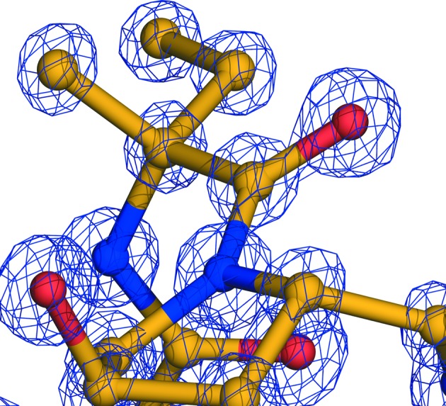
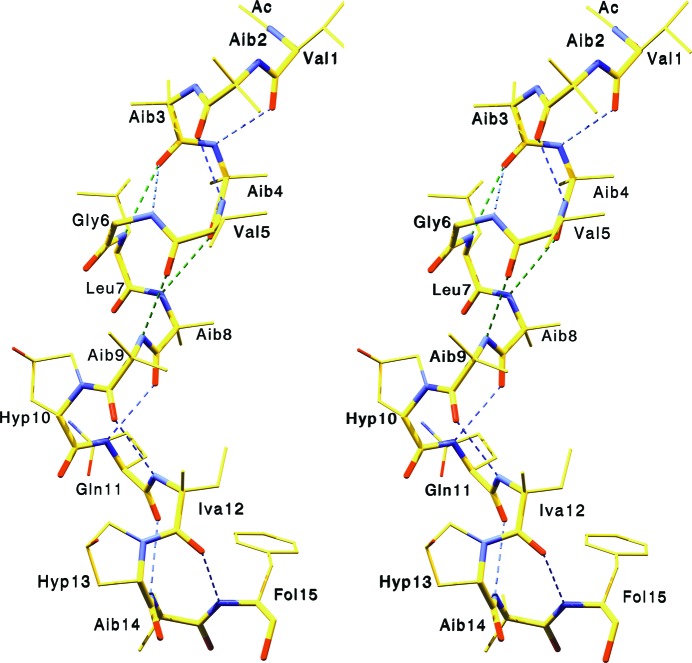
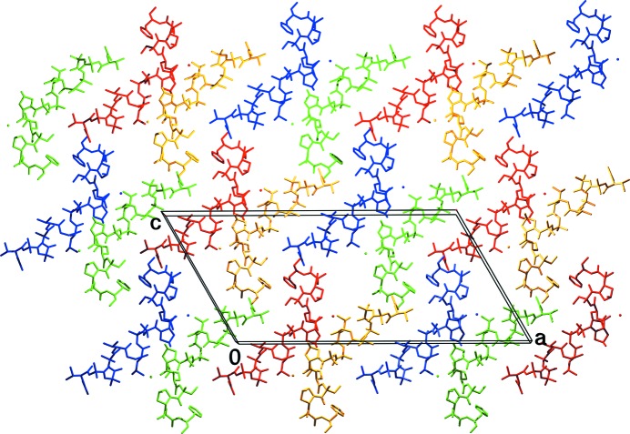
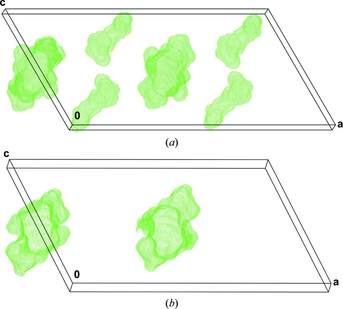
Bergofungin is a peptide antibiotic that is produced by the ascomycetous fungus Emericellopsis donezkii HKI 0059 and belongs to peptaibol subfamily 2. The crystal structure of bergofungin A has been determined and refined to 0.84 Å resolution. This is the second crystal structure of a natural 15-residue peptaibol, after that of samarosporin I. The amino-terminal phenylalanine residue in samarosporin I is exchanged to a valine residue in bergofungin A. According to agar diffusion tests, this results in a nearly inactive antibiotic peptide compared with the moderately active samarosporin I. Crystals were obtained from methanol solutions of purified bergofungin mixed with water. Although there are differences in the intramolecular hydrogen-bonding scheme of samarosporin I, the overall folding is very similar for both peptaibols, namely 310-helical at the termini and α-helical in the middle of the molecules. Bergofungin A and samarosporin I molecules are arranged in a similar way in both lattices. However, the packing of bergofungin A exhibits a second solvent channel along the twofold axis. This latter channel occurs in the vicinity of the N-terminus, where the natural substitution resides.
Keywords: 310-helix; Emericellopsis donezkii; crystal structure; hydrogen bond; peptaibols; peptide antibiotics; α-helix.
Figures
Similar articles
-
The crystal structure of samarosporin I at atomic resolution.J Pept Sci. 2012 Nov;18(11):678-84. doi: 10.1002/psc.2454. Epub 2012 Sep 28. J Pept Sci. 2012. PMID: 23019149
-
Isolation and structure of bergofungin, a new antifungal peptaibol from Emericellopsis donezkii HKI 0059.J Antibiot (Tokyo). 1996 Aug;49(8):817-20. doi: 10.7164/antibiotics.49.817. J Antibiot (Tokyo). 1996. PMID: 8823517 No abstract available.
-
Bergofungin D, a peptaibol template for the introduction of chemical modifications, synthesis of analogs and comparative studies of their structures.J Pept Sci. 2024 Aug;30(8):e3598. doi: 10.1002/psc.3598. Epub 2024 Mar 26. J Pept Sci. 2024. PMID: 38531546
-
Aib residues in peptaibiotics and synthetic sequences: analysis of nonhelical conformations.Chem Biodivers. 2008 Jul;5(7):1238-62. doi: 10.1002/cbdv.200890112. Chem Biodivers. 2008. PMID: 18649312 Review.
-
Peptaibols as a model for the insertions of chemical modifications.Arch Biochem Biophys. 2018 Nov 15;658:16-30. doi: 10.1016/j.abb.2018.09.016. Epub 2018 Sep 20. Arch Biochem Biophys. 2018. PMID: 30243710 Review.
Cited by
-
Genomic and Metabolomic Analyses of the Marine Fungus Emericellopsis cladophorae: Insights into Saltwater Adaptability Mechanisms and Its Biosynthetic Potential.J Fungi (Basel). 2021 Dec 30;8(1):31. doi: 10.3390/jof8010031. J Fungi (Basel). 2021. PMID: 35049971 Free PMC article.
-
Haloalkalitolerant Fungi from Sediments of the Big Tambukan Saline Lake (Northern Caucasus): Diversity and Antimicrobial Potential.Microorganisms. 2023 Oct 19;11(10):2587. doi: 10.3390/microorganisms11102587. Microorganisms. 2023. PMID: 37894245 Free PMC article.
-
Bionectriaceae: a poorly known family of hypocrealean fungi with major commercial potential.Stud Mycol. 2025 Jun;111:115-198. doi: 10.3114/sim.2025.111.04. Epub 2025 Apr 17. Stud Mycol. 2025. PMID: 40371418 Free PMC article.
-
A Novel Lipopeptaibol Emericellipsin A with Antimicrobial and Antitumor Activity Produced by the Extremophilic Fungus Emericellopsis alkalina.Molecules. 2018 Oct 27;23(11):2785. doi: 10.3390/molecules23112785. Molecules. 2018. PMID: 30373232 Free PMC article.
References
-
- Bansal, M., Kumart, S. & Velavan, R. (2000). J. Biomol. Struct. Dyn. 17, 811–819. - PubMed
-
- Berg, A., Ritzau, M., Ihn, W., Schlegel, B., Fleck, W. F., Heinze, S. & Gräfe, U. (1996). J. Antibiot. 49, 817–820. - PubMed
-
- Berman, H. M., Westbrook, J., Feng, Z., Gilliland, G., Bhat, T. N., Weissig, H., Shindyalov, I. N. & Bourne, P. E. (2006). International Tables for Crystallography, Vol. F, edited by M. G. Rossmann & E. Arnold, pp. 675–684. Dordrecht: Kluwer Academic Publishers.
-
- Bunkóczi, G., Schiell, M., Vértesy, L. & Sheldrick, G. M. (2003). J. Pept. Sci. 9, 745–752. - PubMed
-
- Christner, C., Zerlin, M., Gräfe, U., Heinze, S., Küllertz, G. & Fischer, G. (1997). J. Antibiot. 50, 384–389. - PubMed
MeSH terms
Substances
LinkOut - more resources
Full Text Sources
Other Literature Sources
Medical
