Review
. 2017 Jan-Feb;38(1):22-33.
doi: 10.1080/13816810.2016.1266667.

The clinical evaluation of infantile nystagmus: What to do first and why

Affiliations
Review

The clinical evaluation of infantile nystagmus: What to do first and why

Morgan Bertsch et al. Ophthalmic Genet. 2017 Jan-Feb.

Abstract

Introduction: Infantile nystagmus has many causes, some life threatening. We determined the most common diagnoses in order to develop a testing algorithm.

Methods: Retrospective chart review. Exclusion criteria were no nystagmus, acquired after 6 months, or lack of examination.

Data collected: pediatric eye examination findings, ancillary testing, order of testing, referral, and final diagnoses. Final diagnosis was defined as meeting published clinical criteria and/or confirmed by diagnostic testing. Patients with a diagnosis not meeting the definition were "unknown." Patients with incomplete testing were "incomplete." Patients with multiple plausible etiologies were "multifactorial." Patients with negative complete workup were "motor."

Results: A total of 284 charts were identified; 202 met inclusion criteria. The three most common causes were Albinism (19%), Leber Congenital Amaurosis (LCA; 14%), and Non-LCA retinal dystrophy (13%). Anatomic retinal disorders comprised 10%, motor another 10%. The most common first test was MRI (74/202) with a diagnostic yield of 16%. For 28 MRI-first patients, nystagmus alone was the indication; for 46 MRI-first patients other neurologic signs were present. 0/28 nystagmus-only patients had a diagnostic MRI while 14/46 (30%) with neurologic signs did. The yield of ERG as first test was 56%, OCT 55%, and molecular genetic testing 47%. Overall, 90% of patients had an etiology identified.

Conclusion: The most common causes of infantile nystagmus were retinal disorders (56%), however the most common first test was brain MRI. For patients without other neurologic stigmata complete pediatric eye examination, ERG, OCT, and molecular genetic testing had a higher yield than MRI scan. If MRI is not diagnostic, a complete ophthalmologic workup should be pursued.

Keywords: Electroretinogram; magnetic resonance imaging; nystagmus.


Conflict of interest statement

Declaration of interest

The authors report no conflicts of interest. The authors alone are responsible for the content and writing of this article. Arlene V. Drack, MD, is a co-investigator in the Phase III RPE65 gene therapy trial which is funded by a grant from Spark Therapeutics.

Figures

Figure 1
Figure 1
Causes of infantile nystagmus in 202 patients.
Figure 2
Figure 2
Yield of MRI as first test in patients with no neurologic signs, vs. those with neurologic signs. The Y axis represents number of patients. The non-neurologic signs group had solely ocular findings at pediatric eye examination. The neurologic group had abnormal optic nerve appearance, abnormal head circumference, developmental delay, or other neurologic signs at the time of pediatric eye examination.
Figure 3
Figure 3
(a) First test results. (b). All tests results. The Y axis represents number of patients in each category. The number of patients receiving each test ever is shown by the blue bar, and number of patients who had an abnormality found that was related to nystagmus is on the orange bar. MRI = brain magnetic resonance imaging; OCT = optical coherence tomography; ERG = electroretinogram; Genetic testing = all molecular genetic testing including chromosome microarray and karytype; Genetic Eye Tests = specific testing of eye-related genes by gene panels, exome sequencing, and del/dup testing; Eye Examination = complete pediatric eye examination.
Figure 4
Figure 4
Right eye with severe optic nerve hypoplasia, left with mild in a child with vertical and horizontal nystagmus and septo-optic dysplasia.
Figure 5
Figure 5
Frontal encephalocoele presenting with infantile nystagmus. Cerebellar tonsillar ectopia was also present.
Figure 6
Figure 6
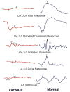
Electroretinogram of patient with CACNA1F mutation and spasmus-nutans-like nystagmus, compared to normal ERG.
Figure 7
Figure 7
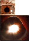
Hazel iris in oblique illumination on the top, with pupillary illumination on the bottom. Note the edge of the crystalline lens visible with surrounding iris transillumination temporally.
Figure 8
Figure 8
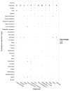
Nystagmus waveform type versus diagnosis by percentage. The y-axis represents the nystagmus waveform types. The x-axis represents a selection of the most common diagnoses. The waveform types may add up to more than 100% per diagnosis due to some patients having more than one waveform type mentioned in the clinical description of their nystagmus in the chart.
Figure 9
Figure 9
Flow chart algorithm for the workup of infantile nystagmus. Used with permission from American Academy of Ophthalmology Knights Templar Pediatric Ophthalmology Education Site(www.aao.org). Key: MRI = magnetic resonance imaging; TIDs= transillumination defects; OCT = optical coherence tomography; LCA=Leber Congenital Amaurosis; ONH= optic nerve hypoplasia; CVI=cortical vision impairment; CSNB=congenital stationary night blindness; JXLR=juvenile Xlinked retinoschisis; Abnl=abnormal; achroma=achromatopsia; RP=retinitis pigmentosa; PAX6=PAX6 gene, responsible for aniridia and related syndromes; FRMD7=FRMD7 gene, an X linked gene associated with motor (idiopathic infantile) nystagmus.

References

    1. Hertle RW, Maldanado VK, Maybodi M, Yang D. Clinical and ocular motor analysis of the infantile nystagmus syndrome in the first 6 months of life. Br J Ophthalmol. 2002 Jun;86(6):670–5. - PMC - PubMed
    1. CEMAS Working Group. The National Eye Institute Publications. Bethesda, MD: The National Eye Institute, The National Institutes of Health; 2001. A National Eye Institute Sponsored Workshop and Publication on The Classification Of Eye Movement Abnormalities and Strabismus (CEMAS) ( www.nei.nih.gov). The National Institutes of Health.
    1. Fu VL, Bilonick RA, Felius J, Hertle RW, Birch EE. Visual acuity development of children with infantile nystagmus syndrome. Invest Ophthalmol Vis Sci. 2011 Mar 14;52(3):1404–11. doi: 10.1167/iovs.09-4686. Print 2011 Mar. - DOI - PMC - PubMed
    1. Brodsky MC, Keating GF. Chiasmal glioma in spasmus nutans: a cautionary note. J Neuroophthalmol. 2014 Sep;34(3):274–5. - PubMed
    1. Delorme C, Gras D, Roze E. Spasmus Nutans: More Than Meets the Eye. Pediatr Neurol. 2015 Oct;53(4):367–8. doi: 10.1016/j.pediatrneurol.2015.06.011. Epub 2015 Jun 20. - DOI - PubMed

MeSH terms