. 2017 Feb 8;12(2):e0170597.
doi: 10.1371/journal.pone.0170597. eCollection 2017.

μCT of ex-vivo stained mouse hearts and embryos enables a precise match between 3D virtual histology, classical histology and immunochemistry

Affiliations

μCT of ex-vivo stained mouse hearts and embryos enables a precise match between 3D virtual histology, classical histology and immunochemistry

Christian Dullin et al. PLoS One. .

Abstract

The small size of the adult and developing mouse heart poses a great challenge for imaging in preclinical research. The aim of the study was to establish a phosphotungstic acid (PTA) ex-vivo staining approach that efficiently enhances the x-ray attenuation of soft-tissue to allow high resolution 3D visualization of mouse hearts by synchrotron radiation based μCT (SRμCT) and classical μCT. We demonstrate that SRμCT of PTA stained mouse hearts ex-vivo allows imaging of the cardiac atrium, ventricles, myocardium especially its fibre structure and vessel walls in great detail and furthermore enables the depiction of growth and anatomical changes during distinct developmental stages of hearts in mouse embryos. Our x-ray based virtual histology approach is not limited to SRμCT as it does not require monochromatic and/or coherent x-ray sources and even more importantly can be combined with conventional histological procedures. Furthermore, it permits volumetric measurements as we show for the assessment of the plaque volumes in the aortic valve region of mice from an ApoE-/- mouse model. Subsequent, Masson-Goldner trichrome staining of paraffin sections of PTA stained samples revealed intact collagen and muscle fibres and positive staining of CD31 on endothelial cells by immunohistochemistry illustrates that our approach does not prevent immunochemistry analysis. The feasibility to scan hearts already embedded in paraffin ensured a 100% correlation between virtual cut sections of the CT data sets and histological heart sections of the same sample and may allow in future guiding the cutting process to specific regions of interest. In summary, since our CT based virtual histology approach is a powerful tool for the 3D depiction of morphological alterations in hearts and embryos in high resolution and can be combined with classical histological analysis it may be used in preclinical research to unravel structural alterations of various heart diseases.


Conflict of interest statement

The authors have declared that no competing interests exist.

Figures

Fig 1
Fig 1. SRμCT results of excised PTA stained and agarose gel embedded mouse hearts.
(A) Virtual cut through a volume rendering representation of a PTA stained and agarose gel embedded excised heart of an adult mouse scanned by SRμCT. The coronal cut displays details of the anatomical structures: the right atrium (ra), the left atrium (la), the right and left ventricle (rv, lv), some coronary arteries (ca) as well as the aorta (a) and the aortic valve (white arrow head) and the pulmonary artery (pa). In addition, the PTA staining allows for identification and representation of the orientation of the muscle fibre bundles. (B) Detailed view of the PTA stained right ventricle shown in (A). The position and orientation of the virtual cut section shown here is indicated by the line labelled B in panel (A) demonstrating that virtual sections are feasible at any position and in any orientation. (C) A representative image of the right ventricle area of an unstained heart, which shows no contrast with the exception of a visible difference between fatty and soft-tissue. Note, due to the absence of contrast, (C) may represent a slightly different region than (B).
Fig 2
Fig 2. Virtual cut sections through the heart region of PTA stained mouse embryos and postnatal mice.
(A) The volume rendering of adult mouse heart and entire mouse embryo at E12 demonstrate that the adult mouse heart has approximately the same size as the entire mouse embryo at E12. Virtual transvers sections are shown for E12 (B), E15 (C) and E18 (D) and P0 (E) displaying details of the anatomical structures: the aorta (a) and lungs (l). As early as E12, the heart shows nearly the final shape and structure, as do other organs like the lung, and later only increase in size. Two different CT systems were used: classical μCT for E12 and SRμCT for E15, E18 and P0, demonstrating that with both CT techniques good and comparable results were obtained.
Fig 3
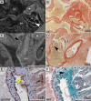
Fig 3. Comparison of CT data sets with histological procedures.
Virtual cross sections through the SRμCT data sets of a PTA stained and (A) agarose gel embedded or (C) paraffin embedded excised mouse heart and their corresponding histological sections (B) and (D) are shown. Notably, even very small structures like the flaps of the aortic valves (indicated by white arrow heads) can be seen by SRμCT. The following structures are clearly depicted: a, aorta; pa, pulmonary artery; la, left atrium and star, heart muscle. HE staining of (B) a section of a heart scanned in agarose gel and later embedded in paraffin and (D) a section from a heart embedded in paraffin before CT acquisition, demonstrates that heart morphology is preserved for classical histology after PTA staining and SRμCT scanning. The pulmonary artery of the heart in the SRμCT image (C) shows a faint PTA contrast, in correlation with the finding of blood residues in the histological section (D). Beside the fact that the CT data of the paraffin embedded heart (C) shows less contrast and more noise, the aortic valves are still clearly depicted (black arrow head). (E) and (F) present results of histological sections adjacent to the one shown in (D) at the position indicated by the white rectangle. (E) Immunostaining with an anti-CD31 antibody of the heart that was CT scanned in paraffin demonstrates the feasibility of IHC on PTA stained heart sections and shows the endothelium indicated by a yellow arrow. (F) MGT staining of a heart embedded in paraffin that was CT scanned visualized collagen in green (black arrow) and smooth muscle cells in violet (white arrow).
Fig 4
Fig 4. Volume measurement of arteriosclerotic plaques.
(A) shows a virtual cross section in the CT data sets of a PTA stained arteriosclerotic heart (ApoE-/- mouse) obtained with classical μCT. A double contour of the vessel wall in the aortic valve (white arrow) can be clearly seen, indicating the fibrotic cap of the plaques as verified by a HE staining of the same heart presented in (B) confirmed the presence of the arteriosclerotic plaque. (C) Three orthogonal slices of the aortic valve region are shown. The arteriosclerotic plaque was segmented in 3D (yellow) and its volume was measured (V = 0.019 mm3). In the pictogram the location of the plaque formations is indicated in red. (D) shows the virtual cross section in the CT data sets of a PTA stained heart of a control mouse obtained with classical μCT and (E) shows the corresponding HE staining. No plaque formation can be observed in the areas corresponding to those shown in (A) and (B).

Similar articles

Cited by

References

    1. Dweck MR, Hyafil F. Can Imaging Improve Our Understanding of Cardiovascular Pathophysiology? Circ Cardiovasc Imaging. 2016;9: e004805 10.1161/CIRCIMAGING.116.004805 - DOI - PubMed
    1. Rastogi V. Artefacts: A Diagnostic Dilemma–A Review. J Clin Diagn Res. 2013; - PMC - PubMed
    1. Bartels M, Krenkel M, Haber J, Wilke RN, Salditt T. X-Ray Holographic Imaging of Hydrated Biological Cells in Solution. Phys Rev Lett. 2015;114. - PubMed
    1. Pauwels E, Van Loo D, Cornillie P, Brabant L, Van Hoorebeke L. An exploratory study of contrast agents for soft tissue visualization by means of high resolution X-ray computed tomography imaging. J Microsc. 2013;250: 21–31. 10.1111/jmi.12013 - DOI - PubMed
    1. Keggin JF. Structure of the Molecule of i2-Phosphotungstic. Nature. 1933;131: 908–909.

LinkOut - more resources