. 2017 Mar 8;17(3):1356-1364.
doi: 10.1021/acs.nanolett.6b03815. Epub 2017 Feb 17.

Tumor-Penetrating Nanosystem Strongly Suppresses Breast Tumor Growth

Affiliations

Tumor-Penetrating Nanosystem Strongly Suppresses Breast Tumor Growth

Shweta Sharma et al. Nano Lett. .

Abstract

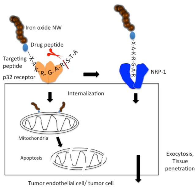
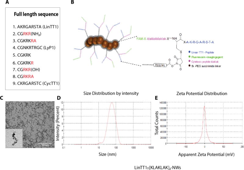
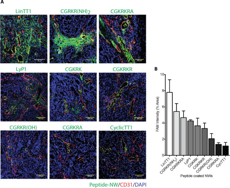
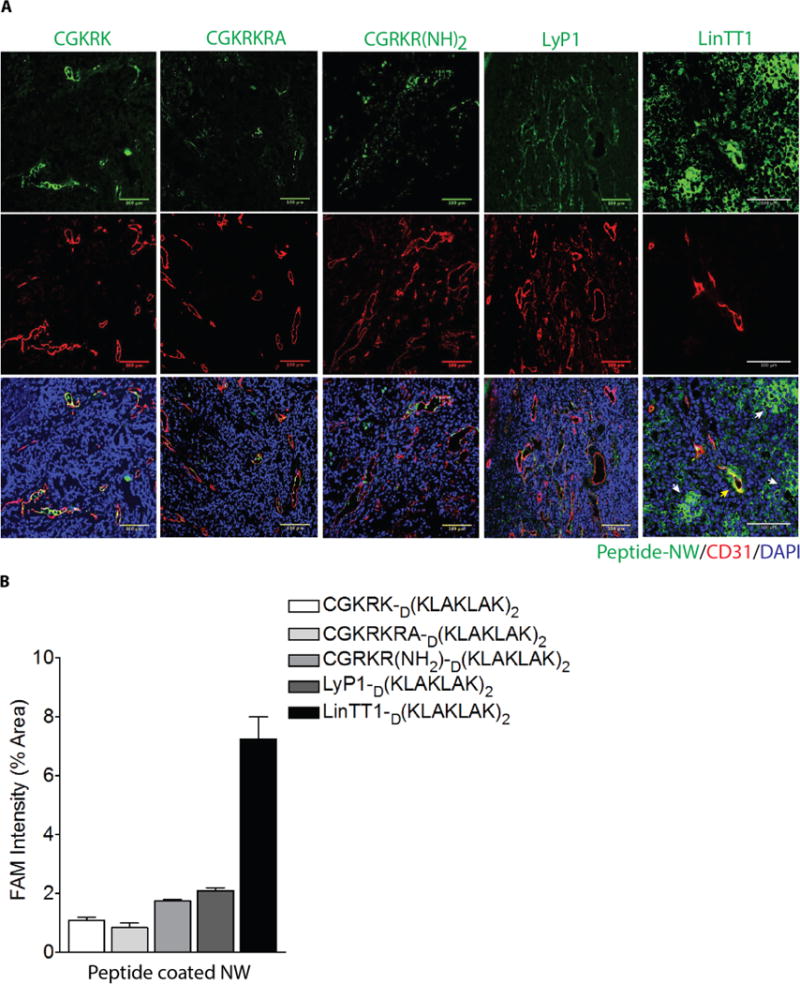
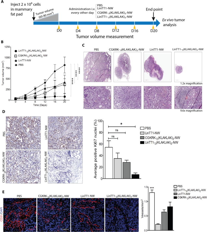
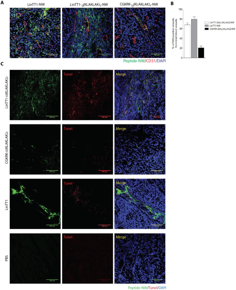
Antiangiogenic and vascular disrupting compounds have shown promise in cancer therapy, but tend to be only partially effective. We previously reported a potent theranostic nanosystem that was highly effective in glioblastoma and breast cancer mouse models, retarding tumor growth and producing some cures [ Agemy , L. et al. Proc. Natl. Acad. Sci. U.S.A. 2011 , 108 , 17450 - 17455 . Agemy , L. et al. Mol. Ther. 2013 , 21 , 2195 - 2204 .]. The nanosystem consists of iron oxide NPs ("nanoworms") coated with a composite peptide with tumor-homing and pro-apoptotic domains. The homing component targets tumor vessels by binding to p32/gC1qR at the surface or tumor endothelial cells. We sought to further improve the efficacy nanosystem by searching for an optimally effective homing peptide that would also incorporate a tumor-penetrating function. To this effect, we tested a panel of candidate p32 binding peptides with a sequence motif that conveys tumor-penetrating activity (CendR motif). We identified a peptide designated as Linear TT1 (Lin TT1) (sequence: AKRGARSTA) as most effective in causing tumor homing and penetration of the nanosystem. This peptide had the lowest affinity for p32 among the peptides tested. The low affinity may have moderated the avidity effect from the multivalent presentation on nanoparticles (NPs), such that the NPs avoid getting trapped by the so-called "binding-site barrier", which can hinder tissue penetration of compounds with a high affinity for their receptors. Treatment of breast cancer mice with the LinTT1 nanosystem showed greatly improved efficacy compared to the original system. These results identify a promising treatment modality and underscore the value of tumor penetration effect in improving the efficacy tumor treatment.

Keywords: C-end Rule; Cancer nanomedicine; iron-oxide nanoparticles; targeted drug delivery; tumor penetrating peptide; vascular disrupting agent.


Conflict of interest statement

Competing interests: ER and VRK are shareholders of EnduRx, a company that hold certain licensing rights on the LyP-1/TT1 technology.

Figures

Figure 1
Figure 1. Schematic representation of the nanosystem and current understanding of its mechanism of action
The system is built on an elongated iron oxide NP (nanoworm; NW) scaffold, with multiple copies of a tandem peptide comprised of a homing peptide and a pro-apoptotic/cytotoxic peptide conjugated onto the surface of the NW. The tumor-homing peptide has two activities: the intact peptide binds to cell surface p32 on tumor vessel endothelium (and on tumor cells and tumor associated macrophages). The peptide is internalized into cells expressing cell surface p32 and transported to mitochondria. A truncated form of the peptide, created by proteolysis at the cell surface, subsequently binds to neuropilin-1 (NRP-1) through its CendR (R/KXXR/K) motif, which triggers a macropinocytosis-related transcytosis and trans-tissue transport pathway (CendR pathway). The NWs are swept into this pathway and out of the vasculature and into extravascular tumor tissue. The pro-apoptotic peptide serves the anti-cancer drug in the construct (see ref. for references).
Figure 2
Figure 2. Design of tumor-homing peptides and NWs
(A) A total of nine p32-binding-peptides were synthesized to test in vivo tumor homing. (B) A chimeric peptide combining a tumor-homing peptide (LinTT1 in this example) with a pro-apoptotic peptide is covalently coupled to NWs; length 40-50 nm). An extra cysteine was added to the N-terminus of the LinTT1 peptide and used for coupling to NWs. The drug peptide and the FAM fluorophore were attached to the free N-terminus of the cysteine residue. (C) Transmission Electron Microscopy (TEM) image of NWs after the amination step. (D) Size of LinTT1-D[KLAKLAK]2 conjugated NWs as determined by dynamic light scattering (DLS). (E) Zeta potential of LinTT1-D[KLAKLAK]2-NWs as measured by DLS.
Figure 3
Figure 3. Tumor homing of TT1-NWs
(A) Mice bearing orthotopic MCF10CA1a breast cancer xenografts were intravenously injected with peptide-coated-NWs (7.5mg iron/kg), and the NWs were allowed to circulate for 5 hours. The mice were perfused through the heart with PBS, and tumors and organs were collected and processed for fluorescence microscopy. A representative confocal microscopy image for each peptide from tumors in 3 mice is shown. Merged image: green, NWs, red, CD31; blue, nuclei. Scale bars 100μm. Striking accumulation of FAM-LinTT1-NWs outside tumor blood vessels is seen. (B) Quantification of tumor accumulation of NWs. Slides were scanned using Scanscope. Ten random areas in each tumor section (n = 3/group) were selected for quantification of NW fluorescence using ImageJ software. Error bars = mean ±SD.
Figure 4
Figure 4. Tumor homing of NWs coated with chimeric peptides consisting of a homing peptide and pro-apototic peptide
(A) Representative confocal images showing NW homing to MCF10CA1a tumors. LinTT1-D[KLAKLAK]2-NWs partially co-localize with tumor blood vessels (CD31), but most of the NWs are outside the blood vessels in the extravascular tumor tissue. (B) Quantification of NW fluorescence in tumors.
Figure 5
Figure 5. Therapeutic efficacy of LinTT1-D[KLAKLAK]2-NWs in MCF10CA1a tumor mice
(A) Schematic representation of treatment approach. (B) Mice bearing MCF10CA1a orthotopic tumor xenografts were intravenously injected with peptide-coated NWs every other day for 3 weeks at a dose of 7.5mg of iron/kg (similar to the daily dose of iron used in anemia treatment). PBS, n = 5; LinTT1-NW, n = 5; CGKRKD[KLAKLAK]2-NWs, n = 6; LinTT1D[KLAKLAK]2-NWs, n = 6. One of two independent experiments with similar results is shown. Error bars, mean ± SD. Statistical analyses were performed with ANOVA; ****P < 0.0001. (C) Representative sections stained with hematoxylin and eosin (H&E). (D) Sections from treated tumors were stained with an antibody specific for proliferating cell nuclear antigen (Ki67). The graph shows percentage of Ki67 positive nuclei. The results are expressed as a mean ±SD (*P < 0.01, one way ANOVA, Kruskal-Wallis test n = 3 mice per group). Scale bar = 200μm. (E) Representative confocal images tumor vasculature stained with anti-CD31 (red). Blood vessels were quantified by counting 10 random fields at 20× magnification, and the results are presented as number of vessels per field (***P < 0.001, one way ANOVA, Tukey’s posthoc test, n = 3 mice per group) S cale bar = 100μm).
Figure 6
Figure 6. Accumulation of peptide-coated NWs and apoptosis in blood vessels of treated tumors
(A) Confocal images showing tumor accumulation of FAM-labeled peptide-NWs after three weeks of treatment. Five hours after the last injection, the mice were perfused through the heart with PBS, and tissues were collected. Tumor sections were stained with anti-CD31 and examined by confocal microscopy. Green, NWs; red, blood vessels; blue, DAPI-stained nuclei. (Scale bars, 100μm.) (B) Quantification of peptide-NWs. Percentage of NW-positive blood vessels was determined in confocal microscopy by analyzing 10 random areas in each tumor section (n = 3/group). Error bars = mean ±SD, scale bar = 100μm. (C) TUNEL staining of treated tumors after three weeks of treatment with the indicated NWs. Merged image: green, NWs; red, TUNEL-positive nuclei; blue, nuclei stained with DAPI. Scale bars, 100μm.

References

    1. Ferrara N, Alitalo K. Nat Med. 1996;5:1359–1364. - PubMed
    1. Hanahan D, Folkman J. Cell. 1996;86:353–364. - PubMed
    1. Ruoslahti E, Bhatia SN, Sailor SJ. J Cell Biol. 2010;188:759–768. - PMC - PubMed
    1. Pham E, Yin M, Peters CG, Lee CR, Brown D, Xu P, Man S, Jayaraman L, Rohde E, Chow A, Lazarus D, Eliasof S, Foster FS, Kerbel RS. Cancer Res. 2016;76:4493–503. - PubMed
    1. Ellerby HM, Arap W, Ellerby LM, Jain R, Andrusiak R, Rio GD, Krajewski S, Lombardo CR, Rao R, Ruoslahti E, Bredesen DE, Pasqualini R. Nat Med. 1999;9:1032–1038. - PubMed

Publication types

MeSH terms