. 2017 Jun 20;8(25):40289-40304.
doi: 10.18632/oncotarget.15041.

Differential sensitivities of bladder cancer cell lines to resveratol are unrelated to its metabolic profile

Affiliations

Differential sensitivities of bladder cancer cell lines to resveratol are unrelated to its metabolic profile

Yang Yang et al. Oncotarget. .

Abstract

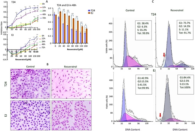
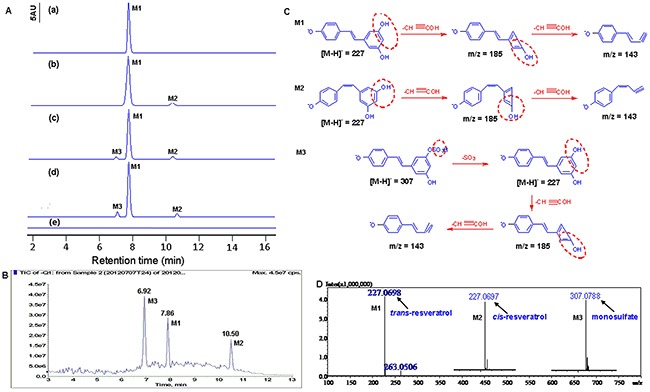
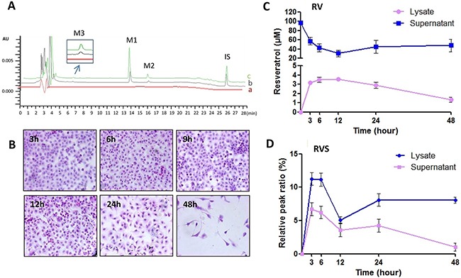
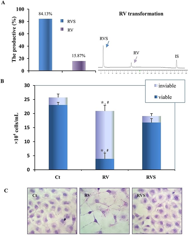
Resveratrol (RV) is a natural polyphenol compound with a wide range of activities, including inhibition of human bladder cancer (HBC) cell growth. Because RV is rapidly metabolized and has poor bioavailability, it is unclear whether the antitumor activity is due to RV or its metabolites. We therefore used liquid chromatography-mass spectroscopy, qRT-PCR, immunocytochemistry and western blotting to evaluate the metabolic profile and biotransformation of RV in the T24 and EJ HBC cell lines. Both T24 and EJ cells generated the same RV metabolite, RV monosulfate (RVS), and both exhibited upregulation of the RV-associated metabolic enzyme SULT1A1 (sulfotransferase). Despite these similarities, T24 cells were more sensitive to RV than EJ cells, yet T24 cells exhibited no sensitivity to an RVS mixture (84.13% RVS). Primary rat bladder epithelial cells showed no adverse effects when exposed to a therapeutic dose (100 μM) of RV. The differences in RV sensitivity between the two HBC cell lines did not reflect differences in the RV metabolic profile or SULT1A1 expression. Because RV exhibited stronger antitumor activity and better safety than RVS, we conclude that RV has significant therapeutic potential for HBC treatment, provided individual differences are considered during clinical research and application.

Keywords: bladder cancer; chemosensitivity; metabolism; resveratrol; sulfotransferase 1A1.


Conflict of interest statement

CONFLICTS OF INTEREST

The authors declare no conflict of interest.

Figures

Figure 1
Figure 1. Chemosensitivity evaluation of resveratrol to T24 and EJ cells
A. Effect of resveratrol treatment on human bladder cancer (HBC) T24 and EJ cells. Cells were incubated with different concentrations (0, 20, 40, 60, 80, 100, 150 and 200μM) resveratrol for different time periods (0, 6, 12, 24, 48 and 72h), respectively, and then the cells number was determined by MTT as described in the Materials and Methods. Data are presented as means ± S.D. of three independent experiments. Bars means standard errors, *P<0.05, **P<0.001 reveal significant difference between RV-treatment and Control HBC cells. #P<0.05, ##P<0.001 show significant different between T24 RV-treatment cells and EJ RV-treatment cells. B. HE morphological staining performed on T24 and EJ cells without (Control) and with 100μM RV (Resveratrol) incubation for 48 hours (100×). Cells at a density of 4×10⁵ cells per well were placed in dishes with coverslips, then T24 and EJ cells were treated without (Control) and with (Resveratrol) 100μM resveratrol treatment for 48h. Cells coverslips were harvested for examination and T24 cells exhibited more obviously spindle-shaped change than EJ cells. C. Flow cytometry analysis on the fractionation of cell cycles and apoptotic cells in T24 and EJ cell populations without (Control) and with (Resveratrol) 100μM resveratrol incubation for 48 hours. Red arrows, indicate the peak of apoptotic cells. Data revealed a presentative experiment in triplicate with similar results.
Figure 2
Figure 2. Identification of resveratrol's metabolites in HBC T24 cells
A. HPLC chromatography analysis. (a) trans-resveratrol standard was dissolved in methanol and analyzed by HPLC (M1, tR=7.86); (b) The culture media incubation with resveratrol without T24 cells for 48h (M1, tR=7.86; M2, tR=7.02); (c) The T24 cells lysate was analyzed after incubation with 100μM resveratrol for 48h (M1, tR=7.89; M2, tR=7.06; M3, tR=10.38); (d) The supernatant of T24 cells was analyzed after incubation with 100μM resveratrol for 48h (M1, tR=7.87; M2, tR=7.01; M3, tR=10.41). (e) The T24 cells treated without resveratrol as Control. B. MS analyses of resveratrol metabolites in T24 cells. Total ion chromatogram (TIC) of the supernatant of T24 cells treated with 100μM resveratrol for 48h. Peak M1, M2 and M3 indicated retention time corresponding to different mass composition of metabolites; C. Proposed mechanism for the decomposition of the m/z 227 [M-H]- ion of resveratrol, the decomposition of the m/z 227 [M-H]- and m/z 307 [M-H]- ion of metabolites. D. Shimadzu LC-MS-IT-TOF-based HRMS analysis of resveratrol metabolites in T24 cells. Arrows labeled as M1, M2 and M3 indicated the exact [M-H]- molecular ion weight of 227.0698 (C14H11O3, calculated m/z 227.0708), 227.0697 (C14H11O3, calculated m/z 227.0708), 307.0788 (C14H11SO6, calculated m/z 307.0276), respectively. In Figure 3, M1 represents trans-resveratrol, M2 represents cis-resveratrol and M3 represents resveratrol monosulfate (RVS), respectively.
Figure 3
Figure 3. RV metabolic pattern in HBC T24 cells
A. Representative HPLC/DAD analysis of resveratrol in T24 cells. (a) HBC T24 cells treated without RV as Control; (b, c) T24 cells treated with 100μM resveratrol for 48h, cell supernatant (b) and cell lysates (c) spiked with 1,8-dihydroxy anthraquinone (internal standard, IS). Peaks: M1. trans-resveratrol, tR=13.82min; M2. cis-resveratrol, tR=15.97min; M3. resveratrol monosulfate (RVS), tR=6.67min; IS. 1,8-dihydroxy anthraquinone, tR=25.93min (Internal standard/IS). B. Morphologic changes were evaluated by HE staining (100×), and T24 cells showed neither observable growth arrest nor morphological change until 24h resveratrol incubation. C & D. Quantification of RV and RVS in T24 cells. Resveratrol concentrations in the cell lysates and supernatant after 100μM resveratrol treatment for 3, 6, 12, 24 and 48h, respectively.
Figure 4
Figure 4. Resveratrol upregulated SULT1A1 expression in T24 and EJ cells
A. Western blots, B. RT-PCR, C. Real-time PCR and D. ICC (100×) all showed that SULT1A1 was upregulated in T24 and EJ cells after resveratrol treatment. Ct, RV represented HBC cells treated without and with 100μM resveratrol incubation for 48h, respectively. * P<0.05, represents statistical significance between RV-treatment HBC cells and the normally cultured HBC cells, respectively.
Figure 5
Figure 5. Biotransformation and bioactivity evaluation of RVS on T24 cells
A. Quantification of the biotransformation efficiency of RVS (Left) by representative HPLC analysis (Right). RV, RVS and IS represent resveratrol, resveratrol monosulfate and 1,8-dihydroxy anthraquinone (Internal standard), respectively. B. Cells number was determined by Trypan Blue exclusion after normal culture (Ct), 100μM trans-resveratrol (RV), and 84.13% resveratrol monosulfate/15.87% trans-resveratrol mixture (RVS) incubation for 48h, respectively. The column indicates the number of viable cells. *, #, RV treatment compared with Ct and RVS treatment, respectively (P<0.01). C. Morphologic evaluation of T24 cells incubated with normally culture (Ct), 100μM trans-resveratrol (RV), and resveratrol monosulfate/trans-resveratrol mixture (RVS) for 48h by HE staining (200×).
Figure 6
Figure 6. The safety evaluation of resveratrol to PBC cells
A. Resveratrol's effect on the cell viability of the primary cultured normal rat bladder epithelial cells (PBC). Cells number was determined by MTT after 48h-100μM resveratrol incubation, data were expressed as means ± S.D. (n=3) (*, P<0.05). B. HE morphological staining performed on PBC cells with 100μM resveratrol treatment for 48h. PBC cells showed neither observable growth arrest nor morphological change. Ct, represented PBC cells were cultured in normal culture media; RV, represented PBC cells were treated with 100μM resveratrol treatment for 48h.

Similar articles

Cited by

References

    1. Jemal A, Bray F, Center MM, Ferlay J, Ward E, Forman D. Global cancer statistics. CA Cancer J Clin. 2011;61:69–90. doi: 10.3322/caac.20107. - DOI - PubMed
    1. Baur JA, Pearson KJ, Price NL, Jamieson HA, Lerin C, Kalra A, Prabhu VV, Allard JS, Lopez-Lluch G, Lewis K, Pistell PJ, Poosala S, Becker KG, et al. Resveratrol improves health and survival of mice on a high-calorie diet. Nature. 2006;444:337–42. doi: 10.1038/nature05354. - DOI - PMC - PubMed
    1. Jang M, Cai L, Udeani GO, Slowing KV, Thomas CF, Beecher CW, Fong HH, Farnsworth NR, Kinghorn AD, Mehta RG, Moon RC, Pezzuto JM. Cancer chemopreventive activity of resveratrol, a natural product derived from grapes. Science. 1997;275:218–20. doi: 10.1126/science.275.5297.218. - DOI - PubMed
    1. Milne JC, Lambert PD, Schenk S, Carney DP, Smith JJ, Gagne DJ, Jin L, Boss O, Perni RB, Vu CB, Bemis JE, Xie R, Disch JS, et al. Small molecule activators of SIRT1 as therapeutics for the treatment of type 2 diabetes. Nature. 2007;450:712–16. doi: 10.1038/nature06261. - DOI - PMC - PubMed
    1. Francioso A, Mastromarino P, Masci A, d’Erme Mosca L. Chemistry, stability and bioavailability of resveratrol. Med Chem. 2014;10:237–45. doi: 10.2174/15734064113096660053. - DOI - PubMed