Review
. 2017 Apr 13;129(15):2070-2082.
doi: 10.1182/blood-2016-10-687830. Epub 2017 Feb 8.

Role of RUNX1 in hematological malignancies

Affiliations
Review

Role of RUNX1 in hematological malignancies

Raman Sood et al. Blood. .

Erratum in

Abstract

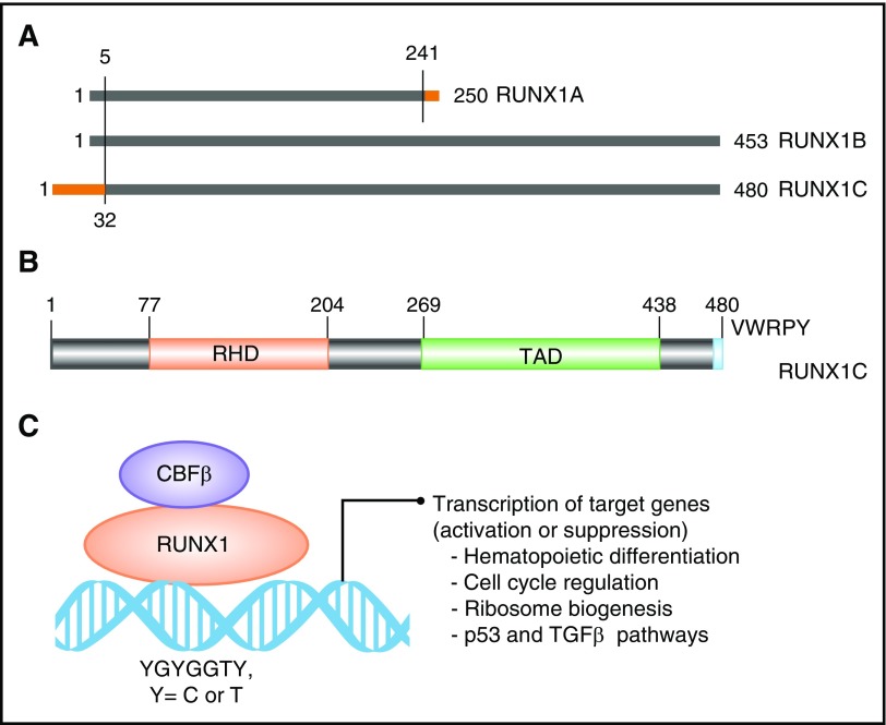
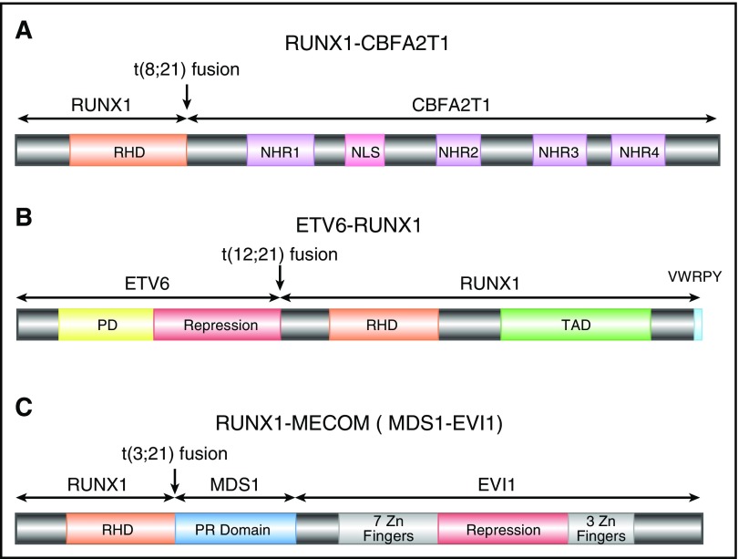
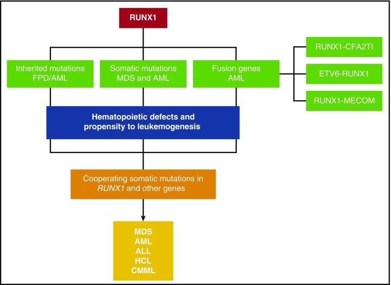
RUNX1 is a member of the core-binding factor family of transcription factors and is indispensable for the establishment of definitive hematopoiesis in vertebrates. RUNX1 is one of the most frequently mutated genes in a variety of hematological malignancies. Germ line mutations in RUNX1 cause familial platelet disorder with associated myeloid malignancies. Somatic mutations and chromosomal rearrangements involving RUNX1 are frequently observed in myelodysplastic syndrome and leukemias of myeloid and lymphoid lineages, that is, acute myeloid leukemia, acute lymphoblastic leukemia, and chronic myelomonocytic leukemia. More recent studies suggest that the wild-type RUNX1 is required for growth and survival of certain types of leukemia cells. The purpose of this review is to discuss the current status of our understanding about the role of RUNX1 in hematological malignancies.


Figures

Figure 1.
Figure 1.
Domain architect of RUNX1 protein and its role as a transcription factor. (A) A schematic depicting the 3 major isoforms of RUNX1 (1A, 1B, and 1C). Isoforms 1A and 1B are transcribed from P1, and isoform 1C is transcribed from P2, thus differing by 32 amino acids at its 5′ end (marked in orange). Isoform 1A contains only the RHD and differs by 9 amino acids at its 3′ end (marked in orange). (B) Schematic of the protein encoded by the largest isoform, 1C, with major functional domains marked: RHD and transactivation domain (TAD). The numbers above the lines represent the amino acid residues. (C) A schematic of RUNX1 heterodimerization with its binding partner, CBFβ, and interaction with DNA at promoters of target genes that carry the specific binding site: YGYGGTY, where Y is C or T.
Figure 2.
Figure 2.
Schematics of RUNX1 fusion proteins. (A) Schematic diagram of full-length RUNX1-CBFA2T1, illustrating the site of fusion between the 2 proteins. RUNX1-CBFA2T1 comprises the RHD from RUNX1 and 4 Nervy homology regions (NHR1–4) from CBFA2T1. The location of the nuclear localization signal (NLS) is also indicated. (B) Schematic diagram of ETV6-RUNX1, illustrating the site of fusion between the 2 proteins. The ETV6-RUNX1 fusion protein contains the N-terminal non-DNA binding moiety of ETV6 fused to almost the entire RUNX1 protein, including its RHD and TAD domains, and the VWRPY motif. (C) Schematic diagram of RUNX1-MECOM, illustrating the site of fusion between the 2 proteins. In the RUNX1-MECOM fusion, RUNX1 fuses with one or both of the “MDS1 and EVI1 complex locus” genes present at chromosome 3q26.
Figure 3.
Figure 3.
A schematic depicting the various mechanisms by which RUNX1 gene is altered in hematological malignancies. Inherited mutations cause FPD/AML. Somatic mutations and fusion genes with cooperating mutations in other genes cause hematological malignancies, such as MDS, AML, ALL, HCL, and CMML.

References

    1. Miyoshi H, Shimizu K, Kozu T, Maseki N, Kaneko Y, Ohki M. t(8;21) breakpoints on chromosome 21 in acute myeloid leukemia are clustered within a limited region of a single gene, AML1. Proc Natl Acad Sci USA. 1991;88(23):10431-10434. - PMC - PubMed
    1. Wang S, Wang Q, Crute BE, Melnikova IN, Keller SR, Speck NA. Cloning and characterization of subunits of the T-cell receptor and murine leukemia virus enhancer core-binding factor. Mol Cell Biol. 1993;13(6):3324-3339. - PMC - PubMed
    1. Bae SC, Yamaguchi-Iwai Y, Ogawa E, et al. . Isolation of PEBP2 alpha B cDNA representing the mouse homolog of human acute myeloid leukemia gene, AML1. Oncogene. 1993;8(3):809-814. - PubMed
    1. Ogawa E, Maruyama M, Kagoshima H, et al. . PEBP2/PEA2 represents a family of transcription factors homologous to the products of the Drosophila runt gene and the human AML1 gene. Proc Natl Acad Sci USA. 1993;90(14):6859-6863. - PMC - PubMed
    1. Gergen JP, Wieschaus EF. The localized requirements for a gene affecting segmentation in Drosophila: analysis of larvae mosaic for runt. Dev Biol. 1985;109(2):321-335. - PubMed

Publication types

MeSH terms

Substances