Review
. 2017 Feb 8;81(1):e00055-16.
doi: 10.1128/MMBR.00055-16. Print 2017 Mar.

The Antigenic Structure of Zika Virus and Its Relation to Other Flaviviruses: Implications for Infection and Immunoprophylaxis

Affiliations
Review

The Antigenic Structure of Zika Virus and Its Relation to Other Flaviviruses: Implications for Infection and Immunoprophylaxis

Franz X Heinz et al. Microbiol Mol Biol Rev. .

Abstract

Zika virus was discovered ∼70 years ago in Uganda and maintained a low profile as a human disease agent in Africa and Asia. Only recently has it caused explosive outbreaks in previously unaffected regions, first in Oceania and then in the Americas since 2015. Of special concern is the newly identified link between congenital malformations (especially microcephaly) and Zika virus infections during pregnancy. At present, it is unclear whether Zika virus changed its pathogenicity or whether the huge number of infections allowed the recognition of a previously cryptic pathogenic property. The purpose of this review is to discuss recent data on the molecular antigenic structure of Zika virus in the context of antibody-mediated neutralization and antibody-dependent enhancement (ADE) of infection, a phenomenon that has been implicated in the development of severe disease caused by the related dengue viruses. Emphasis is given to epitopes of antibodies that potently neutralize Zika virus and also to epitopes that provide antigenic links to other important human-pathogenic flaviviruses such as dengue, yellow fever, West Nile, Japanese encephalitis, and tick-borne encephalitis viruses. The antigenic cross talk between Zika and dengue viruses appears to be of special importance, since they cocirculate in many regions of endemicity and sequential infections are likely to occur frequently. New insights into the molecular antigenic structure of Zika virus and flaviviruses in general have provided the foundation for great progress made in developing Zika virus vaccines and antibodies for passive immunization.

Keywords: Zika virus; flavivirus antigenic structure.


Figures

FIG 1
FIG 1
Schematics and structures of flavivirus particles and E proteins. (A) Schematic representation of immature (left) and mature (right) virions. C, capsid protein; prM, precursor of membrane protein (M); E, envelope protein; sE, soluble envelope protein. (B and C) Cryo-EM structures of immature and mature dengue 1 virus, respectively (reprinted from reference with permission). (D and E) Surface representations of the herringbone arrangement of E dimers at the surface of mature dengue (D) and Zika (E) viruses. Images were constructed by using data reported under PDB accession numbers 3J27 and 5IZ7 (46, 64). A set of 3 parallel dimers forming one of the 30 “rafts” in the herringbone structure is highlighted by white contours. (F and G) Ribbon diagrams of the dengue 2 and Zika virus E protein dimers in their side and top views, respectively. Images were constructed by using data reported under PDB accession numbers 3J27 and 5IZ7 (46, 64). Asn-linked carbohydrates are shown as light blue spheres. TM, transmembrane. In all representations, the three E protein domains are displayed in red (DI), yellow (DII), and blue (DIII), and the DI-DIII as well as DIII-stem linkers are shown in purple. The fusion loop at the tip of domain II is shown in orange.
FIG 2
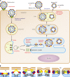
FIG 2
Flavivirus life cycle and fusion mechanism. (A) The virus enters cells by receptor-mediated endocytosis and fuses its membrane by an acidic-pH-triggered mechanism in the endosome to release the viral RNA. The positive-stranded genomic RNA serves as the only viral mRNA and leads to the synthesis of a polyprotein that is co- and posttranslationally processed into three structural and seven nonstructural proteins. Virus assembly takes place at the ER membrane and leads to the formation of immature virions, which are further transported through the exocytic pathway. The acidic pH in the TGN causes structural changes that allow the cleavage of prM by the cellular protease furin and lead to the herringbone-like arrangement of E (Fig. 1D and E). At acidic pH, the cleaved pr part remains associated with the particles (preventing premature fusion in the TGN) and falls off at the neutral pH of the extracellular environment. Subviral particles (SVPs) are formed as a by-product of virion assembly and contain a lipid membrane and prM-E complexes but lack a capsid. SVPs are transported, processed, and released like whole virions. (B) Schematic of flavivirus membrane fusion. The acidic pH in the endosome causes the dissociation of the E dimer and exposure of the FL, which mediates the interaction with endosomal membranes. Further structural changes lead to the relocation of DIII and the formation of a trimer in a hairpin-like structure in which the FL and the TM regions are juxtaposed. These rearrangements provide the energy for membrane fusion and result in the merger of the two lipid bilayers. Color codes are as described in the legend of Fig. 1.
FIG 3
FIG 3
Relationships of flaviviruses based on percent amino acid sequence divergence of E proteins. Sequences were obtained from the ViPR data bank (https://www.viprbrc.org/). GenBank accession numbers are as follows: L06436 for Powassan virus, U27495 for tick-borne encephalitis virus, AY640589 for yellow fever virus, JN226796 for Wesselsbron virus, DQ211652 for West Nile virus, D90194 for Japanese encephalitis virus, DQ859064 for Spondweni virus, KJ776791 for Zika virus, AF226687 for dengue 1 virus, DQ863638 for dengue 3 virus, M29095 for dengue 2 virus, and GQ398256 for dengue 4 virus.
FIG 4
FIG 4
Display of variable surface-exposed amino acids highlighted in red on the background of the Zika virus E dimer (gray). (A) All amino acids that differ in a comparison between Zika virus and the other mosquito-borne flaviviruses displayed in Fig. 3. Circles highlight the conserved patch of amino acids around the fusion loop. (B) All amino acids that differ in a comparison between Zika virus and dengue viruses 1 to 4. (C) Amino acids that differ between Zika virus strains H/PF/2013 and MR766. (D) All amino acids that differ between 111 Zika virus strains. Circles highlight the variability around the glycan loop in domain I. Sequences were obtained from the ViPR data bank (https://www.viprbrc.org/) on 23 September 2016 and analyzed with the Protein Variability Server (PVS) (166). Only Zika virus E sequences from complete genomes were used for the comparisons. Images were constructed by using data reported under PDB accession number 5IZ7 (64).
FIG 5
FIG 5
X-ray structures of Zika virus E dimers (A and C) and the E monomer (D) (side views) in complex with fragments of MAbs (green) (A, C, and D). (A) EDE-specific MAb C8 (PDB accession number 5LBS) (66). (B) Footprints of C8 in the Zika virus and dengue 2 virus E dimers in their top views (PDB accession number 5LCV [66] and PDB accession number 4UTC [77]). (C) DIII-specific MAb ZV67 (PDB accession number 5KVG [74] and PDB accession number 5LCV [66]). Only one Fab is shown on DIII on the right. In DIII of the second subunit (left), the loops forming the epitope (light blue) are highlighted by arrows. (D) FL-specific MAb 2A10G6 (PDB accession number 5JHL [65]).
FIG 6
FIG 6
Schematic diagram of extrinsic antibody-dependent enhancement of infection in Fcγ receptor-positive cells. (Left) Virus-antibody complexes are internalized through Fcγ receptor-mediated endocytosis. Because of incomplete neutralization, the virus can fuse in the endosome and initiate virus production. (Right) Immune complexes containing completely neutralized virus can also be taken up through Fcγ receptor interactions but fail to fuse in the endosome and therefore do not lead to the production of progeny virus.

References

    1. Pierson TC, Diamond MS. 2013. Flaviviruses, p 747–794. In Knipe DM, Howley PM, Cohen JI, Griffin DE, Lamb RA, Martin MA, Racaniello VR, Roizman B (ed), Fields virology, 6th ed Lippincott Williams & Wilkins, Philadelphia, PA.
    1. Simmonds P, Becher P, Collett MS, Gould EA, Heinz FX, Meyers G, Monath T, Pletnev A, Rice CM, Stiasny K, Thiel HJ, Weiner A, Bukh J. 2011. Family Flaviviridae, p 1003–1020. In King AMQ, Lefkowitz E, Adams MJ, Carstens EB (ed), Virus taxonomy. Ninth report of the International Committee on Taxonomy of Viruses. Elsevier Academic Press, San Diego, CA.
    1. Weaver SC, Costa F, Garcia-Blanco MA, Ko AI, Ribeiro GS, Saade G, Shi P-Y, Vasilakis N. 2016. Zika virus: history, emergence, biology, and prospects for control. Antiviral Res 130:69–80. doi:10.1016/j.antiviral.2016.03.010. - DOI - PMC - PubMed
    1. Kindhauser MK, Allen T, Frank V, Santhana R, Dye C. 9 February 2016. Zika: the origin and spread of a mosquito-borne virus. Bull World Health Organ doi:10.2471/BLT.16.171082. - DOI - PMC - PubMed
    1. Samarasekera U, Triunfol M. 2016. Concern over Zika virus grips the world. Lancet 387:521–524. doi:10.1016/S0140-6736(16)00257-9. - DOI - PubMed

Publication types