. 2017 Mar 24;292(12):4789-4800.
doi: 10.1074/jbc.M116.763821. Epub 2017 Feb 8.

Structural Insights into the Broad Substrate Specificity of a Novel Endoglycoceramidase I Belonging to a New Subfamily of GH5 Glycosidases

Affiliations

Structural Insights into the Broad Substrate Specificity of a Novel Endoglycoceramidase I Belonging to a New Subfamily of GH5 Glycosidases

Yun-Bin Han et al. J Biol Chem. .

Abstract

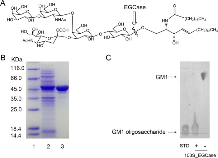
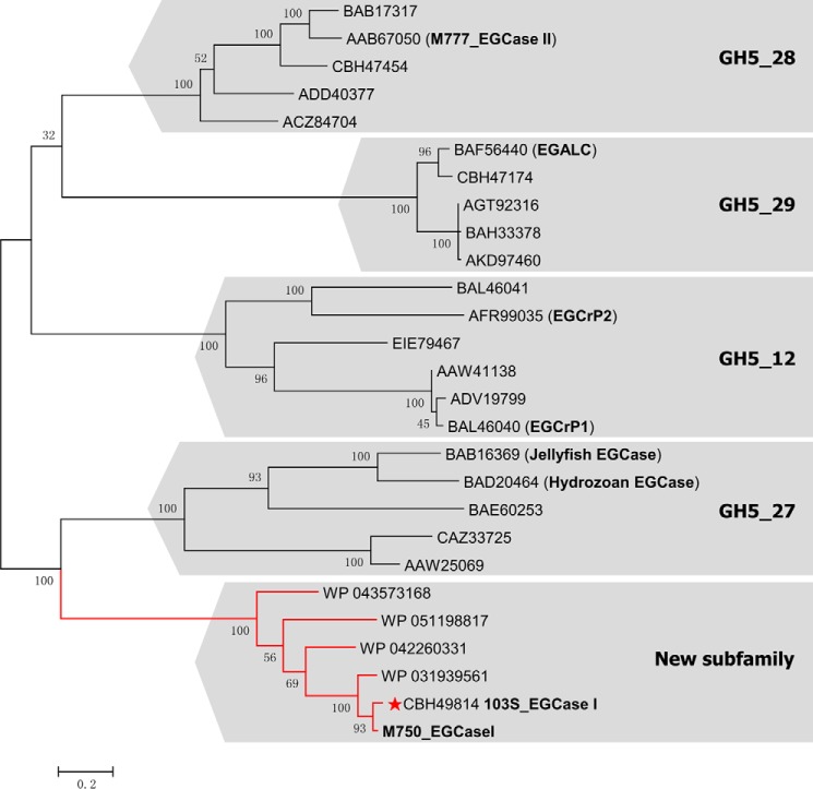
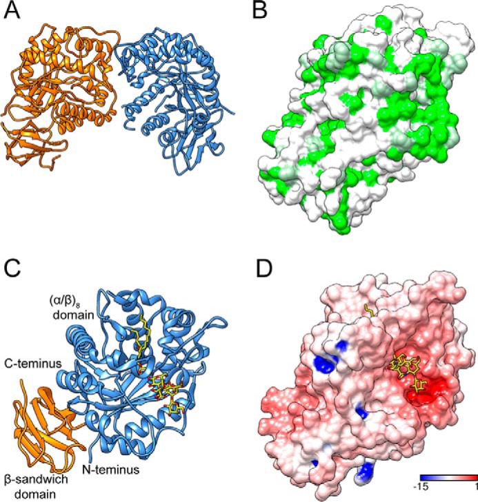
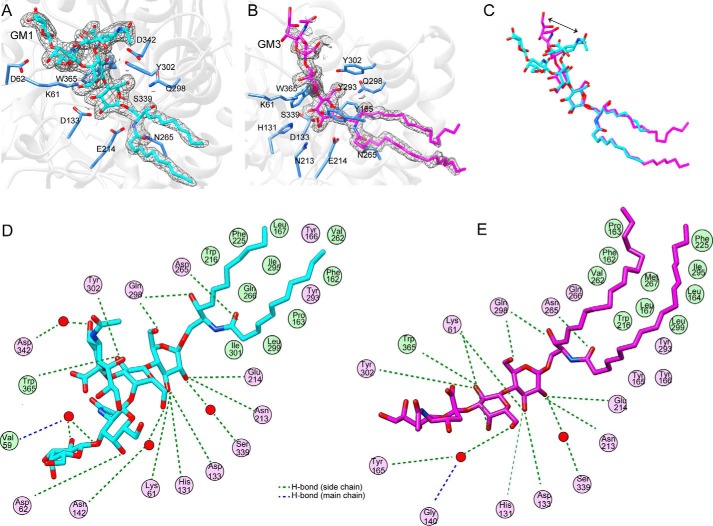
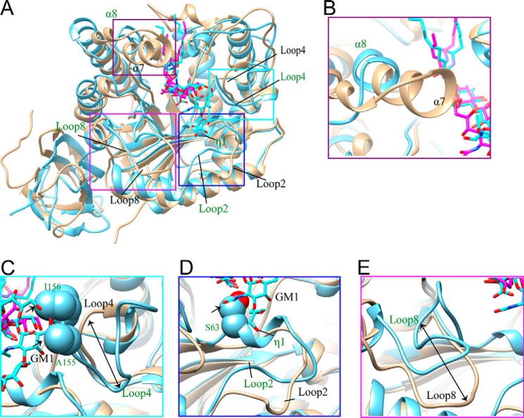
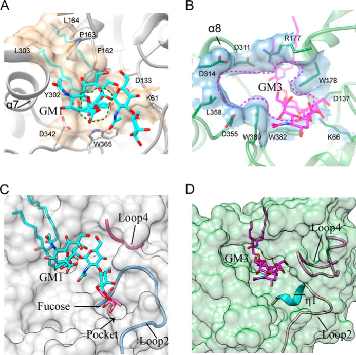
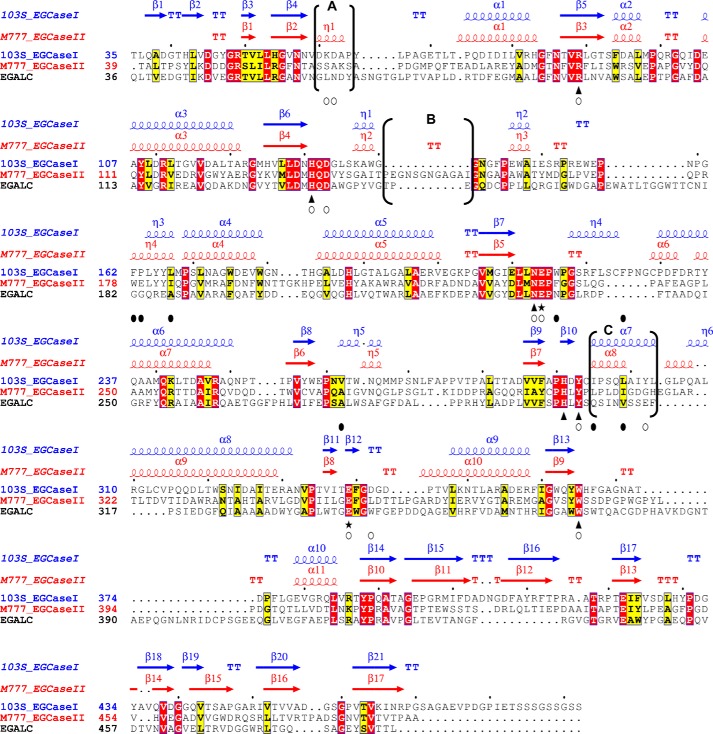
Endoglycoceramidases (EGCases) specifically hydrolyze the glycosidic linkage between the oligosaccharide and the ceramide moieties of various glycosphingolipids, and they have received substantial attention in the emerging field of glycosphingolipidology. However, the mechanism regulating the strict substrate specificity of these GH5 glycosidases has not been identified. In this study, we report a novel EGCase I from Rhodococcus equi 103S (103S_EGCase I) with remarkably broad substrate specificity. Based on phylogenetic analyses, the enzyme may represent a new subfamily of GH5 glycosidases. The X-ray crystal structures of 103S_EGCase I alone and in complex with its substrates monosialodihexosylganglioside (GM3) and monosialotetrahexosylganglioside (GM1) enabled us to identify several structural features that may account for its broad specificity. Compared with EGCase II from Rhodococcus sp. M-777 (M777_EGCase II), which possesses strict substrate specificity, 103S_EGCase I possesses a longer α7-helix and a shorter loop 4, which forms a larger substrate-binding pocket that could accommodate more extended oligosaccharides. In addition, loop 2 and loop 8 of the enzyme adopt a more open conformation, which also enlarges the oligosaccharide-binding cavity. Based on this knowledge, a rationally designed experiment was performed to examine the substrate specificity of EGCase II. The truncation of loop 4 in M777_EGCase II increased its activity toward GM1 (163%). Remarkably, the S63G mutant of M777_EGCase II showed a broader substrate spectra and significantly increased activity toward bulky substrates (up to >1370-fold for fucosyl-GM1). Collectively, the results presented here reveal the exquisite substrate recognition mechanism of EGCases and provide an opportunity for further engineering of these enzymes.

Keywords: crystal structure; glycoside hydrolase; protein engineering; sphingolipid; substrate specificity.


Conflict of interest statement

The authors declare that they have no conflicts of interest with the contents of this article

Figures

FIGURE 1.
FIGURE 1.
Purification and activity assessment of recombinant 103S_EGCase I. A, a typical substrate (GM1 ganglioside) for EGCase. The position of EGCase endohydrolysis is indicated by the wavy line. B, SDS-PAGE showing the purification of the recombinant 103S_EGCase I. Lane 1, protein marker; lane 2, sample corresponding to 20 μl of the culture of E. coli BL21 (DE3) pLysS transformed with pET28a-103S_EGCase I after 12 h of induction; lane 3, 10 μg of purified recombinant 103S_EGCase I. C, TLC showing the oligosaccharides released from GM1 by 103S_EGCase I. GM1 (10 nmol) was incubated with 1 μg of the recombinant 103S_EGCase I at 37 °C for 60 min. The reaction was terminated by heating the reaction mixture in a boiling water bath for 5 min. After high speed centrifugation, the supernatants were loaded onto a TLC plate and developed with chloroform, methanol, 0.02% CaCl2 (5:4:1, v/v/v). GSLs and oligosaccharides were visualized with orcinol-H2SO4 reagent. STD, GM1 oligosaccharide standard; −, without 103S_EGCase I; +, with 103S_EGCase I.
FIGURE 2.
FIGURE 2.
Phylogenetic tree of EGCase and its homologs in the GH5 family based on the maximum likelihood method with 100 bootstrap replications conducted by MEGA6. The sequence of M750_EGCase I was obtained from the literature (15), whereas the other sequences included in the analysis were obtained from the NCBI database. All bootstrap values are displayed. Scale bar, 0.2 amino acid substitutions/site. The three-dimensional structure of 103S_EGCase I (GenBankTM accession number CBH49814) from R. equi 103S solved in this study is marked with a red star. M777_EGCase II (GenBankTM accession number AAB67050) was from Rhodococcus sp. strain M-777, EGALC (GenBankTM accession number BAF56440) was from R. equi, EGCrP1 (GenBankTM accession number BAL46040) was from C. neoformans, and EGCrP2 (GenBankTM accession number AFR99035) was from C. neoformans. The GH5 subfamily number for each branch is shown.
FIGURE 3.
FIGURE 3.
Overall structures of 103S_EGCase I and its GM1 substrate complex. A, ribbon diagram of 103S_EGCase I structure dimer. B, hydrophobic surface potential of 103S_EGCase I chain A (green, hydrophobic; white, polar). The molecular surface was colored by amino acid hydrophobicity using the KD hydrophobicity scale (43). C, ribbon representation of the structure of the 103S_EGCase I-GM1 complex. The N-terminal domain (cornflower blue) adopts an (α/β)8 fold, and the C-terminal domain (orange) adopts a β-sandwich fold. D, electrostatic surface potential of 103S_EGCase I (red, electronegative; blue, electropositive; contoured from −15 to 1 kT/e).
FIGURE 4.
FIGURE 4.
Ligand-binding mode of 103S_EGCase I. A, close-up view of the GM1-binding site in 103S_EGCase I/E339S. The 2FoFc electron density map was contoured at 1σ around GM1 in gray mesh. B, close-up view of the GM3-binding site in 103S_EGCase I/E339S. The 2FoFc electron density map was contoured at 1σ around GM3 in gray mesh. C, comparison of the conformations of GM1 (cyan) and GM3 (magenta). The arrow shows the different portions of the sialic acid residue. D, schematic view of the interaction of 103S_EGCase I with GM1. E, schematic view of the interaction of 103S_EGCase I with GM3. This panel was generated using MOE.
FIGURE 5.
FIGURE 5.
Major structural differences in the sugar-binding sites of 103S_EGCase I and M777_EGCase II. A, the structures of 103S_EGCase I (tan) and M777_EGCase II (light blue) are superimposed, and the differences are highlighted with boxes. B, the α7-helix of 103S_EGCase I was longer than the α8-helix in M777_EGCase II. C, M777_EGCase II had a longer loop 4 than 103S_EGCase I, in which Ala155 and Ile156 may clash with GM1, as indicated by the arrow. D, the conformations of loop 2 in 103S_EGCase I and M777_EGCase II are different, and Ser63 located in loop 2 of M777_EGCase II may clash with GM1. E, loop 8 in 103S_EGCase I and M777_EGCase II had a large difference in conformation.
FIGURE 6.
FIGURE 6.
Comparison of the active site pockets of 103S_EGCase I and M777_EGCase II. A, Phe162, Pro163, Leu164, Tyr302, and Leu303 in 103S_EGCase I form a large cap over the active site. Lys61, Asp133, Phe162, Tyr302, Asp342, and Trp365 form a small opening for the active site pocket. B, Arg177 and Asp311 in M777_EGCase II form a small cap over the active site. Lys66, Asp137, Arg177, Trp178, Asp311, Asp314, Asp355, Leu358, Trp382, and Trp389 in M777_EGCase II form a large opening for the active site pocket. C, loop 2 and the short loop 4 of 103S_EGCase I define a broad sugar-binding cavity. The model of fucosyl-GM1 in the binding site was created by superimposing its structure onto GM1 and then adjusting the fucosyl unit to a reasonable conformation using Coot. The small pocket was able to accommodate the fucosyl unit. D, loop 2 and the long loop 4 of M777_EGCase II define a crowded sugar-binding cavity.
FIGURE 7.
FIGURE 7.
Structure-based sequence alignment of 103S_EGCase I, M777_EGCase II and EGALC. The amino acid sequences of 103S_EGCase I (GenBankTM accession number CBH49814), M777_EGCase II (GenBankTM accession number AAB67050), and EGALC (GenBankTM accession number BAF56440) were aligned using PROMALS3D and shaded in ESPript 3.0. Identical residues are shown in open boxes with white letters on a red background. Similar residues are shown in open boxes with black letters on a yellow background. Conserved amino acid residues in the GH5 family of glycosidases are indicated by triangles. Two glutamates, functioning as an acid/base catalyst and nucleophile, respectively, are indicated by stars. Residues that form hydrogen bonds or hydrophobic interactions with the sugar moiety are indicated by empty circles. Residues that form the hydrophobic tunnel are indicated by black filled circles. The secondary structural elements are shown above the amino acid residues in blue (103S_EGCase I, PDB code 5J7Z) and red (M777_EGCase II, PDB code 2OSX). The major differences in the secondary structures of 103S_EGCase I and M777_II are indicated in brackets and marked as regions A, B, and C.

References

    1. Fujitani N., Takegawa Y., Ishibashi Y., Araki K., Furukawa J., Mitsutake S., Igarashi Y., Ito M., and Shinohara Y. (2011) Qualitative and quantitative cellular glycomics of glycosphingolipids based on rhodococcal endoglycosylceramidase-assisted glycan cleavage, glycoblotting-assisted sample preparation, and matrix-assisted laser desorption ionization tandem time-of-flight mass spectrometry analysis. J. Biol. Chem. 286, 41669–41679 - PMC - PubMed
    1. Furukawa J., Sakai S., Yokota I., Okada K., Hanamatsu H., Kobayashi T., Yoshida Y., Higashino K., Tamura T., Igarashi Y., and Shinohara Y. (2015) Quantitative GSL-glycome analysis of human whole serum based on an EGCase digestion and glycoblotting method. J. Lipid Res. 56, 2399–2407 - PMC - PubMed
    1. Ito M., and Yamagata T. (1986) A novel glycosphingolipid-degrading enzyme cleaves the linkage between the oligosaccharide and ceramide of neutral and acidic glycosphingolipids. J. Biol. Chem. 261, 14278–14282 - PubMed
    1. Ashida H., Yamamoto K., Kumagai H., and Tochikura T. (1992) Purification and characterization of membrane-bound endoglycoceramidase from Corynebacterium sp. Eur. J. Biochem. 205, 729–735 - PubMed
    1. Li S.-C., DeGasperi R., Muldrey J. E., and Li Y.-T. (1986) A unique glycosphingolipid-splitting enzyme (ceramide-glycanase from leech) cleaves the linkage between the oligosaccharide and the ceramide. Biochem. Biophys. Res. Commun. 141, 346–352 - PubMed

Publication types

LinkOut - more resources