. 2017 Apr 7;292(14):6014-6026.
doi: 10.1074/jbc.M117.777714. Epub 2017 Feb 8.

SERINC5 protein inhibits HIV-1 fusion pore formation by promoting functional inactivation of envelope glycoproteins

Affiliations

SERINC5 protein inhibits HIV-1 fusion pore formation by promoting functional inactivation of envelope glycoproteins

Chetan Sood et al. J Biol Chem. .

Abstract

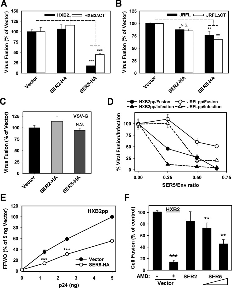
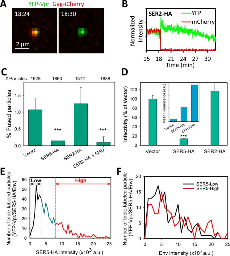
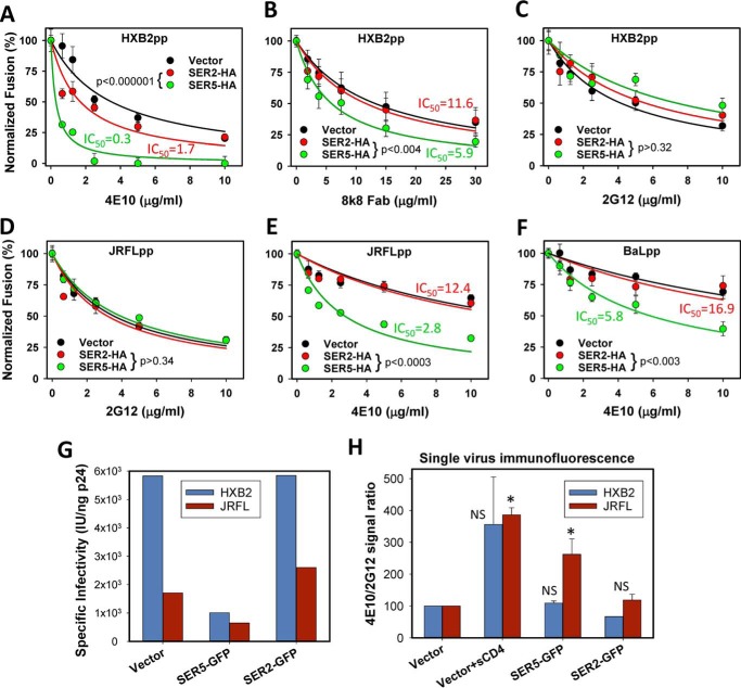
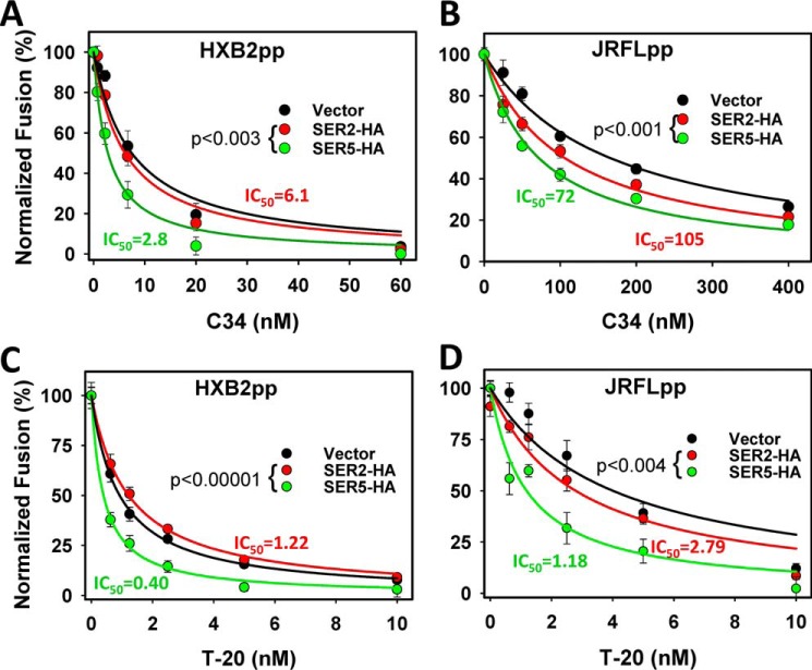
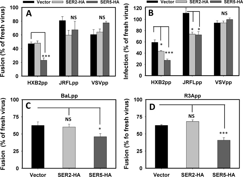
The host proteins, SERINC3 and SERINC5, have been recently shown to incorporate into HIV-1 particles and compromise their ability to fuse with target cells, an effect that is antagonized by the viral Nef protein. Envelope (Env) glycoproteins from different HIV-1 isolates exhibit a broad range of sensitivity to SERINC-mediated restriction, and the mechanism by which SERINCs interfere with HIV-1 fusion remains unclear. Here, we show that incorporation of SERINC5 into virions in the absence of Nef inhibits the formation of small fusion pores between viruses and cells. Strikingly, we found that SERINC5 promotes spontaneous functional inactivation of sensitive but not resistant Env glycoproteins. Although SERINC5-Env interaction was not detected by co-immunoprecipitation, incorporation of this protein enhanced the exposure of the conserved gp41 domains and sensitized the virus to neutralizing antibodies and gp41-derived inhibitory peptides. These results imply that SERINC5 restricts HIV-1 fusion at a step prior to small pore formation by selectively inactivating sensitive Env glycoproteins, likely through altering their conformation. The increased HIV-1 sensitivity to anti-gp41 antibodies and peptides suggests that SER5 also delays refolding of the remaining fusion-competent Env trimers.

Keywords: Env inactivation; HIV neutralization; conformational changes; fluorescence; hemifusion; host defense; membrane fusion; membrane-proximal extracellular domain; peptides; virus entry.


Conflict of interest statement

The authors declare that they have no conflicts of interest with the contents of this article

Figures

Figure 1.
Figure 1.
SER5 inhibits HIV-1 fusion. A–C, Nef-negative HIV-1 pseudoviruses bearing full-length or cytoplasmic tail-deleted (ΔCT) HXB2 Env, JRFL Env, or VSV-G, either lacking or containing SER5-HA, were allowed to enter TZM-bl cells, and the resulting fusion was measured by the BlaM assay. N.S., not significant. D, comparison of virus fusion and infection in TZM-bl cells for HXB2pp and JRFLpp produced in cells transfected with increasing amounts of the SER5-HA plasmid and constant amount of the Env plasmid. E, effect of SER5 on FFWO mediated by HXB2pp. Increasing p24 quantities of HXB2pp produced in the presence or absence of SER5-HA were added to a mixed confluent monolayer of N4X4-DSP-1 and N4X4-DSP-2 cells by spinoculation. Fusion was allowed to proceed for 2 h at 37 °C. Data are mean and S.E. of two independent experiments in triplicate. F, inhibition of HXB2 Env-mediated cell-cell fusion by SER5. The N4X4-DSP-2 cells were overlaid with 293-DSP-1 cells transiently transfected with equal amounts of full-length HXB2 Env and SER2-GFP, SER5-GFP, or empty vector or 4:1 ratio of Env/SER5 (penultimate bar). The fusion efficiency was measured after 2 h by dual-split luciferase assay, as in E. Viruses lacking SER5 (Vector) were additionally treated with 100 μm AMD3100 as negative control. Data are mean and S.D. of two independent experiment in triplicates.
Figure 2.
Figure 2.
SER5 does not considerably promote lysosomal degradation of HIV-1 or stall fusion at a hemifusion stage. A, fusion of HXB2pp containing or lacking SERINCs with TZM-bl cells was synchronized by pre-binding the virus in the cold and shifting to 37 °C. After 30 min at 37 °C, cells were treated with 0.5 mm CPZ for 30 s and washed, and incubation was continued for 60 min. BMS-529 (10 μm) was added to control wells to block HXB2pp fusion. B, CPZ treatment promotes VSVpp fusion independent of the presence of SERINCs. The VSVpp fusion protocol and the CPZ treatment step were as in A. Negative control included NH4Cl (70 mm). Data are mean and S.E. of two independent experiments in triplicate. C, cell viability for the experiments in A and B. D, SER5-HA viruses are marginally more degraded compared with control viruses. HXB2pp containing or lacking SERINCs were pre-bound to TZM-bl cells in the cold and incubated at 37 °C for 90 min in the presence or absence of 50 nm BafA1 or 10 μm of the HIV-1 fusion inhibitor BMS-529. Virus-cell fusion was measured by the BlaM assay. Data are mean and S.E. of two independent experiments in triplicate.
Figure 3.
Figure 3.
SER5 restricts fusion pore formation between single HXB2pp and target cell. HXB2pp co-labeled with YFP-Vpr and Gag-iCherry were produced in cells transfected with SER5-HA, SER2-HA, or an empty vector. Virions were pre-bound to CD4/CXCR4-expressing CV1 cells in the cold, and their fusion was initiated by shifting to 37 °C. A and B, images and single particle tracking results for fusion of SER2-HA containing HXB2pp showing the release of the mCherry marker. C, analysis of the effect of SER5 and SER2 on single HXB2pp fusion in the absence or in the presence of 10 μm AMD3100. Data are means and S.D. from 4 to 5 independent experiments. The numbers of double-labeled particles analyzed for each condition are shown above the graph. D, infectivity of the viral preparations analyzed in C in TZM-bl cells. Inset, percent fluorescent virions after immunostaining for HA tag to control for SER2/SER5 incorporation. E, SER5-HA fluorescence intensity distribution from imaging single pseudoviruses immunostained with anti-HA and 2G12 antibodies. Arbitrary classification of particles based on their low and high SER5 content is shown (black and red lines). F, Env content (2G12 staining intensity) distribution for low versus high SER5 particles (black and red lines, respectively).
Figure 4.
Figure 4.
SER5 and CCR5 display similar long range mobility in the plasma membrane. SER5-GFP or CCR5-GFP was transiently expressed in CV-1 cells. A, small circular regions within the basal plasma membrane of a CV-1 cell transfected with SER5-GFP were photobleached, and recovery of fluorescence over time was measured. B, top, small section of the plasma membrane scanned during FRAP acquisition shows circular areas used for measuring background, fluorescence recovery, and control intensity (circled white, red, and green, respectively) immediately after bleaching. B, bottom, corrected trace of sum intensity in the FRAP region was fit to an exponential rise function to calculate the half-time of intensity recovery (t½) and immobile fraction. C and D, comparison of the diffusion coefficient (C) and immobile fraction (D) of CCR5-GFP and SER5-GFP expressed in the plasma membrane. Data are mean and S.D. of two experiments (>10 measurements for each condition).
Figure 5.
Figure 5.
SER5 potentiates the neutralizing activity of antibodies and peptides against cryptic gp41 epitopes exposed during fusion. A–F, HIV-1 particles pseudotyped with HXB2, JRFL, or BaL26 Env, lacking or containing SER5-HA or SER2-HA, were used to inoculate TZM-bl cells, and the resulting fusion was measured by the BlaM assay. Indicated concentrations of neutralizing antibodies were added after virus pre-binding in the cold, immediately before raising the temperature. Data are means of two independent experiments performed in triplicate. The IC50 values obtained by curve-fitting and statistical significance of the effects of SER5 on HIV-1 sensitivity to neutralization (sum of squares reduction test) are shown on the plots. G and H, specific infectivity (G) and 4E10/2G12 median single-particle staining intensity ratio (H) for HXB2pp and JRFLpp. Data are means and S.D. from two independent experiments. The sCD4-induced increase in 4E10 binding was statistically significant for JRFLpp but not HXB2 particles due to a large variance between the immunofluorescence experiments. See also supplemental Fig. S6.
Figure 6.
Figure 6.
Enhanced sensitivity of HXB2 and JRFL pseudoviruses to inhibition by gp41-derived peptides. A and B, dose-dependent inhibition of control and SER2- or SER5-containing HXB2pp and JRFLpp by C34. C and D, effect of SER5 on sensitivity of HXB2pp and JRFLpp to T-20 peptide. Virus fusion with TZM-bl cells was measured by the BlaM assay. Data are means of two independent experiments performed in triplicate. The IC50 values obtained by curve-fitting and statistical significance of the SER5 effects on the HIV-1 sensitivity to inhibitory peptides, as determined by the sum of squares reduction test, are shown on the plots.
Figure 7.
Figure 7.
SER5 selectively promotes functional inactivation of HXB2 Env. A, Nef-negative HIV-1 particles pseudotyped with HXB2 Env, JRFL Env, or VSV-G, containing or lacking SER5-HA or SER2-HA, were preincubated in growth medium containing 10 mm HEPES (pH 7.3) for 4 h at 37 °C or used immediately after thawing the samples (fresh). Viruses were bound to TZM-bl cells in the cold by spinoculation, and cells were washed and incubated at 37 °C for 90 min. The fusion efficiency measured by the BlaM assay was normalized to the respective freshly initiated virus. B, pseudovirus infectivity was measured using conditions described in A. C and D, SER5 promotes spontaneous inactivation of pseudoviruses bearing BaL and R3A Env, as measured by a virus-cell fusion assay after a 4-h preincubation at 37 °C, as above. Data are mean and S.E. of two independent experiments in triplicates.

References

    1. Aiken C., and Trono D. (1995) Nef stimulates human immunodeficiency virus type 1 proviral DNA synthesis. J. Virol. 69, 5048–5056 - PMC - PubMed
    1. Schwartz O., Maréchal V., Danos O., and Heard J. M. (1995) Human immunodeficiency virus type 1 Nef increases the efficiency of reverse transcription in the infected cell. J. Virol. 69, 4053–4059 - PMC - PubMed
    1. Day J. R., Münk C., and Guatelli J. C. (2004) The membrane-proximal tyrosine-based sorting signal of human immunodeficiency virus type 1 gp41 is required for optimal viral infectivity. J. Virol. 78, 1069–1079 - PMC - PubMed
    1. Cavrois M., Neidleman J., Yonemoto W., Fenard D., and Greene W. C. (2004) HIV-1 virion fusion assay: uncoating not required and no effect of Nef on fusion. Virology 328, 36–44 - PubMed
    1. Campbell E. M., Nunez R., and Hope T. J. (2004) Disruption of the actin cytoskeleton can complement the ability of Nef to enhance human immunodeficiency virus type 1 infectivity. J. Virol. 78, 5745–5755 - PMC - PubMed

Publication types

MeSH terms

Substances

LinkOut - more resources