

Severe Chromoblastomycosis-Like Cutaneous Infection Caused by Chrysosporium keratinophilum
- PMID: 28179902
- PMCID: PMC5264138
- DOI: 10.3389/fmicb.2017.00083
Severe Chromoblastomycosis-Like Cutaneous Infection Caused by Chrysosporium keratinophilum
Abstract
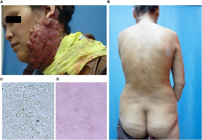
Chrysosporium species are saprophytic filamentous fungi commonly found in the soil, dung, and animal fur. Subcutaneous infection caused by this organism is rare in humans. We report a case of subcutaneous fungal infection caused by Chrysosporium keratinophilum in a 38-year-old woman. The patient presented with severe chromoblastomycosis-like lesions on the left side of the jaw and neck for 6 years. She also got tinea corporis on her trunk since she was 10 years old. Chrysosporium keratinophilum was isolated from the tissue on the neck and scales on the trunk, respectively. The patient showed satisfactory response to itraconazole therapy, although she discontinued the follow-up.
Keywords: Chrysosporium keratinophilum; cutaneous infection; diagnosis; fungal infection; treatment.
Figures


References
-
- De Hoog G. S., Guarro J., Figueras M. J., Gené J., Hubalek Z. (2000). Atlas of Clinical Fungi 2nd Edn Spain: Reus.
-
- Hocquette A., Grondin M., Bertout S., Mallié M. (2005). Acremonium, Beauveria, Chrysosporium, Fusarium, Onychocola, Paecilomyces, Penicillium, Scedosporium and Scopulariopsis fungi responsible for hyalohyphomycosis. J. Mycol. Méd. 15 136–149.
-
- Hubalek Z. (2000). Keratinophilic fungi associated with free-living mammals and birds: revista iberoamericana de micologia. Rev. Iberoam. Micol. 17 93–103.
LinkOut - more resources
Full Text Sources
Other Literature Sources
Research Materials