

Cancer stem-like cells can be induced through dedifferentiation under hypoxic conditions in glioma, hepatoma and lung cancer
- PMID: 28179999
- PMCID: PMC5253691
- DOI: 10.1038/cddiscovery.2016.105
Cancer stem-like cells can be induced through dedifferentiation under hypoxic conditions in glioma, hepatoma and lung cancer
Abstract
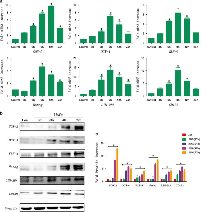
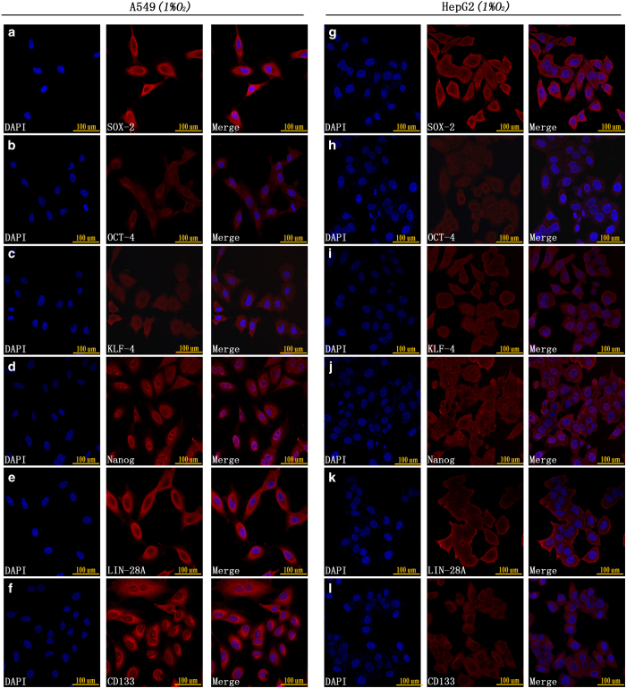
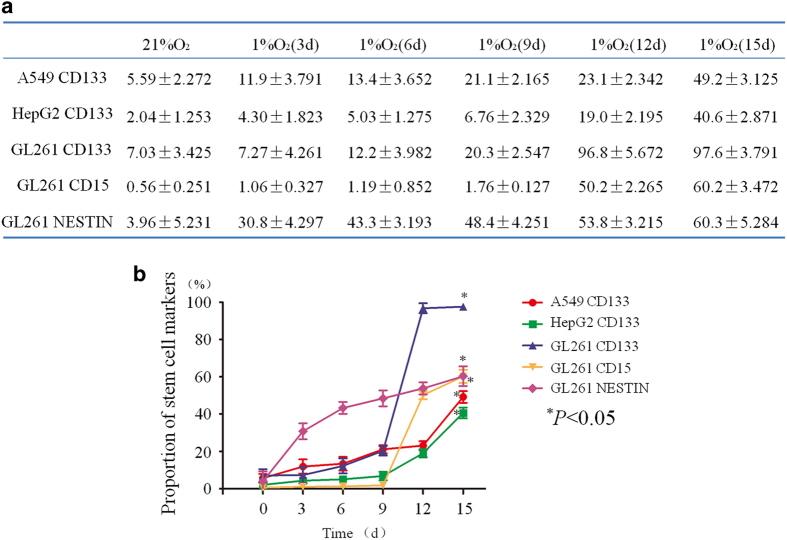
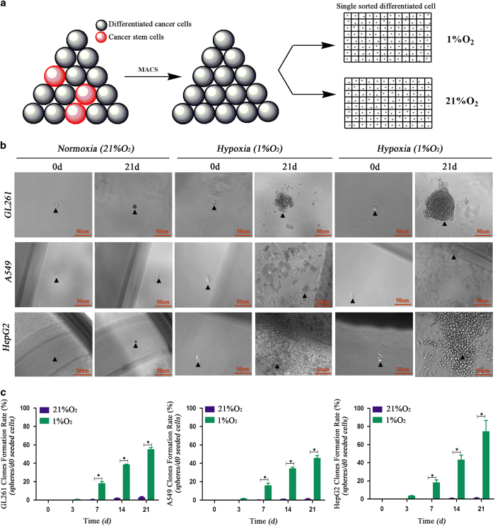
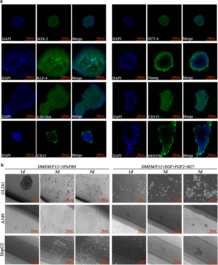
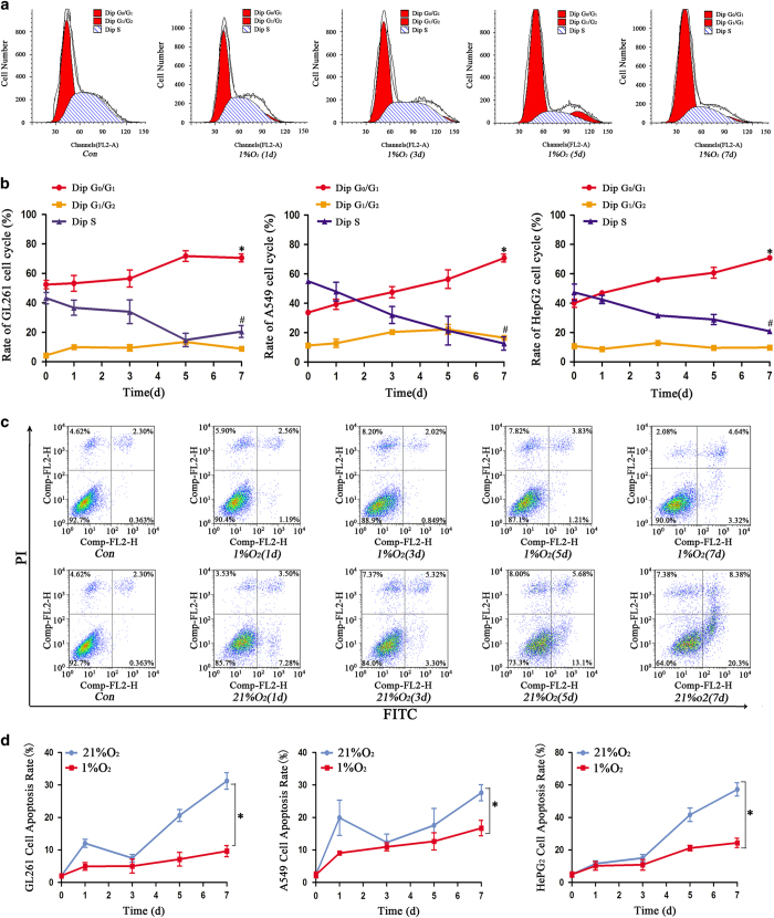
Traditional studies have shown that transcription factors, including SOX-2, OCT-4, KLF-4, Nanog and Lin-28A, contribute to the dedifferentiation and reprogramming process in normal tissues. Hypoxia is a physiological phenomenon that exists in tumors and promotes the expression of SOX-2, OCT-4, KLF-4, Nanog and Lin-28A. Therefore, an interesting question is whether hypoxia as a stimulating factor promotes the process of dedifferentiation and induces the formation of cancer stem-like cells. Studies have shown that OCT-4 and Nanog overexpression induced the formation of cancer stem cell-like cells through dedifferentiation and enhanced malignancy in lung adenocarcinoma, and reprogramming SOX-2 in pancreatic cancer cells also promoted the dedifferentiation process. Therefore, we investigated this phenomenon in glioma, lung cancer and hepatoma cells and found that the transcription factors mentioned above were highly expressed under hypoxic conditions and induced the formation of spheres, which exhibited asymmetric division and cell cycle arrest. The dedifferentiation process induced by hypoxia highlights a new pattern of cancer development and recurrence, demonstrating that all kinds of cancer cells and the hypoxic microenvironment should be taken into consideration when developing tumor therapies.
Figures
Similar articles
-
HIF1α/HIF2α induces glioma cell dedifferentiation into cancer stem cells through Sox2 under hypoxic conditions.J Cancer. 2022 Jan 1;13(1):1-14. doi: 10.7150/jca.54402. eCollection 2022. J Cancer. 2022. PMID: 34976166 Free PMC article.
-
HIF1α regulates single differentiated glioma cell dedifferentiation to stem-like cell phenotypes with high tumorigenic potential under hypoxia.Oncotarget. 2017 Apr 25;8(17):28074-28092. doi: 10.18632/oncotarget.15888. Oncotarget. 2017. PMID: 28427209 Free PMC article.
-
HIF1α regulates glioma chemosensitivity through the transformation between differentiation and dedifferentiation in various oxygen levels.Sci Rep. 2017 Aug 11;7(1):7965. doi: 10.1038/s41598-017-06086-2. Sci Rep. 2017. PMID: 28801626 Free PMC article.
-
Reprogramming cancer cells in endocrine-related tumors: open issues.Curr Med Chem. 2014;21(9):1146-51. doi: 10.2174/0929867321666131129125624. Curr Med Chem. 2014. PMID: 24304280 Review.
-
Hypoxia-induced dedifferentiation of tumor cells--a mechanism behind heterogeneity and aggressiveness of solid tumors.Semin Cell Dev Biol. 2005 Aug-Oct;16(4-5):554-63. doi: 10.1016/j.semcdb.2005.03.007. Epub 2005 Apr 26. Semin Cell Dev Biol. 2005. PMID: 16144692 Review.
Cited by
-
Prognostic value of differentiation status in gastric cancer.BMC Cancer. 2018 Sep 3;18(1):865. doi: 10.1186/s12885-018-4780-0. BMC Cancer. 2018. PMID: 30176846 Free PMC article.
-
Targeting hypoxic cancer stem cells (CSCs) with Doxycycline: Implications for optimizing anti-angiogenic therapy.Oncotarget. 2017 Jun 12;8(34):56126-56142. doi: 10.18632/oncotarget.18445. eCollection 2017 Aug 22. Oncotarget. 2017. PMID: 28915578 Free PMC article.
-
The HIF1α/HIF2α-miR210-3p network regulates glioblastoma cell proliferation, dedifferentiation and chemoresistance through EGF under hypoxic conditions.Cell Death Dis. 2020 Nov 18;11(11):992. doi: 10.1038/s41419-020-03150-0. Cell Death Dis. 2020. PMID: 33208727 Free PMC article.
-
Targeting GSTP1 as Therapeutic Strategy against Lung Adenocarcinoma Stemness and Resistance to Tyrosine Kinase Inhibitors.Adv Sci (Weinh). 2023 Mar;10(7):e2205262. doi: 10.1002/advs.202205262. Epub 2023 Jan 29. Adv Sci (Weinh). 2023. PMID: 36709476 Free PMC article.
-
Tumour Microenvironment Stress Promotes the Development of Drug Resistance.Antioxidants (Basel). 2021 Nov 11;10(11):1801. doi: 10.3390/antiox10111801. Antioxidants (Basel). 2021. PMID: 34829672 Free PMC article. Review.
References
-
- Takahashi K, Tanabe K, Ohnuki M, Narita M, Ichisaka T, Tomoda K et al. Induction of pluripotent stem cells from adult human fibroblasts by defined factors. Cell 2007; 131: 861–872. - PubMed
-
- Lee HK, Morin P, Wells J, Hanlon EB, Xia W. Induced pluripotent stem cells (iPSCs) derived from frontotemporal dementia patient's peripheral blood mononuclear cells. Stem Cell Res 2015; 15: 325–327. - PubMed
LinkOut - more resources
Full Text Sources
Other Literature Sources
Research Materials
Miscellaneous
