

Review
doi: 10.1055/s-0036-1593840.
Epub 2016 Nov 10.
An Update on Molecular Diagnostic Testing of Human Imprinting Disorders
Affiliations
- PMID: 28180023
- PMCID: PMC5288000
- DOI: 10.1055/s-0036-1593840
Item in Clipboard
Review
An Update on Molecular Diagnostic Testing of Human Imprinting Disorders
J Pediatr Genet.
2017 Mar.
Abstract
Imprinted genes are expressed in a parent of origin manner. Dysregulation of imprinted genes expression causes various disorders associated with abnormalities of growth, neurodevelopment, and metabolism. Molecular mechanisms leading to imprinting disorders and strategies for their diagnosis are discussed in this review article.
Keywords: DNA methylation; epigenetics; genomic imprinting.
Figures
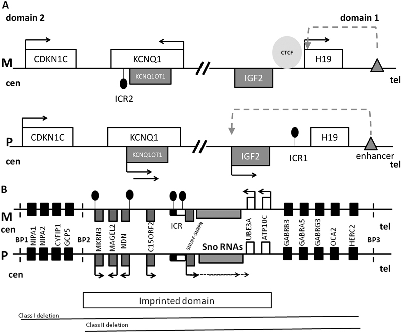
(
A
) The map of chromosome 11p15.5 imprinted cluster consisting of domain 1 and 2. M is maternal chromosome, P is paternal chromosome, cen is centromere, tel is telomere, white rectangles are maternally expressed genes, gray rectangles are paternally expressed genes, black arrows indicate gene expression, black circles indicate DNA methylation, gray triangle is enhancer, dashed arrow indicates accessibility of IGF2 promoter to the enhancer, and light gray circle is CCCTC-binding factor (CTCF). In domain 1, imprinting control region 1 (ICR1) is located upstream of H19 noncoding RNA and is methylated on the paternal allele. Methylation prevents CTCF binding to the paternal ICR1, permitting access of the IGF2 promoter to the downstream enhancer. Thus, IGF2 is expressed, whereas H19 is silenced. On the maternal allele, CTCF binds to the unmethylated ICR1, blocking IGF2 promoter access to the enhancer. Thus, IGF2 is silenced and H19 uses the enhancer and is transcribed. In domain 2, ICR2 is located at the 5′ end of the noncoding RNA KCNQ1OT1 (KCNQ1 opposite strand transcript) and is methylated on the maternal allele. When ICR2 is unmethylated, KCNQOT1 is transcribed, KCNQ1 and CDKN1C are silenced in
cis
. Methylation of ICR2 results in KCNQOT1 silencing and expression of KCNQ1 and CDKN1C. IGF2 promotes growth, and CDKN1C suppresses growth. Overexpression of IGF2 and/or loss of expression of CDKN1C is associated with Beckwith–Wiedemann syndrome (overgrowth), whereas overexpression of CDKN1C and/or loss of expression of IGF2 is associated with Russell–Silver syndrome (growth restriction). (
B
) Map of human chromosomal region 15q11-q13 involved in Prader-Willi (PWS), Angelman (AS), and 15q11q13 maternal duplication syndromes, containing an imprinted domain as well as biallelically expressed genes. M is maternal chromosome, P is paternal chromosome, cen is centromere, tel is telomere, white rectangles are maternally expressed genes, gray rectangles are paternally expressed genes, black arrows indicate gene expression in a parent of origin-specific manner, and black rectangles are biallelically expressed genes. Imprinted control region (ICR) is shown as rounded rectangle with the black half being the PWS critical element and the white part the AS critical element; black circles indicate DNA methylation. SNURF/SNRPN gene expression is regulated by ICR methylation and it is expressed in multiple splice forms, including the UBE3A antisense transcript which is paternally expressed in brain and silences paternal UBE3A transcription in brain. Clusters of C/D box small nucleolar RNAs (snoRNAs) are encoded within introns of SNURF-SNRPN and are regulated by its promoter. Promoters of MKRN3 and NDN are methylated on the maternal allele. Dashed vertical lines show the regions of common break points (BP) located within regions of low copy repeats. The most common causes of PWS and AS are paternal and maternal deletions, respectively, occurring between BP1 and BP3 or between BP2 and BP3. It is known that the critical gene for AS is UBE3A which is expressed from the maternal allele in human brain and biallelically in other tissues. The relative contributions of single genes to PWS and maternal 15q11-q13 duplication syndrome remain unknown. The drawing is not to scale.
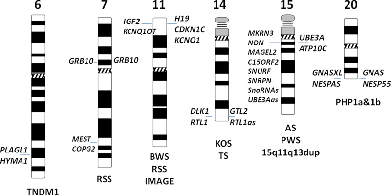
Map of human imprinted domains known to be associated with disease. Paternally expressed genes are shown on the left side of the chromosome and maternally expressed genes are shown on the right side of the chromosome (parent of origin-specific expression of GRB10 is different in different tissues). Associated conditions are shown under the chromosome. TNDM1 is transient neonatal diabetes mellitus (TNDM1), RSS is Russell–Silver syndrome, BWS is Beckwith–Wiedemann syndrome, KOS is Kagami-Ogata syndrome, TS is Temple syndrome, AS is Angelman syndrome, PWS is Prader-Willi syndrome, and PHP1a&b are pseudo-hypoparathyroidism types 1a&b.
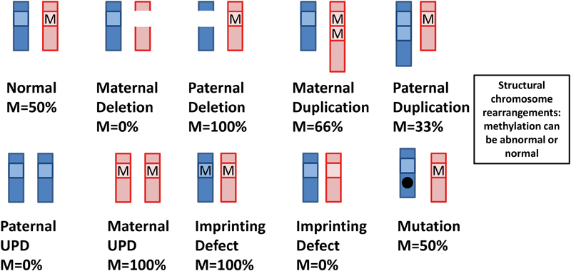
Schematic drawing of types of molecular alterations observed in imprinting disorders and their effect on DNA methylation at imprinting control regions (ICRs) in a scenario when methylation occurs on the maternal allele. The paternal chromosome is shown in light gray, and the maternal chromosome is shown in dark gray. M- is DNA methylation at the ICR. Imprinting defect can result either from idiopathic DNA methylation change or microdeletion/microduplication at ICR.
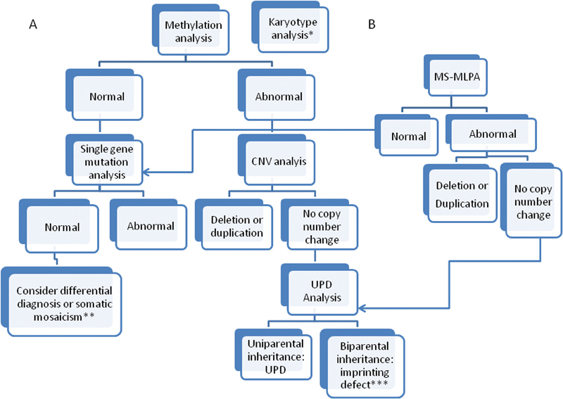
Testing strategies for imprinting disorders (ID) based on initial methylation analysis (
A
) or simultaneous methylation and CNV analysis by MS-MLPA (
B
). *Karyotype analysis to test for structural chromosome rearrangements can be initiated at the same time as methylation analysis if this is a known cause for an ID. **If all testing is negative, but the phenotype is highly suggestive of a specific ID, testing of another cell type (skin fibroblasts, buccal cells) can be considered to detect somatic mosaicism. ***In the majority of cases, an ID will be due to an idiopathic methylathion change, however depending on the resolution of previous CNV analysis, testing for ICR microdeletion/microduplication can be performed to rule out inherited genomic alterations.
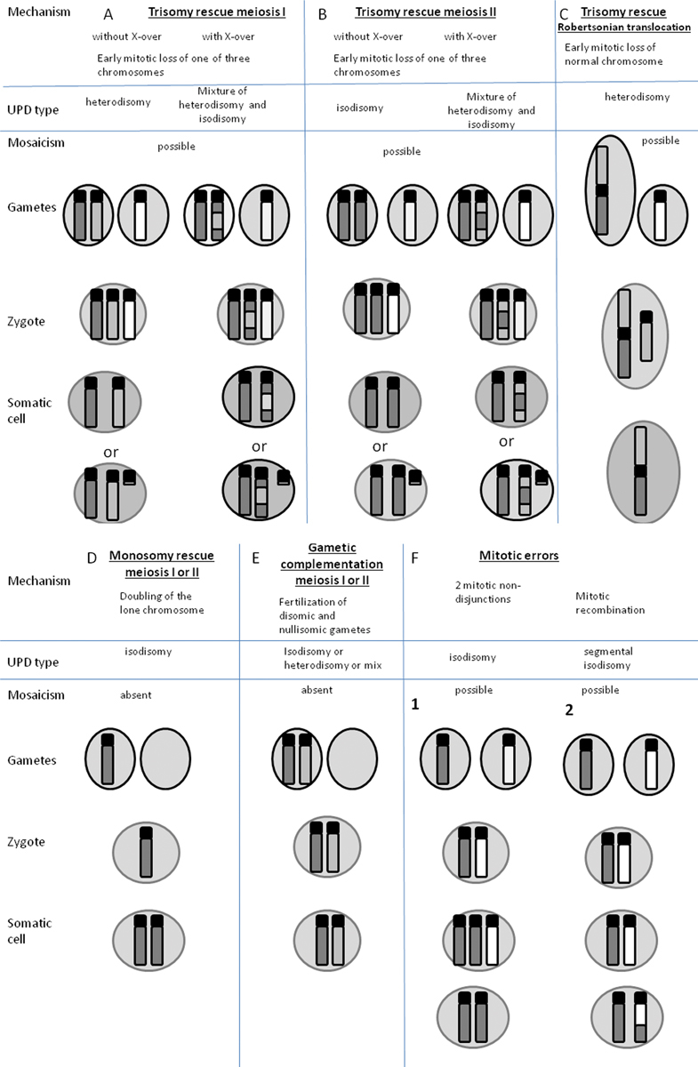
Possible mechanisms of uniparental disomy (UPD) formation. Dark gray and light gray are maternal nonhomologous chromosomes, white is paternal chromosomes. The examples of UPD are shown for maternal chromosomes, however paternal UPD can occur by the same mechanism. Where mosaicism is shown as a possible outcome, there is a possibility of the presence of trisomic cells in the placenta or the fetus for (
A
), (
B
), (
C
), and (
F1)
, in the case of segmental isodisomy (
F2
), mosaicism is present for a biparental cell line. (
A
,
B
,
C
) Trisomy can result from meiosis I or II nondisjunction in a parent with a normal karyotype. Rescue can occur by complete loss of one of the parental chromosomes or formation of a residual small supernumary marker chromosome. Trisomy rescue and UPD can also occur if the egg or sperm carries a balanced translocation and nondisjunction occurs. (
C
) Oocyte carriers of a homologous Robertsonian translocation or isochromosome. (
D
) Monosomic fertilized zygote has to undergo immediate endoreduplication of monosomic chromosomes, otherwise the embryo won't survive. (
E
) Gamete complementation is a very rare event as it assumes nondisjunction of the same chromosome in both oocyte and sperm. (
F1
) Two sequential mitotic nondisjunction events can lead to isodisomy. (
F2
) Segmental UPD can result from homologous chromatid exchange during mitosis, resulting in segmental loss of heterozygosity and mosaicism for a UPD cell line and a biparental cell line. (This figure was adapted with modifications from Yamazawa K, Ogata T, Ferguson-Smith AC. 2010).
References
-
- Peters J. The role of genomic imprinting in biology and disease: an expanding view. Nat Rev Genet. 2014;15(08):517–530. - PubMed
-
- Kaneda M, Okano M, Hata Ket al.Essential role for de novo DNA methyltransferase Dnmt3a in paternal and maternal imprinting Nature 2004429(6994):900–903. - PubMed
-
- Bourc'his D, Xu G L, Lin C S, Bollman B, Bestor T H.Dnmt3L and the establishment of maternal genomic imprints Science 2001294(5551):2536–2539. - PubMed
-
- Messerschmidt D M, de Vries W, Ito M, Solter D, Ferguson-Smith A, Knowles B B.Trim28 is required for epigenetic stability during mouse oocyte to embryo transition Science 2012335(6075):1499–1502. - PubMed
Publication types
LinkOut - more resources
Full Text Sources
Other Literature Sources
