. 2017 Feb 17;45(3):1529-1538.
doi: 10.1093/nar/gkw1224.

Structural basis for the dimerization of Nab2 generated by RNA binding provides insight into its contribution to both poly(A) tail length determination and transcript compaction in Saccharomyces cerevisiae

Affiliations

Structural basis for the dimerization of Nab2 generated by RNA binding provides insight into its contribution to both poly(A) tail length determination and transcript compaction in Saccharomyces cerevisiae

Shintaro Aibara et al. Nucleic Acids Res. .

Abstract

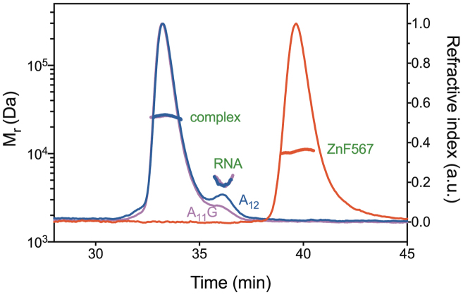
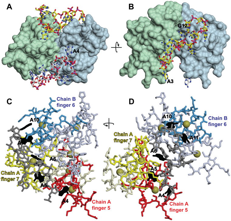
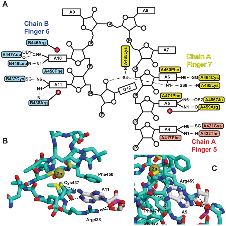
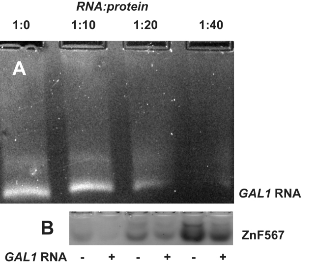
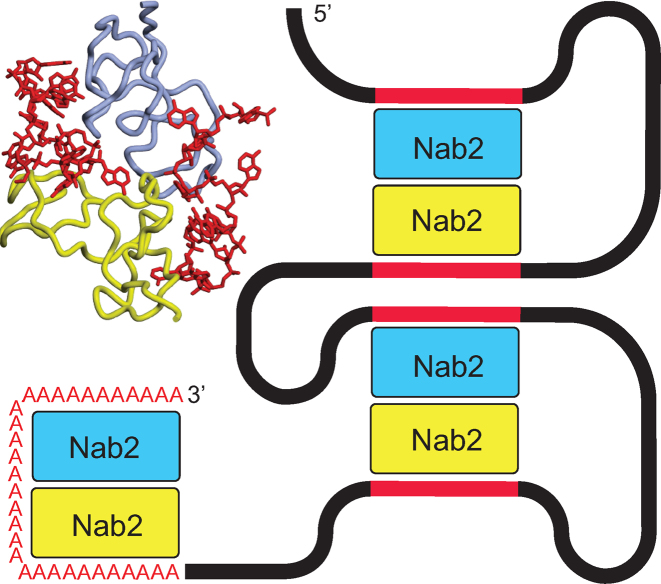
In Saccharomyces cerevisiae generation of export-competent mRNPs terminates the nuclear phase of the gene expression pathway and facilitates transport to the cytoplasm for translation. Nab2 functions in this process to control both mRNP compaction that facilitates movement through nuclear pore complexes and the length of transcript poly(A) tails. Nab2 has a modular structure that includes seven CCCH Zn fingers that bind to A-rich RNAs and fingers 5–7 are critical for these functions. Here, we demonstrate, using both biophysical and structural methods, that binding A11G RNA induces dimerization of Zn fingers 5–7 mediated by the novel spatial arrangement of the fingers promoting each RNA chain binding two protein chains. The dimerization of Nab2 induced by RNA binding provides a basis for understanding its function in both poly(A) tail length regulation and in the compaction of mature transcripts to facilitate nuclear export.


Figures

Figure 1.
Figure 1.
Schematic illustration of the domain structure of S. cerevisiae Nab2. The protein is based an N-terminal domain (40), an RGG domain, and seven Zn fingers, joined by flexible linkers. The Zn fingers are clustered into three groups (1+2, 3+4 and 567) and in each group the fingers have defined orientations to one another (17,18).
Figure 2.
Figure 2.
Multiangle light scattering data. Nab2 Zn fingers 567 (red) and the complexes formed with A11G (magenta) and A12 (blue) RNA that had an Mr consistent with the formation of a heterotetramer containing two protein and two RNA chains. A small excess of RNA was added to ensure complete complex formation. In contrast to the complexes formed with RNA that contained two protein chains, Nab2 Zn fingers 567 alone was monomeric.
Figure 3.
Figure 3.
Crystal structure of the ZnF567:A11G complex. (A, B) Overview of a heterotetramer containing two protein and two RNA chains showing how the adenine purine rings are buried into cavities in the surface of the Zn fingers. The dimerization of the two Nab2 Zn finger protein chains is mediated by each RNA chain binding to both protein chains (shown here as green and light blue), thereby linking them together. (C, D) Each RNA chain (gray) was bound to fingers 5 (red) and 7 (yellow) of one protein chain and finger 6 (blue) of the other protein chain. The interacting adenines are black. A4 was bound to finger 5 of chain A; A5 and A6 to finger 7 of chain A; and A10 and A11 to finger 6 of chain B (shown schematically in Figure 4A). Zn atoms are represented by gold spheres.
Figure 4.
Figure 4.
Interactions between Nab2 Zn fingers 5–7 and adenine bases. (A) Schematic illustration of the interactions between A11G RNA and Zn fingers 5–7. On one protein chain, Zn finger 5 (red) binds to base A4 while Zn finger 7 (yellow) binds bases A5 and A6, whereas in the second, protein chain in the dimer Zn finger 6 (blue) binds bases A10 and A11. In addition to hydrophobic interactions with an aromatic side chain and putative π interactions with a basic side chain, the specificity of the interaction involves H-bonds formed by the adenine N6 to either a SG of the finger or an appropriately positioned acidic side chain. Two different types of interaction between the purine ring and the protein mediate specific recognition of adenine. In one interaction, illustrated by A11 (B), the purine stacks against an aromatic side chain (Phe450) and forms π interactions with a basic side chain (Arg438) together with N6 forming a H-bond to the SG of one of the cysteines (Cys437) that is coordinated to Zn. In the second interaction (C), illustrated by A5, the purine ring is stacked against Phe450 and Leu449 on one side and forms π interactions with Arg445 on the other, as well as forming H-bonds between its N6 and the carbonyl and OD1 of Asp447 as well as between N1 and the amide of Leu449.
Figure 5.
Figure 5.
Binding of Nab2 ZnF567 to GAL1 RNA. (A) Adding progressively larger amounts of ZnF567 depleted the amount of GAL1 RNA that migrated to the positive electrode in native agarose gels. (B) Similarly, in native PAGE gels the Nab2 protein was depleted by GAL1 RNA. At a 10:1 ratio most of the protein was depleted and there was substantial depletion even when it was added at a 40:1 ratio, consistent with each RNA binding multiple copies of the protein.
Figure 6.
Figure 6.
Compaction of GAL1 RNA by Nab2 ZnF567. (A) Electron micrograph of complexes formed between GAL1 RNA and ZnF567 negatively stained with uranyl acetate following GraFix fixation. Fields contained roughly spherical stain-excluding particles of diameter of the order of 120 Å, consistent with the RNA becoming compacted following binding of the protein. (B) By contrast, rather than excluding uranyl acetate, in the absence of ZnF567 GAL1 RNA bound the stain and so appeared dark as is typically seen with nucleic acids (37). (C) When ZnF567F450A was bound to GAL1 RNA, the particles were less compact than those obtained with the wild-type protein.
Figure 7.
Figure 7.
Schematic illustration of Nab2 function in polyadenylation and compaction. A poly(A) tail length of ∼60 nt (red As) could be generated by RNA-binding mediated Nab2 dimerization with each Nab2 binding ∼30 nt. Similarly, Nab2 dimers generated by binding A-rich regions of the coding sequence (red sections) would link different parts of the transcript resulting in compaction.

References

    1. Singh G., Pratt G., Yeo G.W., Moore M.J.. The clothes make the mRNA: past and present trends in mRNP fashion. Annu. Rev. Biochem. 2015; 84:325–354. - PMC - PubMed
    1. Niño C.A., Hérissant L., Babour A., Dargemont C.. mRNA nuclear export in yeast. Chem. Rev. 2013; 113:8523–8545. - PubMed
    1. Katahira J. Nuclear export of messenger RNA. Genes. 2015; 6:163–184. - PMC - PubMed
    1. Stewart M. Nuclear export of mRNA. Trends Biochem. Sci. 2010; 35:609–617. - PubMed
    1. Oeffinger M., Zenklusen D.. To the pore and through the pore: a story of mRNA export kinetics. Biochim. Biophys. Acta. 2012; 1819:494–506. - PMC - PubMed

MeSH terms