

Dermal phospho-alpha-synuclein deposits confirm REM sleep behaviour disorder as prodromal Parkinson's disease
- PMID: 28180961
- PMCID: PMC5348554
- DOI: 10.1007/s00401-017-1684-z
Dermal phospho-alpha-synuclein deposits confirm REM sleep behaviour disorder as prodromal Parkinson's disease
Abstract
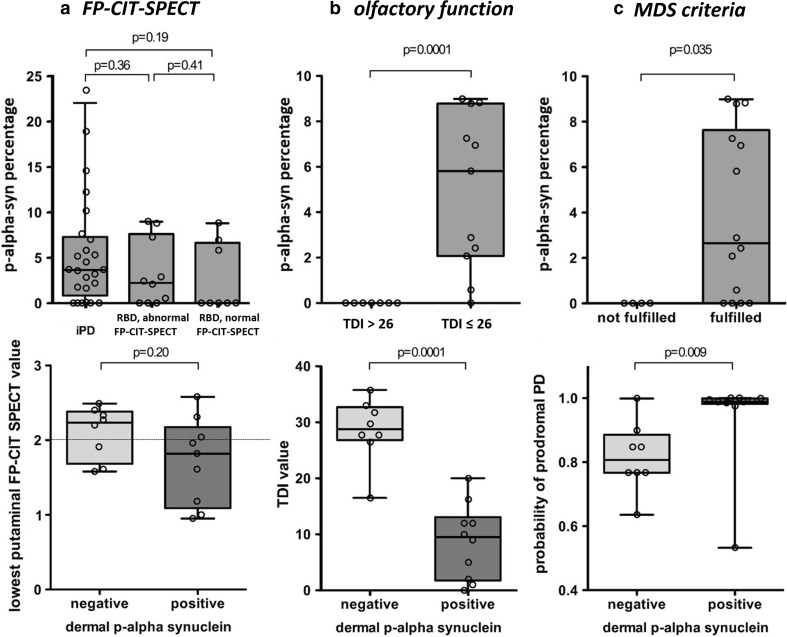
Phosphorylated alpha-synuclein (p-alpha-syn) deposits, one of the neuropathological hallmarks of Parkinson's disease (PD), have recently been detected in dermal nerve fibres in PD patients with good specificity and sensitivity. Here, we studied whether p-alpha-syn may serve as a biomarker in patients with a high risk of developing PD, such as those with REM sleep behaviour disorder (RBD). We compared the presence and distribution of p-alpha-syn deposits in dermal nerve fibres in 18 patients with RBD, 25 patients with early PD and 20 normal controls. Skin biopsy was taken at C7, Th10, and the upper and lower leg. Presynaptic dopamine transporter imaging using FP-CIT-SPECT was performed in all patients with RBD and in 11 patients with PD. All RBD patients underwent olfactory function testing. The likelihood ratio (LR) for prodromal PD was calculated for each patient based on published research criteria. Skin serial sections were assessed by double-immunofluorescence labelling with antibodies to pSer129-alpha-syn under blinded conditions. P-alpha-syn was visualized in 10/18 patients with RBD (sensitivity of 55.6%) and in 20/25 early PD patients (sensitivity of 80%) but in none of the controls (specificity of 100%). The percentage of dermal structures innervated by p-alpha-syn-positive fibres was negatively correlated with dopamine transporter binding in the FP-CIT-SPECT (ρ = -0.377, p = 0.048), with olfactory function (ρ = -0.668, p = 0.002), and positively correlated with the total LR for RBD to present prodromal PD (ρ = 0.531, p = 0.023). Dermal p-alpha-syn can be considered a peripheral histopathological marker of synucleinopathy and can be detected in a subgroup of RBD patients presumably representing prodromal PD. Dermal p-alpha-syn is detectable in RBD patients without PD motor symptoms, thereby stratifying a patient group that is of great interest for clinical trials testing disease-modifying drugs.
Keywords: Alpha-synuclein; FP-CIT-SPECT; Parkinson’s disease; REM sleep behaviour disorder; Skin biopsy.
Conflict of interest statement
The study was funded by a grant of Internationaal Parkinson Fonds, The Netherlands, to CS, WHO and KD. K. Doppler received personal fees for educational talks from Baxter/Baxalta and for educational material from Grifols. J. Booij received grants and personal fees from GE Healthcare. T. Musacchio received non-financial support from Merz, Allergan and Medtronic. J. Volkmann received grants and personal fees from Medtronic Inc. and Boston Scientific and personal fees from St. Jude. C. Sommer received personal fees from Baxter/Baxalta, Genzyme, Grifols, Air Liquide, CSL Behring, Kedrion, Novartis, Astellas and Pfizer for educational talks and/or consultancy and grant from Kedrion grants and is funded by the European Union's Seventh Framework Programme ("ncRNAPain", grant agreement number 602133), by Deutsche Forschungsgemeinschaft and the German ministry of education and research (CMT-Net). W.H. Oertel received personal fees for educational talks and/or consultancy from Abbvie, Adamas, Bristol-Myer-Squibb, Desitin, Mundipharma, Neuropore, Novartis, Roche and UCB Pharma and grants from the Deutsche Forschungs-Gemeinschaft, the Internationaal Parkinson-Fonds The Netherlands, the Michael J. Fox Foundation, USA, the National Research Fond Luxembourg and from Novartis Pharma, Germany.
Figures
References
-
- Beck A, Steer R, Brown G. The Beck depression inventory-second edition manual. San Antonio: The Psychological Corporation; 1996.
-
- Berg D, Postuma RB, Adler CH, Bloem BR, Chan P, Dubois B, Gasser T, Goetz CG, Halliday G, Joseph L, Lang AE, Liepelt-Scarfone I, Litvan I, Marek K, Obeso J, Oertel W, Olanow CW, Poewe W, Stern M, Deuschl G. MDS research criteria for prodromal Parkinson’s disease. Mov Disord. 2015;30:1600–1611. doi: 10.1002/mds.26431. - DOI - PubMed
-
- Boeve BF, Silber MH, Ferman TJ, Lin SC, Benarroch EE, Schmeichel AM, Ahlskog JE, Caselli RJ, Jacobson S, Sabbagh M, Adler C, Woodruff B, Beach TG, Iranzo A, Gelpi E, Santamaria J, Tolosa E, Singer C, Mash DC, Luca C, Arnulf I, Duyckaerts C, Schenck CH, Mahowald MW, Dauvilliers Y, Graff-Radford NR, Wszolek ZK, Parisi JE, Dugger B, Murray ME, Dickson DW. Clinicopathologic correlations in 172 cases of rapid eye movement sleep behavior disorder with or without a coexisting neurologic disorder. Sleep Med. 2013;14:754–762. doi: 10.1016/j.sleep.2012.10.015. - DOI - PMC - PubMed
-
- Booij J, Hemelaar TG, Speelman JD, de Bruin K, Janssen AG, van Royen EA. One-day protocol for imaging of the nigrostriatal dopaminergic pathway in Parkinson’s disease by [123I]FPCIT SPECT. J Nucl Med. 1999;40:753–761. - PubMed
Publication types
MeSH terms
Substances
LinkOut - more resources
Full Text Sources
Other Literature Sources
Medical
Miscellaneous
