. 2017 Jun;368(3):615-627.
doi: 10.1007/s00441-017-2575-2. Epub 2017 Feb 8.

Comprehensive functional analysis of Rab GTPases in Drosophila nephrocytes

Affiliations

Comprehensive functional analysis of Rab GTPases in Drosophila nephrocytes

Yulong Fu et al. Cell Tissue Res. 2017 Jun.

Abstract

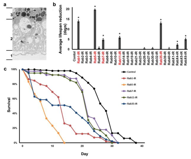
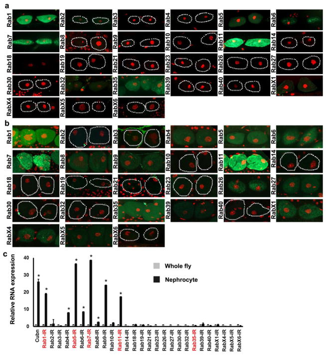
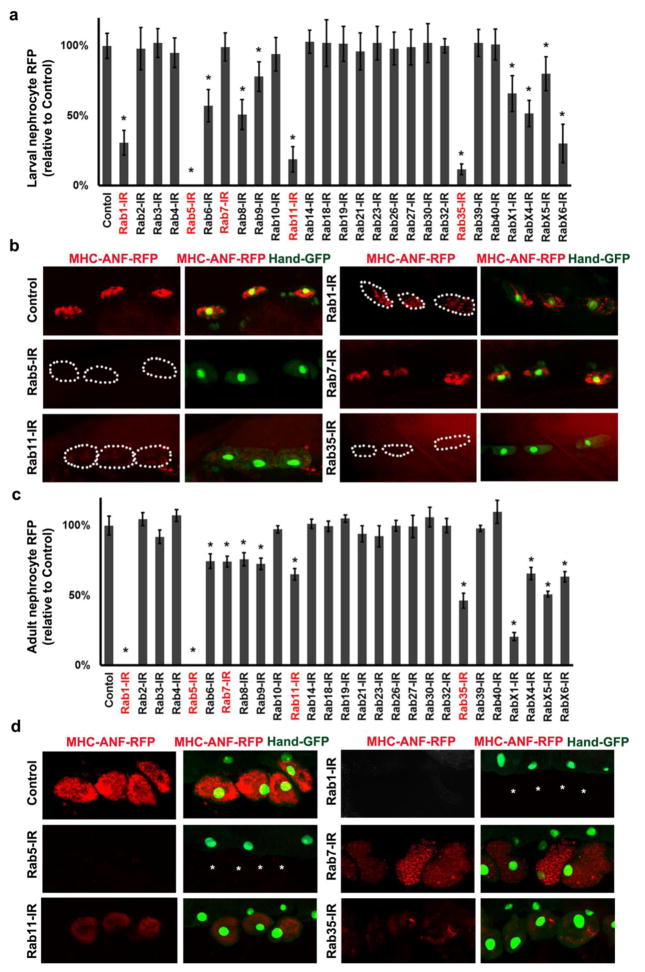
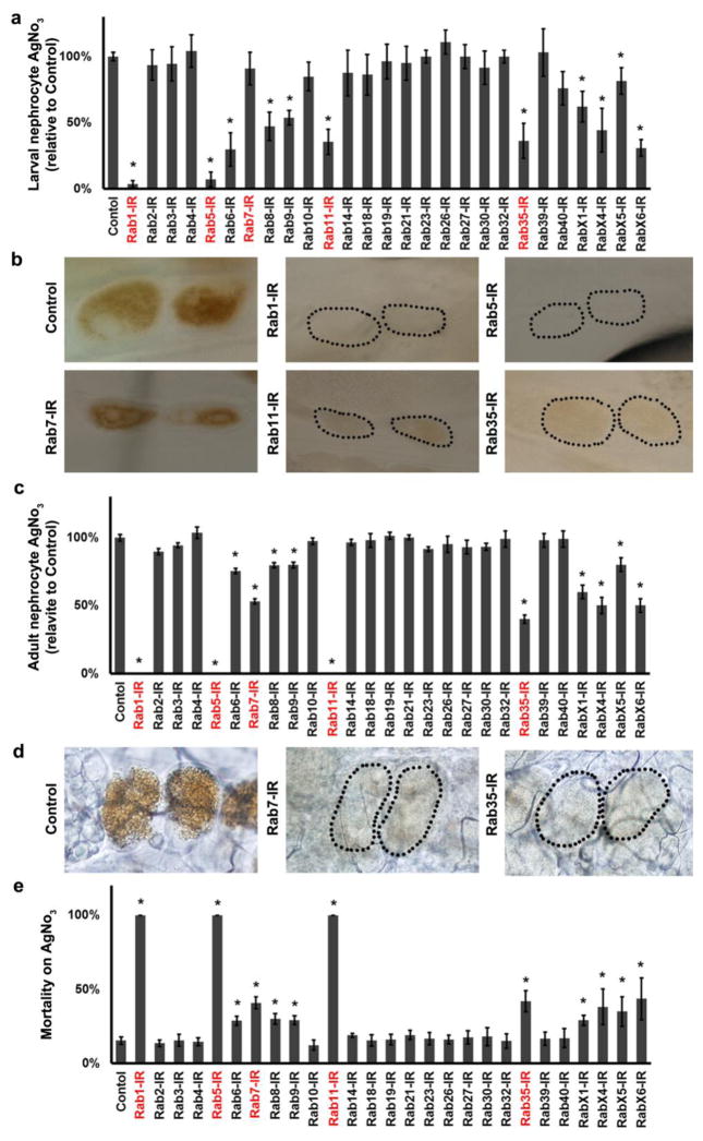
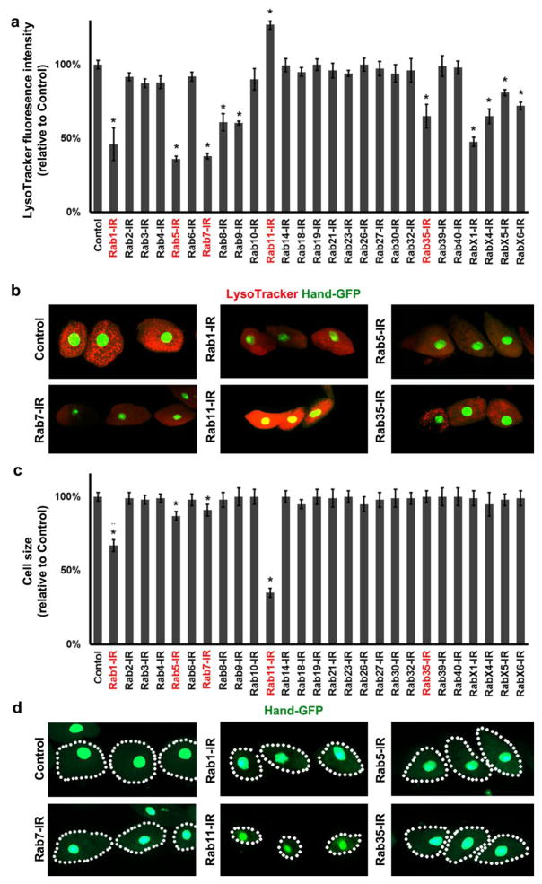
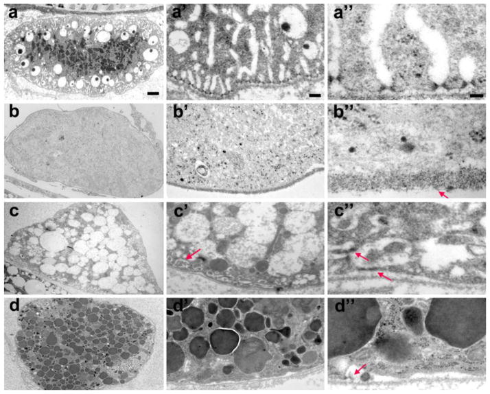
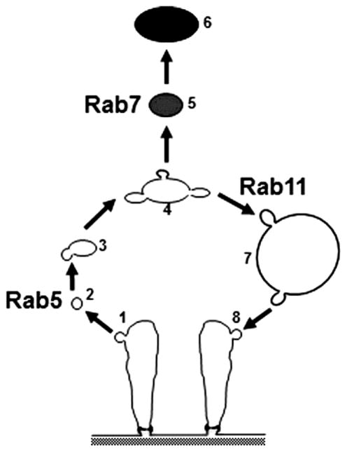
The Drosophila nephrocyte is a critical component of the fly renal system and bears structural and functional homology to podocytes and proximal tubule cells of the mammalian kidney. Investigations of nephrocyte cell biological processes are fundamental to understanding the insect renal system. Nephrocytes are highly active in endocytosis and vesicle trafficking. Rab GTPases regulate endocytosis and trafficking but specific functions of nephrocyte Rabs remain undefined. We analyzed Rab GTPase expression and function in Drosophila nephrocytes and found that 11 out of 27 Drosophila Rabs were required for normal activity. Rabs 1, 5, 7, 11 and 35 were most important. Gene silencing of the nephrocyte-specific Rab5 eliminated all intracellular vesicles and the specialized plasma membrane structures essential for nephrocyte function. Rab7 silencing dramatically increased clear vacuoles and reduced lysosomes. Rab11 silencing increased lysosomes and reduced clear vacuoles. Our results suggest that Rab5 mediates endocytosis that is essential for the maintenance of functionally critical nephrocyte plasma membrane structures and that Rabs 7 and 11 mediate alternative downstream vesicle trafficking pathways leading to protein degradation and membrane recycling, respectively. Elucidating molecular pathways underlying nephrocyte function has the potential to yield important insights into human kidney cell physiology and mechanisms of cell injury that lead to disease. The Drosophila nephrocyte is emerging as a useful in vivo model system for molecular target identification and initial testing of therapeutic approaches in humans.

Keywords: Drosophila; Endosome; Nephrocyte; Rab; Vesicle trafficking.


Conflict of interest statement

Conflict of interest

The authors declare that they have no conflict of interest.

Figures

Fig. 1
Fig. 1. Functional and cytoarchitectural domains of the Drosophila pericardial nephrocyte, adult fly mortality associated with Rab gene silencing, adult fly survival curves resulting from nephrocyte-specific silencing of genes encoding Rabs 1, 5, 7, 11, and 35
a TEM of pericardial nephrocyte showing the three functional and cytoarchitectural domains: domain 1 composed of basement membrane, and “foot processes” spanned by nephrocyte diaphragms; domain 2 containing lacunar channels formed by invaginations of the plasma membrane, and clear vesicles; domain 3 composed largely of lysosomes. b Effect of nephrocyte-specific Rab gene silencing on average lifespan reduction (in days, relative to wild type control) of adult flies maintained under standard conditions. Three replicates were analyzed per genotype. Values are mean ± s.d. of 3 separate samples. Results were analyzed by Student’s t-test. Statistical significance was defined as P<0.05. c Adult fly survival curves associated with nephrocyte-specific silencing of Rabs 1, 5, 7, 11, and 35.
Fig. 2
Fig. 2. Rab protein expression in nephrocytes, and relative RNA abundance in heart tissue
Fluorescence micrographs showing a third instar larvae and b adult nephrocyte expression of endogenously tagged YFP-Rab proteins (green). Nephrocyte nuclei labeled with DAPI (red). Dotted lines indicate outlines of nephrocytes. c qRT-PCR analysis of Rab RNA enrichment in dissected adult fly heart tissue (nephrocytes and cardiomyocytes) relative to whole fly. Cubilin (Cubn) was previously shown to be expressed predominantly in nephrocytes (Zhang, Zhao, Chao, Muir and Han, 2013a) and was thus used as a positive control. Values are mean ± s.d. of 3 separate samples. Results were analyzed by Student’s t-test. Statistical significance was defined as P<0.05.
Fig. 3
Fig. 3. Nephrocyte uptake of hemolymph protein
a Second instar larvae nephrocytes in which the indicated Rab gene was silenced were quantitatively assessed for uptake of ANF-RFP fusion protein from the hemolymph. Red fluorescent protein (RFP) fused to rat atrium natriuretic factor (ANF) is expressed in and secreted by muscle cells from a transgene driven by the MHC enhancer. ANF-RFP fluorescence in nephrocytes expressed as percent of WT control. b Fluorescence microscopy showing ANF-RFP uptake (red) by nephrocytes (GFP nuclear expression) in which the indicated Rab gene expression was silenced. Dotted lines indicate outlines of nephrocytes. c Adult flies in which the indicated Rab gene was silenced were quantitatively assessed for uptake of ANF-RFP fusion protein from the hemolymph. ANF-RFP in nephrocytes expressed as percent of WT control. d Fluorescence microscopy showing ANF-RFP (red) uptake by nephrocytes (GFP nuclear expression) in which the indicated Rab gene expression was silenced. Asterisks indicate missing nephrocytes as a result of Rab1 and Rab5 silencing. For quantification, 20 nephrocytes were analyzed from each of 3 larvae or flies per genotype. The results are presented as mean±s.d. Results were analyzed by Student’s t-test. Statistical significance was defined as P<0.05.
Fig. 4
Fig. 4. AgNO3 Uptake and Developmental Toxicity Assays
a Third instar larvae nephrocytes in which the indicated Rab gene was silenced were quantitatively assessed for sequestration of ingested AgNO3. AgNO3 in nephrocytes expressed as percent of WT control. b Micrographs showing sequestration of ingested AgNO3 in larval pericardial nephrocytes in which the indicated Rab gene expression was silenced. Dotted lines indicate outlines of nephrocytes. c Adult nephrocytes in which the indicated Rab gene was silenced were quantitatively assessed for sequestration of ingested AgNO3. AgNO3 in nephrocytes expressed as percent of WT control (note that Rab1 and Rab5 silencing result in absence of adult nephrocytes). d Micrographs showing sequestration of ingested AgNO3 in adult pericardial nephrocytes in which the indicated Rab gene expression was silenced. Dotted lines indicate outlines of nephrocytes. e Developmental toxicity from ingestion of dietary AgNO3. Mortality index is expressed as the % of second instar larvae of the indicated Rab silencing genotype that fail to develop into adult flies. For quantification, 20 nephrocytes were analyzed from each of 3 larvae or flies per genotype. The results are presented as mean±s.d. Results were analyzed by Student’s t-test. Statistical significance was defined as P<0.05.
Fig. 5
Fig. 5. Nephrocyte-specific gene silencing effects on lysosomes and nephrocyte cell size
a Quantitative analysis of Rab gene silencing effects on LysoTracker (lysosome marker) fluorescence intensity in nephrocytes of third instar larvae. Fluorescence intensity expressed as percent of WT control. b Micrographs showing LysoTracker dye fluorescence (red) in pericardial nephrocytes (green, labeled with nuclear GFP fluorescence from expression of Hand-GFP transgene) of larvae in which the indicated Rab gene was silenced. c Quantitation of larval nephrocyte cell size as a function of Rab gene silencing. Bar graphs shows cell area as percent of WT control nephrocytes. d Micrographs showing cell size of Rab-silenced larval nephrocytes (labeled by nuclear GFP fluorescence from expression of Hand-GFP transgene). Dotted lines delineate cell boundaries. For quantification, 20 nephrocytes were analyzed from each of 3 larvae per genotype. The results are presented as mean±s.d. Results were analyzed by Student’s t-test. Statistical significance was defined as P<0.05.
Fig. 6
Fig. 6. Rab5, Rab7 and Rab11 gene expression is required for normal third instar larvae nephrocyte ultrastructure and vesicle composition
a-a” In wild type control nephrocytes the lysosomes are concentrated in the interior of the cell, surrounded by clear vacuoles. NSDs and lacunar channels are precisely arrayed around the cell periphery. b-b” Rab5 gene silencing eliminated NSDs, lacunar channels, vacuoles, and lysosomes. The basement membrane was substantially thickened (arrow, b”). c-c” Rab7 gene silencing led to the accumulation of large clear vacuoles, and absence of lysosomes. Lacunar channels were greatly reduced in size and morphologically abnormal (arrow, c’), while NSDs were mislocalized and structurally deformed (arrows, c”). d-d” Rab11 gene silencing dramatically increased the number of lysosomes, at the expense of large clear vacuoles. Lacunar channels and NSDs were virtually absent, except for occasional remnants (arrow, d”). Scale bars: 2 microns (a–d), 0.2 micron (a’–d’), and 0.05 micron (a”–d”).
Fig. 7
Fig. 7. A model highlighting the central roles of Rab5, Rab7, and Rab11 in endocytic membrane trafficking in pericardial nephrocytes
Rab5 regulates the endocytosis of Clathrin-coated vesicles (1) from the plasma membrane of lacunar channels containing hemolymph components that have been filtered by the NSD. Vesicle uncoating (2) is followed by fusion of early endosomes (3) and formation of late and sorting endosomes (4). Rab7 directs vesicle cargo targeted for degradation (5) to the lysosomes (6). Rab11 regulates recycling of membrane lipids and membrane receptors via clear vacuoles (7) and recycling vesicles (8) back to the lacunar channel PM. In the absence of Rab5 the normal vesicle organization of the nephrocyte was abolished. In addition, the highly ordered cell surface structures featuring NSDs and lacunar channels were no longer present, indicating that endocytosis is essential for the formation and/or maintenance of the ultrastructural features required for nephrocyte hemolymph filtration, reabsorption of proteins, and sequestration of toxins. Silencing of Rab7 prevented the formation of lysosomes and promoted the proliferation and expansion of clear vacuoles. This was associated with disruption of the normal NSD and lacunar channel ultrastructure, though components of these structures were still visible in a disordered and compacted state at the cell periphery. Knockdown of Rab11, by contrast, led to superabundance of lysosomes and disappearance of clear vacuoles. This phenotype was associated with loss of NSDs and lacunar channels, presumably reflecting the requirement for Rab11 in recycling of materials to maintain these structures.

Similar articles

Cited by

References

    1. Akilesh S, Huber TB, Wu H, Wang G, Hartleben B, Kopp JB, Miner JH, Roopenian DC, Unanue ER, Shaw AS. Podocytes use FcRn to clear IgG from the glomerular basement membrane. Proceedings of the National Academy of Sciences of the United States of America. 2008;105:967–972. - PMC - PubMed
    1. Bechtel W, Helmstadter M, Balica J, Hartleben B, Kiefer B, Hrnjic F, Schell C, Kretz O, Liu S, Geist F, Kerjaschki D, Walz G, Huber TB. Vps34 deficiency reveals the importance of endocytosis for podocyte homeostasis. Journal of the American Society of Nephrology : JASN. 2013;24:727–743. - PMC - PubMed
    1. Cagan RL. The Drosophila nephrocyte. Current Opinion in Nephrology and Hypertension. 2011;20:409–415. - PubMed
    1. Carson JM, Okamura K, Wakashin H, McFann K, Dobrinskikh E, Kopp JB, Blaine J. Podocytes degrade endocytosed albumin primarily in lysosomes. PloS One. 2014;9:e99771. - PMC - PubMed
    1. Christensen EI, Birn H. Megalin and cubilin: multifunctional endocytic receptors. Nature Reviews Molecular Cell Biology. 2002;3:256–266. - PubMed

Substances

LinkOut - more resources