

Characteristic MR image finding of squatting exercise-induced rhabdomyolysis of the thigh muscles
- PMID: 28181821
- PMCID: PMC5605064
- DOI: 10.1259/bjr.20160740
Characteristic MR image finding of squatting exercise-induced rhabdomyolysis of the thigh muscles
Abstract
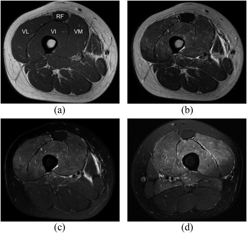
Objective: To describe the characteristic MRI appearance of squatting-induced rhabdomyolysis involving the thigh muscles.
Methods: This study consisted of 10 cases obtained at 3 institutions from 2005 to 2015. A retrospective review was performed to obtain clinical information and MR scans for rhabdomyolysis of the thigh muscles. MRI was analyzed according to the distribution and degree of muscle involvement; the degree was assessed and graded as normal, mild or prominent.
Results: The mean patient age was 20.2 years (range, 15-24 years), and 7 of the 10 patients were male. All patients had history of excessive squatting action, suffered clinically from bilateral thigh pain and were confirmed to have rhabdomyolysis through analysis of serum creatine kinase (CK) levels. All of the patients (10/10) exhibited diffuse mild to prominent degree involvement of the anterior thigh muscles according to fluid-sensitive MR sequences. Among the anterior thigh muscles, the rectus femoris was spared in 8 patients (8/10) and mild degree involved in 2 patients (2/10). Thus, no cases exhibited prominent degree involvement of the rectus femoris muscle.
Conclusion: Preservation of the rectus femoris muscle on MRI in squatting-induced rhabdomyolysis may be useful for differentiating rhabdomyolysis from other aetiologies. Advances in knowledge: Preservation of rectus femoris on MRI is distinguishable finding in squatting-induced rhabdomyolysis and reflects the functional anatomy of anterior thigh muscles.
Figures


Similar articles
-
Whole-body muscle MRI in McArdle disease.Neuromuscul Disord. 2022 Jan;32(1):5-14. doi: 10.1016/j.nmd.2021.07.397. Epub 2021 Aug 5. Neuromuscul Disord. 2022. PMID: 34711478
-
Magnetic resonance imaging changes of thigh muscles in myopathy with antibodies to signal recognition particle.Rheumatology (Oxford). 2015 Jun;54(6):1017-24. doi: 10.1093/rheumatology/keu422. Epub 2014 Nov 21. Rheumatology (Oxford). 2015. PMID: 25417246
-
Muscle magnetic resonance imaging in spinal muscular atrophy type 3: Selective and progressive involvement.Muscle Nerve. 2017 May;55(5):651-656. doi: 10.1002/mus.25385. Epub 2017 Jan 5. Muscle Nerve. 2017. PMID: 27543937
-
Exertional compartment syndrome of the thigh: a rare diagnosis and literature review.J Emerg Med. 2010 Aug;39(2):e93-9. doi: 10.1016/j.jemermed.2007.10.081. Epub 2008 Jul 2. J Emerg Med. 2010. PMID: 18597970 Review.
-
[Ultrasound findings in rhabdomyolysis].Cir Cir. 2016 Nov-Dec;84(6):518-522. doi: 10.1016/j.circir.2015.06.036. Epub 2016 Jan 6. Cir Cir. 2016. PMID: 26772896 Review. Spanish.
References
-
- Khan FY. Rhabdomyolysis: a review of the literature. Neth J Med 2009; 67: 272–83. - PubMed
-
- Vanholder R, Sever MS, Erek E, Lameire N. Rhabdomyolysis. J Am Soc Nephrol 2000; 11: 1553–61. - PubMed
-
- Warren JD, Blumbergs PC, Thompson PD. Rhabdomyolysis: a review. Muscle Nerve 2002; 25: 332–47. - PubMed
-
- Guis S, Mattei JP, Cozzone PJ, Bendahan D. Pathophysiology and clinical presentations of rhabdomyolysis. Joint Bone Spine 2005; 72: 382–91. doi: https://doi.org/10.1016/j.jbspin.2004.04.010 - DOI - PubMed
Publication types
MeSH terms
LinkOut - more resources
Full Text Sources
Other Literature Sources
Medical
Research Materials
