. 2017 Feb 9;8(2):e2598.
doi: 10.1038/cddis.2017.23.

TAK1 regulates resident macrophages by protecting lysosomal integrity

Affiliations

TAK1 regulates resident macrophages by protecting lysosomal integrity

Yosuke Sakamachi et al. Cell Death Dis. .

Abstract

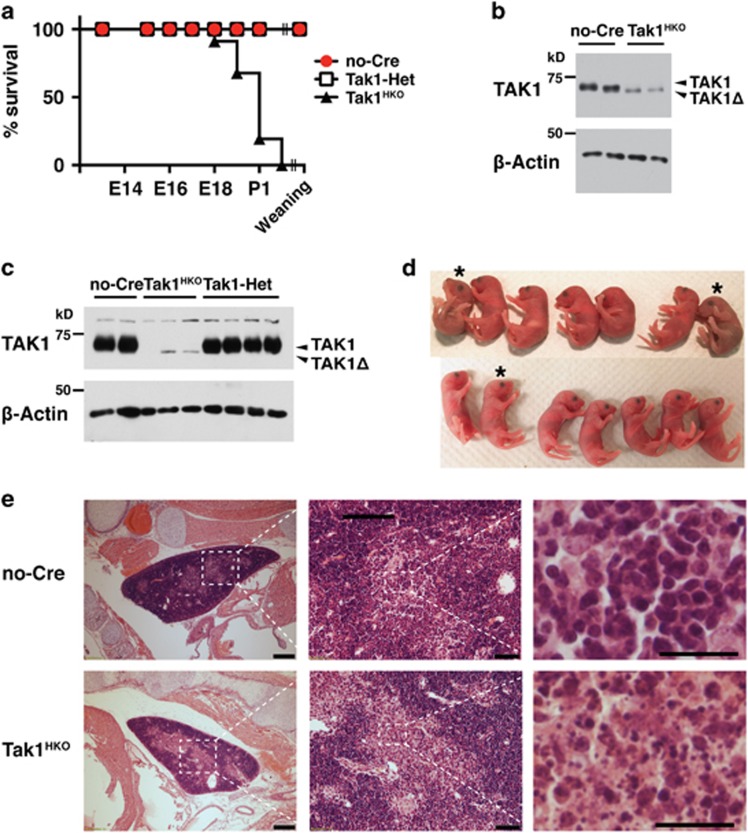
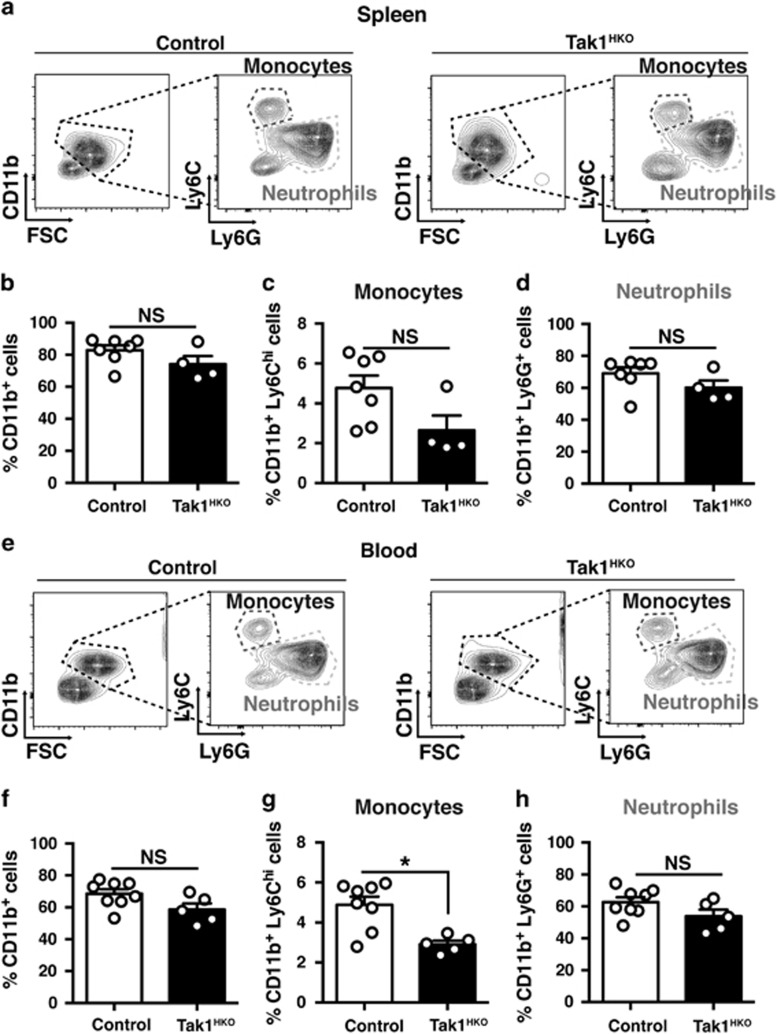
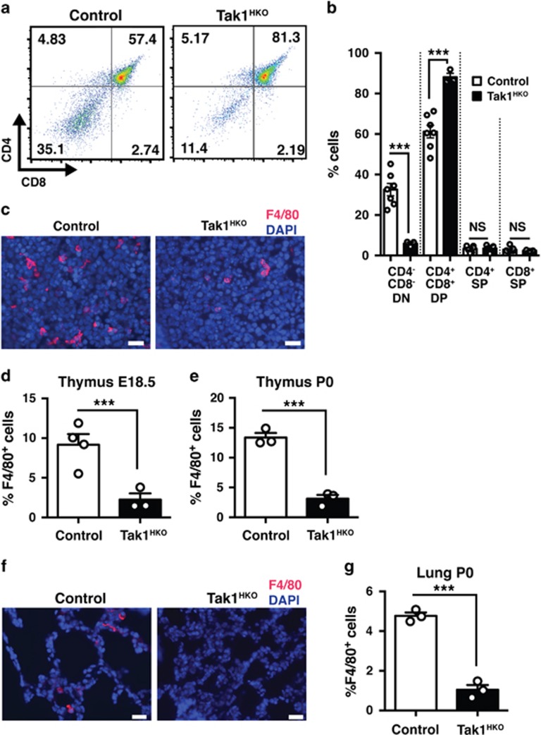
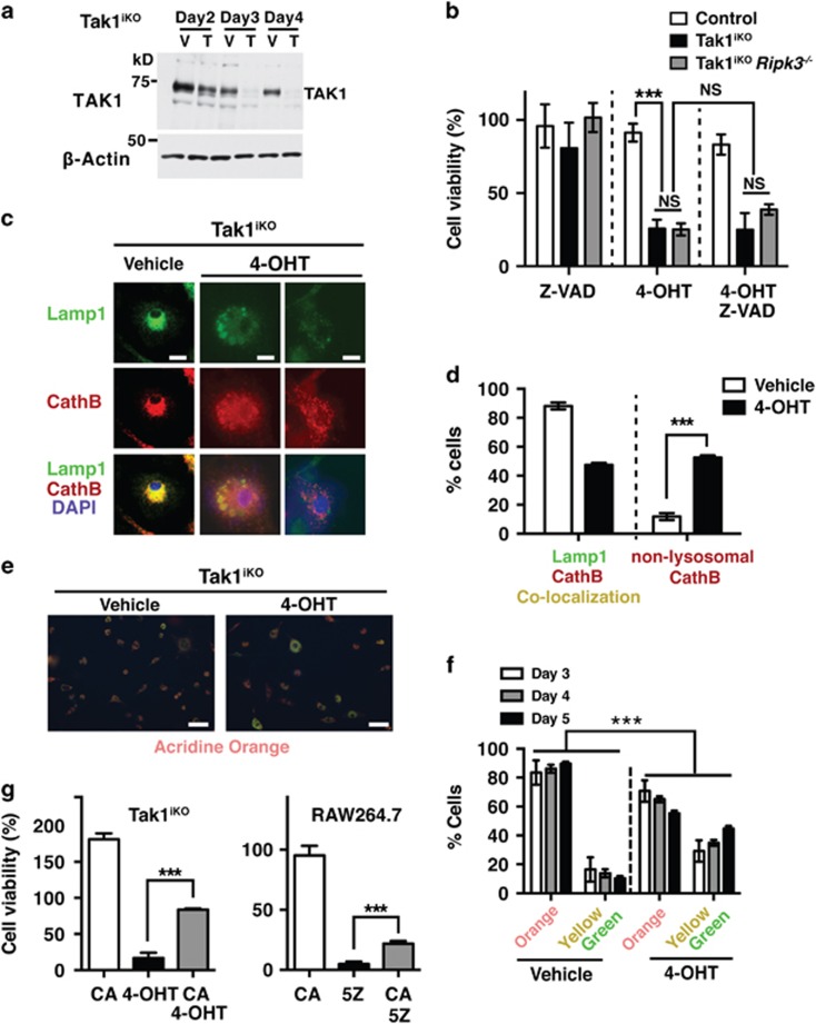
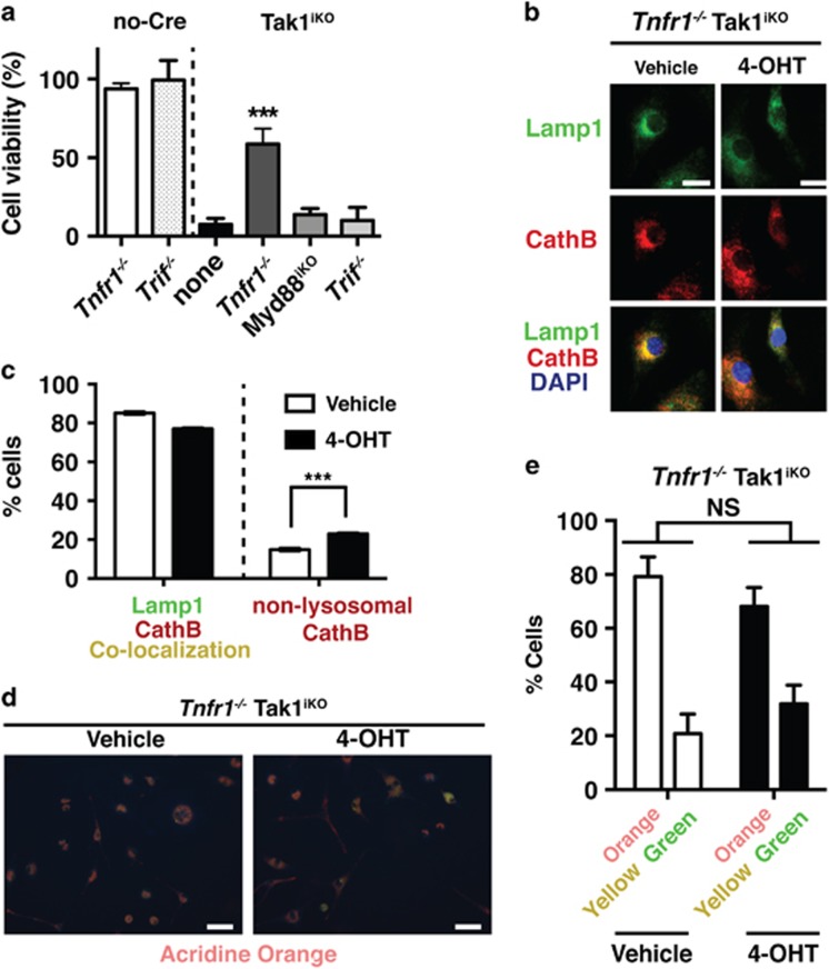
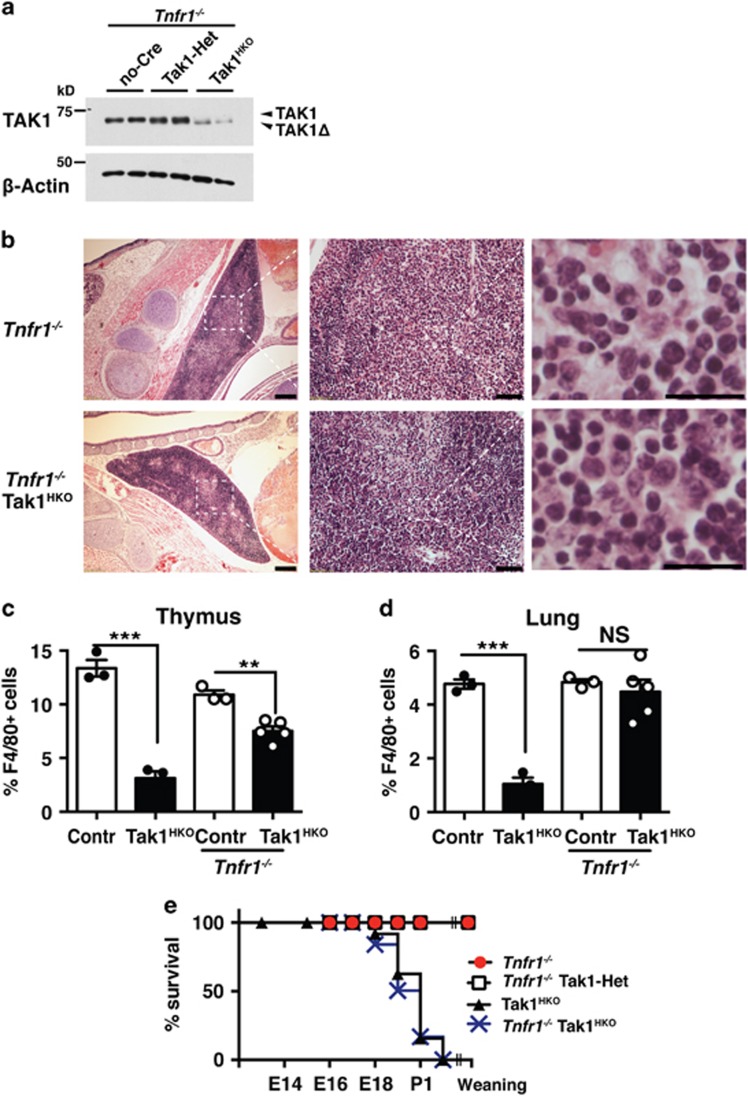
Hematopoietic cell survival and death is critical for development of a functional immune system. Here, we report that a protein kinase, TAK1, is selectively required for resident macrophage integrity during embryogenesis. Hematopoietic lineage-specific deletion of Tak1 gene (Tak1HKO) caused accumulation of cellular debris in the thymus in perinatal mice. Although no overt alteration in thymocytes and blood myeloid populations was observed in Tak1HKO mice, we found that thymic and lung macrophages were diminished. In the in vitro setting, Tak1 deficiency caused profound disruption of lysosomes and killed bone marrow-derived macrophages (BMDMs) without any exogenous stressors. Inhibition of the lysosomal protease, cathepsin B, partially blocked Tak1-deficient BMDM death, suggesting that leakage of the lysosomal contents is in part the cause of cell death. To identify the trigger of this cell death, we examined involvement of TNF and Toll-like receptor pathways. Among them, we found that deletion of Tnfr1 partially rescued cell death. Finally, we show that Tnfr1 deletion partially restored thymic and lung macrophages in vivo. These results suggest that autocrine and potentially paracrine TNF kills Tak1-deficient macrophages during development. Our results reveal that TAK1 signaling maintains proper macrophage populations through protecting lysosomal integrity.


Conflict of interest statement

The authors declare no conflict of interest.

Figures

Figure 1
Figure 1
Hematopoietic-specific Tak1 deficiency causes perinatal lethality. (a) Percentages of viable no-Cre (Tak1flox/flox or Tak1flox/+), Tak1-Het (vav-Cre Tak1flox/+), and Tak1HKO (vav-Cre Tak1flox/flox) embryos. The total numbers of animals analyzed including live and dead were no-Cre, 178; Tak1-Het, 110, Tak1HKO, 94. P<0.001 (log-rank test). (b) no-Cre control and Tak1HKO thymus were isolated from E16.5 mice and analyzed by immunoblotting for the indicated targets. Recombined Tak1 gene expressed a truncated and non-functional TAK1 protein (TAK1Δ). β-actin is shown as a loading control. (c) no-Cre, Tak1-Het, and Tak1HKO blood were isolated from E18.5 mice and analyzed by immunoblotting for the indicated targets. (d) P0 littermates from the mating of Tak1flox/floxand Tak1flox/+ Vav-Cre parents. Asterisks indicate Tak1HKO mice. (e) H&E staining of E18.5 no-Cre and Tak1HKO thymus. Scale bars, 200 μm (left panels), 50 μm (middle panels), 20 μm (right panels)
Figure 2
Figure 2
Hematopoietic-specific Tak1 deletion does not impair the development of splenocytes and circulating myeloid cells. (a) Gating strategy and representative flow cytometry data of control (no-Cre and Tak1-Het) and Tak1HKO splenocytes at P0. (b) Total CD11b+ cells in P0 control (n=7) and Tak1HKO (n=4) spleen as a percent of total live cells. (c) CD11b+ Ly6Chi cells (monocytes) and (d) CD11b+ Ly6G+ cells (neutrophils) in P0 control (n=7) and Tak1HKO (n=4) spleen as a percent of total live cells. (e) Gating strategy and representative flow cytometry data of control (no-Cre and Tak1-Het) and Tak1HKO blood at P0. (f) Total CD11b+ cells in P0 control (n=8) and Tak1HKO (n=5) blood as a percent of total live cells. (g) CD11b+ Ly6Chi cells (monocytes) and (H) CD11b+ Ly6G+ cells (neutrophils) in P0 control (n=8) and Tak1HKO (n=4) blood as a percent of total live cells. Means±S.E. and all data points are shown. *P<0.05; NS, not significant (two-tailed Student's t-test with equal distributions)
Figure 3
Figure 3
Hematopoietic-specific deletion of Tak1 diminishes thymic and pulmonary macrophages. (a) Thymocytes were isolated from E18.5 control (no-Cre and Tak1-Het, n=7) and Tak1HKO (n=3), and analyzed by flow cytometry. Gating strategy and representative data of control and Tak1HKO including percent of cells in each quadrant are shown. (b) Quantification of percent DN, CD4 CD8; DP, CD4+ CD8+; SPCD4, CD4+ CD8; and SPCD8, CD4 CD8+ thymocytes of samples shown in a. (c) E18.5 control and Tak1HKO thymus was analyzed by immunofluorescence staining using anti-F4/80 antibody (red) and DAPI (blue). Scale bars, 20 μm. (d) Quantification of F4/80+ cells in in total DAPI stained cells of the thymus from E18.5 control (n=4) and Tak1HKO (n=3). (e) P0 thymus control (n=3) and Tak1HKO (n=3). (f) P0 control and Tak1HKO lung was analyzed by immunofluorescence staining using anti-F4/80 antibody (red) and DAPI (blue). Scale bars, 20 μm. (g) Quantification of F4/80+ cells in total DAPI stained cells. P0 control (n=3); Tak1HKO (n=3). Means±S.E. and all data points are shown. ***P<0.001; NS, not significant (two-tailed Student's t-test with equal distributions)
Figure 4
Figure 4
Tak1 deficiency impairs lysosomes and kills BMDMs. (a) Inducible Tak1-deficient (Tak1iKO) BMDM treated with vehicle alone (ethanol, V) or 0.3 μM 4-OHT (T) for 2, 3 and 4 days and analyzed by immunoblotting for TAK1. β-actin is shown as a loading control. (b) Measurement of percent cell viability of control (n=3), Tak1iKO (n=3), and Tak1iKO Ripk3−/− (n=4) BMDMs 5 days post 0.3 μM 4-OHT treatment using crystal violet viability assay. Some cells were incubated with Z-VAD-FMK (Z-VAD, 20 μM) for 3 days prior to fixation. (c and d) Tak1iKO BMDMs were treated with vehicle or 0.3 μM 4-OHT for 5 days. Lysosomal architecture disruption was observed in Tak1iKO BMDMs 5 days post 0.3 μM 4-OHT treatment. Lysosomal architecture was visualized by staining using anti-lamp1 (green) and anti-cathepsin B (CathB; red) antibodies. Scale bars, 10 μm. (e and f) Tak1iKO BMDMs were treated with vehicle or 0.3 μM 4-OHT. Lysosomal function was determined by incubating cells in acridine orange at 3, 4 and 5 days post 4-OHT treatment. Orange staining indicates normal functional lysosomal pH (around 3.5), and yellow or green staining indicates increased lysosomal pH. Scale bars, 50 μm. (g, Left) Viability of Tak1iKO BMDMs treated 30 μM CA-074Me (cathepsin B inhibitor, CA), 0.3 μM 4-OHT, or 30 μM CA+0.3 μM 4-OHT. Percent live Tak1iKO cells relative to the vehicle-treated same genotype cells are shown. (Right) RAW264.7 cells were treated with 30 μM CA, 200 nM 5Z-7-oxozeaenol (5Z), or 30 μM CA+200 nM 5Z. All graphs, means±S.D.; ***P<0.001; NS, not significant (one-way ANOVA)
Figure 5
Figure 5
Tnfr1 deletion partially protects lysosome and blocks cell death in Tak1-deficient BMDMs. (a) Percent cell viability of Tak1iKO BMDMs in Tnfr1−/− (n=3), Myd88iKO(n=3), or Trif−/−(n=3) backgrounds. Crystal violet stained 4-OHT-treated cells relative to those of the vehicle-treated same genotype cells were calculated and shown as percentages. Means±S.D.; ***P<0.001 (one-way ANOVA). (b and c) Tak1iKO Tnfr1−/− and no-Cre Tnfr1−/− BMDMs were treated with vehicle or 0.3 μM 4-OHT for 5 days. Lysosomal architecture was visualized by staining using anti-lamp1 (green) and anti-cathepsin B (CathB; red) antibodies. Scale bar, 10 μm. Means±S.D.; ***P<0.001 (one-way ANOVA). (d and e) Lysosomal function was determined by incubating cells in acridine orange at 4 days post 4-OHT treatment. Orange staining indicates normal functional lysosomal pH (around 3.5), and yellow or green staining indicates increased lysosomal pH. Scale bar, 50 μm. Means±S.D.; ***P<0.001 (one-way ANOVA)
Figure 6
Figure 6
Tnfr1 deletion partially restores tissue-resident macrophages and developmental abnormalities. (a) no-Cre Tnfr1−/−, Tak1-Het Tnfr1−/−, and Tak1HKO Tnfr1−/− thymocytes were isolated from E18.5 mice and analyzed by immunoblotting. Recombined Tak1 gene expressed a truncated and non-functional TAK1 protein (TAK1Δ). β-actin is shown as a loading control. (b) H&E staining of E18.5 Tnfr1−/− and Tak1HKO Tnfr1−/− thymus. Scale bars, 200 μm (left panels), 50 μm (middle panels), 20 μm (right panels). (c and d) Quantification of F4/80+ cells in DAPI stained cells in control (Contr) and Tak1HKO in wild type (left two bars) and Tnfr1−/−background (right two bars). The left two bars are the same data shown in Figures 3e and g. (c) P0 Tnfr1−/− (n=3) and Tak1HKO Tnfr1−/− (n=3) thymus; and (d) P0 Tnfr1−/− (n=3) and Tak1HKO Tnfr1−/− (n=5) lungs. Means±S.E. and all data points are shown. ***P<0.001; NS, not significant (two-tailed Student's t-test with equal distributions). (e) Percentages of viable Tnfr1−/−, Tak1-Het Tnfr1−/−, and Tak1HKO Tnfr1−/− mice. Viability data of Tak1HKO (Figure 1a) are also included as a comparison. The total numbers of animals analyzed including live and dead were Tnfr1−/−, 100; Tak1-Het Tnfr1−/−, 51, Tak1HKO Tnfr1−/−, 48; P<0.0001 (log-rank test)

Similar articles

Cited by

References

    1. Ginhoux F, Guilliams M. Tissue-resident macrophage ontogeny and homeostasis. Immunity 2016; 44: 439–449. - PubMed
    1. Lavin Y, Mortha A, Rahman A, Merad M. Regulation of macrophage development and function in peripheral tissues. Nat Rev Immunol 2015; 15: 731–744. - PMC - PubMed
    1. Chen GY, Nunez G. Sterile inflammation: sensing and reacting to damage. Nat Rev Immunol 2010; 10: 826–837. - PMC - PubMed
    1. Lavin Y, Winter D, Blecher-Gonen R, David E, Keren-Shaul H, Merad M et al. Tissue-resident macrophage enhancer landscapes are shaped by the local microenvironment. Cell 2014; 159: 1312–1326. - PMC - PubMed
    1. Mihaly SR, Ninomiya-Tsuji J, Morioka S. TAK1 control of cell death. Cell Death Differ 2014; 21: 1667–1676. - PMC - PubMed

Publication types

Substances

LinkOut - more resources