

Duodenal GLP-1 signaling regulates hepatic glucose production through a PKC-δ-dependent neurocircuitry
- PMID: 28182013
- PMCID: PMC5386475
- DOI: 10.1038/cddis.2017.28
Duodenal GLP-1 signaling regulates hepatic glucose production through a PKC-δ-dependent neurocircuitry
Abstract
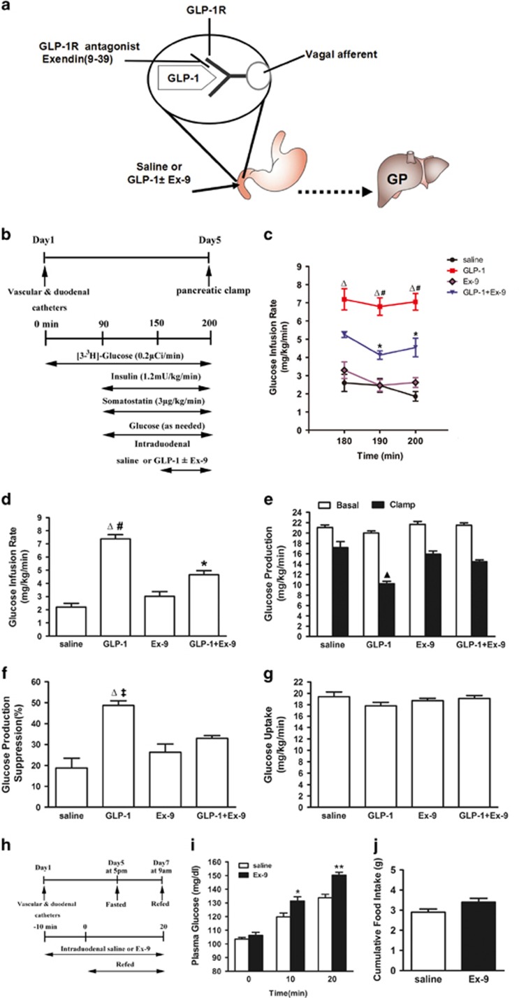
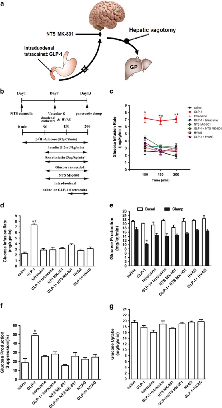
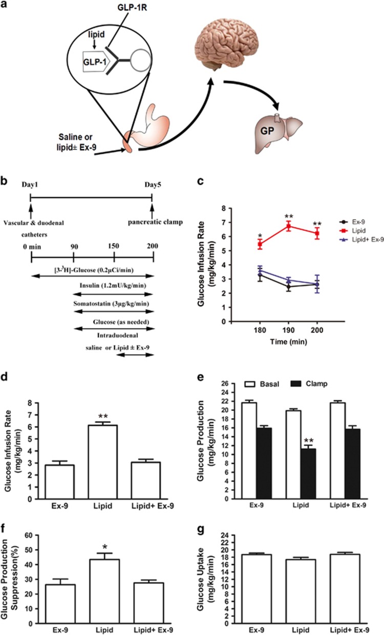
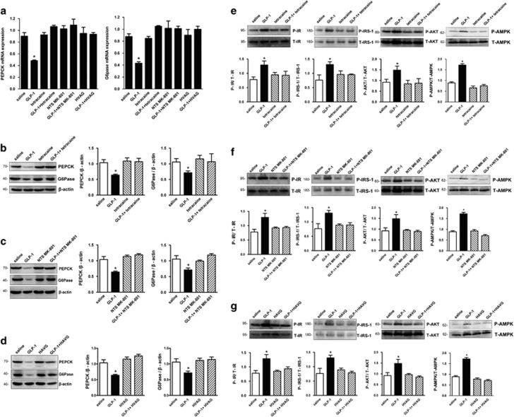
Intestinal glucagon-like peptide-1 (GLP-1) is a hormone that stimulates insulin secretion and acts as a neuropeptide to control glucose homeostasis, but little is known whether intestinal GLP-1 has any effect in the control of hepatic glucose production (HGP). Here we found that intraduodenal infusion of GLP-1 activated duodenal PKC-δ, lowered HGP and was accompanied by a decrease in hepatic expression of gluconeogenic enzymes and an increase in hepatic insulin signaling in rats. However, gut co-infusion of either the GLP-1 receptor antagonist Ex-9, or the PKC-δ inhibitor rottlerin with GLP-1, negated the ability of gut GLP-1 to lower HGP and to increase hepatic insulin signaling during clamps. The metabolic and molecular signal effects of duodenal GLP-1 were also negated by co-infusion with tetracaine, pharmacologic inhibition of N-methyl-d-aspartate receptors within the dorsalvagal complex, or hepatic vagotomy in rats. In summary, we identified a neural glucoregulatory function of gut GLP-1 signaling.
Conflict of interest statement
The authors declare no conflict of interest.
Figures

Similar articles
-
Gut ghrelin regulates hepatic glucose production and insulin signaling via a gut-brain-liver pathway.Cell Commun Signal. 2019 Jan 25;17(1):8. doi: 10.1186/s12964-019-0321-y. Cell Commun Signal. 2019. PMID: 30683114 Free PMC article.
-
Duodenal mucosal protein kinase C-δ regulates glucose production in rats.Gastroenterology. 2011 Nov;141(5):1720-7. doi: 10.1053/j.gastro.2011.06.042. Epub 2011 Jun 23. Gastroenterology. 2011. PMID: 21704002
-
Duodenal activation of cAMP-dependent protein kinase induces vagal afferent firing and lowers glucose production in rats.Gastroenterology. 2012 Apr;142(4):834-843.e3. doi: 10.1053/j.gastro.2011.12.053. Epub 2012 Jan 12. Gastroenterology. 2012. PMID: 22245844
-
Brain GLP-1 and insulin sensitivity.Mol Cell Endocrinol. 2015 Dec 15;418 Pt 1:27-32. doi: 10.1016/j.mce.2015.02.017. Epub 2015 Feb 24. Mol Cell Endocrinol. 2015. PMID: 25724479 Free PMC article. Review.
-
Hepatic functions of GLP-1 and its based drugs: current disputes and perspectives.Am J Physiol Endocrinol Metab. 2016 Sep 1;311(3):E620-7. doi: 10.1152/ajpendo.00069.2016. Epub 2016 Aug 9. Am J Physiol Endocrinol Metab. 2016. PMID: 27507553 Review.
Cited by
-
Pancreatic GLP-1r binding potential is reduced in insulin-resistant pigs.BMJ Open Diabetes Res Care. 2020 Nov;8(2):e001540. doi: 10.1136/bmjdrc-2020-001540. BMJ Open Diabetes Res Care. 2020. PMID: 33132211 Free PMC article.
-
An extended minimal model of OGTT: estimation of α- and β-cell dysfunction, insulin resistance, and the incretin effect.Am J Physiol Endocrinol Metab. 2024 Feb 1;326(2):E182-E205. doi: 10.1152/ajpendo.00278.2023. Epub 2023 Dec 13. Am J Physiol Endocrinol Metab. 2024. PMID: 38088864 Free PMC article.
-
Hypothalamic P62 (SQSTM1) regulates energy balance by modulating leptin signaling.Theranostics. 2024 Oct 7;14(17):6605-6624. doi: 10.7150/thno.96480. eCollection 2024. Theranostics. 2024. PMID: 39479445 Free PMC article.
-
Brunner's Gland Hyperplasia in a Patient after Roux-Y Gastric Bypass: An Important Pitfall in GLP-1 Receptor Imaging.Case Rep Endocrinol. 2020 Apr 3;2020:4510910. doi: 10.1155/2020/4510910. eCollection 2020. Case Rep Endocrinol. 2020. PMID: 32313706 Free PMC article.
-
Genetic analysis of the correlation between GLP1 action and metabolic liver disease: Insights from Mendelian randomization analysis.J Diabetes Investig. 2025 Aug;16(8):1409-1419. doi: 10.1111/jdi.70087. Epub 2025 Jun 1. J Diabetes Investig. 2025. PMID: 40452205 Free PMC article.
References
-
- Murphy KG, Bloom SR. Gut hormones and the regulation of energy homeostasis. Nature 2006; 444: 854–859. - PubMed
-
- Eissele R, Goke R, Willemer S, Harthus HP, Vermeer H, Arnold R et al. Glucagon-like peptide-1 cells in the gastrointestinal tract and pancreas of rat, pig and man. Eur J Clin Invest 1992; 22: 283–291. - PubMed
-
- Baggio LL, Drucker DJ. Biology of incretins: GLP-1 and GIP. Gastroenterology 2007; 132: 2131–2157. - PubMed
Publication types
MeSH terms
Substances
LinkOut - more resources
Full Text Sources
Other Literature Sources
