. 2017 Apr 21;12(4):979-988.
doi: 10.1021/acschembio.7b00066. Epub 2017 Feb 17.

Evolution and Distribution of C7-Cyclitol Synthases in Prokaryotes and Eukaryotes

Affiliations

Evolution and Distribution of C7-Cyclitol Synthases in Prokaryotes and Eukaryotes

Andrew R Osborn et al. ACS Chem Biol. .

Abstract

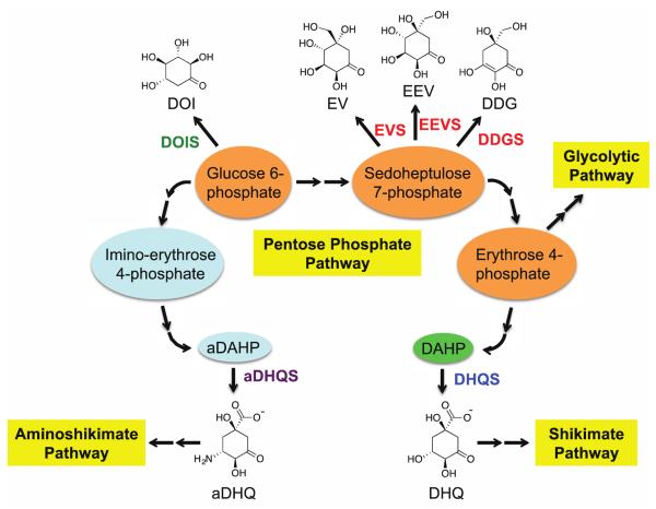
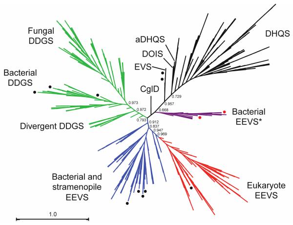
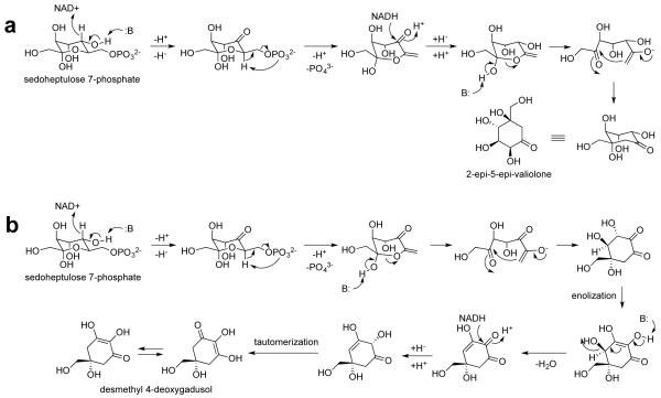
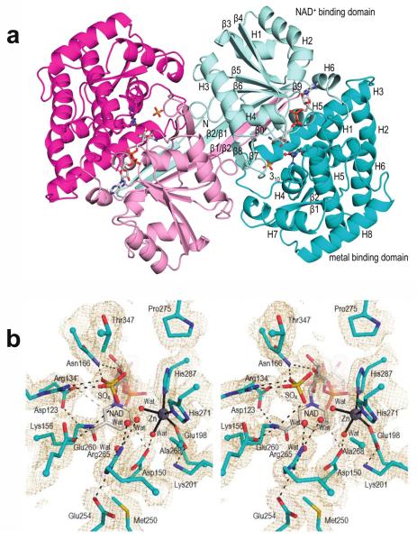
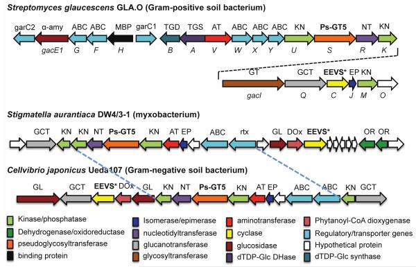
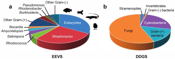
2-Epi-5-epi-valiolone synthase (EEVS), a C7-sugar phosphate cyclase (SPC) homologous to 3-dehydroquinate synthase (DHQS), was discovered during studies of the biosynthesis of the C7N-aminocyclitol family of natural products. EEVS was originally thought to be present only in certain actinomycetes, but analyses of genome sequences showed that it is broadly distributed in both prokaryotes and eukaryotes, including vertebrates. Another SPC, desmethyl-4-deoxygadusol synthase (DDGS), was later discovered as being involved in the biosynthesis of mycosporine-like amino acid sunscreen compounds. Current database annotations are quite unreliable, with many EEVSs reported as DHQS, and most DDGSs reported as EEVS, DHQS, or simply hypothetical proteins. Here, we identify sequence features useful for distinguishing these enzymes, report a crystal structure of a representative DDGS showing the high similarity of the EEVS and DDGS enzymes, identify notable active site differences, and demonstrate the importance of two of these active site residues for catalysis by point mutations. Further, we functionally characterized two representatives of a distinct clade equidistant from known EEVS and known DDGS groups and show them to be authentic EEVSs. Moreover, we document and discuss the distribution of genes that encode EEVS and DDGS in various prokaryotes and eukaryotes, including pathogenic bacteria, plant symbionts, nitrogen-fixing bacteria, myxobacteria, cyanobacteria, fungi, stramenopiles, and animals, suggesting their broad potential biological roles in nature.


Figures

Figure 1
Figure 1
Six sugar phosphate cyclase family members, their substrates and products, and their connections to the Pentose Phosphate Pathway. DOIS, 2-deoxy-scyllo-inosose synthase; EVS, 2-epi-valiolone synthase; EEVS, 2-epi-5-epi-valiolone synthase; DDGS, desmethyl-4-deoxygadusol synthase; aDHQS, aminodehydroquinate synthase; DHQS, 3-dehydroquinate synthase.
Figure 2
Figure 2
Radial cladogram of the sugar phosphate cyclase superfamily. A group of putative bacterial EEVS (drawn in purple and designated “EEVS*”) occurs as a separate clade from the known EEVS clades (drawn in blue and red). Numbers show local support values. Black circles show sedoheptulose 7-phosphate cyclases (EEVS, EVS, and DDGS) that have been biochemically characterized. Red circles identify the two bacterial EEVS* (GacC and Staur_1386) that are characterized in this study.
Figure 3
Figure 3
Comparisons of partial amino acid sequences of the sugar phosphate cyclases. Conserved amino acid residues in two fingerprint segments are shown from bacterial DHQS, aminoDHQS, DOIS, and EVS and various EEVS and DDGS groups as labeled. Black circles above the sequences indicate residues found in the catalytic pocket of the enzymes. Stars above the EEVS and DDGS sequence groups indicates residues that differentiate DDGS from EEVS.
Figure 4
Figure 4
Proposed catalytic mechanisms for EEVS (a) and DDGS (b).
Figure 5
Figure 5
Overall structure and active site of AvDDGS. a. Ribbon diagram of the two chains of the AvDDGS dimer are show in cyan and magenta tones, respectively, with the N-terminal NAD+-binding domains in light hues and the C-terminal metal-binding domains in dark hues. The NAD+, Zn2+ with its coordinating residues, and sulfate bound in the active site are shown. Secondary structural elements and domains of one monomer are labeled. The extended β-strands involved in the domain-swapped interaction (labeled β1/β2 and β2/β1) represent β1 and β2, respectively, in the labeled monomer. b. Stereoview of the AvDDGS active site residues (cyan carbons), waters (red spheres), sulfate bound in the active site (yellow), NAD+ (grey carbons), and Zn2+ (silver sphere). Coordination bonds (black lines), select hydrogen bonds (black dashes), or approaches of interest (grey dashes) and 2Fo-Fc electron density (orange, contoured at 1ρrms) are also shown.
Figure 6
Figure 6
Comparisons of the AvDDGS active site region with the DHQS•CBP complex and with ShEEVS. a. Stereoview of select active site residues in AvDDGS (cyan carbons) with NAD+ (grey carbons) overlaid on DHQS in complex with CBP (salmon carbons; PDB 1DQS). Hydrogen bonding interactions (dashed lines) and coordination bonds with Zn2+ (solid lines) in the AvDDGS (black) and DHQS (grey) active sites are shown. The sulfate (yellow) and waters (red spheres) in the active site of AvDDGS in agreement with components of CBP or water (salmon sphere) in the active site of DHQS as well as the zinc ions (silver spheres) are shown. Labels for Arg265 (AvDDGS) and Arg264 (DHQS) are separated due to their disparate positions in the active site and in the sequence alignment. A prime on a residue number means it is from the other subunit of the dimer. View of active site is rotated roughly 90° counter-clockwise with respect to the view in Figure 5b and 6b. b. Stereoview of active site residues in AvDDGS (cyan carbons) with NAD+ (grey carbons), sulfate bound in the active site (yellow), and select waters (red spheres) overlaid on ShEEVS with NAD+ (both purple carbons) and select waters (purple spheres). Hydrogen bonds (dashed) and coordination bonds (solid) are shown in the AvDDGS active site (black lines) and ShEEVS active site (light pink lines). A prime on a residue number means it is from the other subunit of the dimer.
Figure 7
Figure 7
Biosynthetic gene clusters in Streptomyces glaucescens GLA.O, Stigmatella aurantiaca DW4/3–1, and Cellvibrio japonicus Ueda107 (previously known as Pseudomonas fluorescens subsp. cellulosa).
Figure 8
Figure 8
Distribution of EEVS and DDGS in the sequenced microorganisms. With the exception of a fungus and seven stramenopiles, all eukaryotic EEVS are from vertebrates (fish, amphibians, reptiles, and birds).

References

    1. Wu X, Flatt PM, Schlorke O, Zeeck A, Dairi T, Mahmud T. A comparative analysis of the sugar phosphate cyclase superfamily involved in primary and secondary metabolism. ChemBioChem. 2007;8:239–248. - PMC - PubMed
    1. Mahmud T. Progress in aminocyclitol biosynthesis. Curr. Opin. Chem. Biol. 2009;13:161–170. - PMC - PubMed
    1. Nango E, Kumasaka T, Hirayama T, Tanaka N, Eguchi T. Structure of 2-deoxy-scyllo-inosose synthase, a key enzyme in the biosynthesis of 2-deoxystreptamine-containing aminoglycoside antibiotics, in complex with a mechanism-based inhibitor and NAD+ Proteins. 2008;70:517–527. - PubMed
    1. Kean KM, Codding SJ, Asamizu S, Mahmud T, Karplus PA. Structure of a sedoheptulose 7-phosphate cyclase: ValA from Streptomyces hygroscopicus. Biochemistry. 2014;53:4250–4260. - PMC - PubMed
    1. Floss HG. Natural products derived from unusual variants of the shikimate pathway. Nat. Prod. Rep. 1997;14:433–452. - PubMed

Publication types

LinkOut - more resources