. 2017 Feb 9;13(2):e1006613.
doi: 10.1371/journal.pgen.1006613. eCollection 2017 Feb.

RNA-seq analysis of Drosophila clock and non-clock neurons reveals neuron-specific cycling and novel candidate neuropeptides

Affiliations

RNA-seq analysis of Drosophila clock and non-clock neurons reveals neuron-specific cycling and novel candidate neuropeptides

Katharine C Abruzzi et al. PLoS Genet. .

Abstract

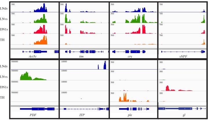
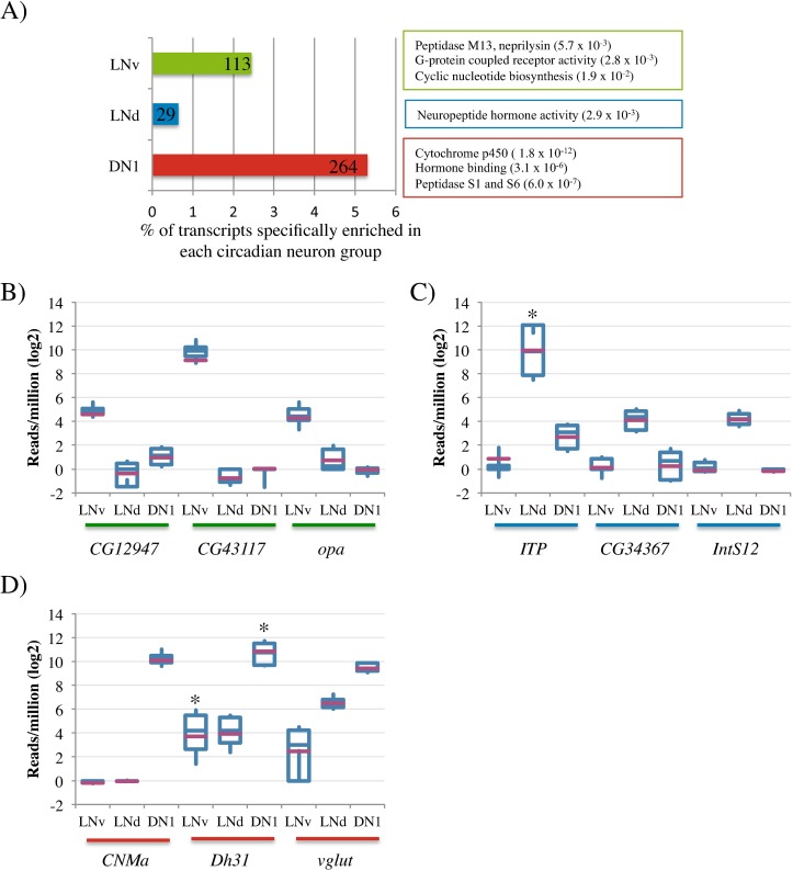
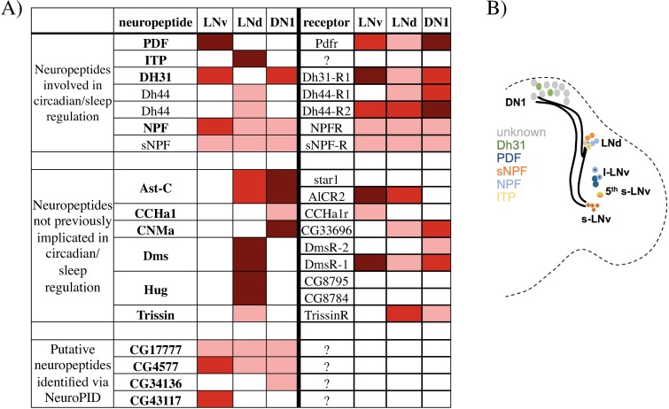
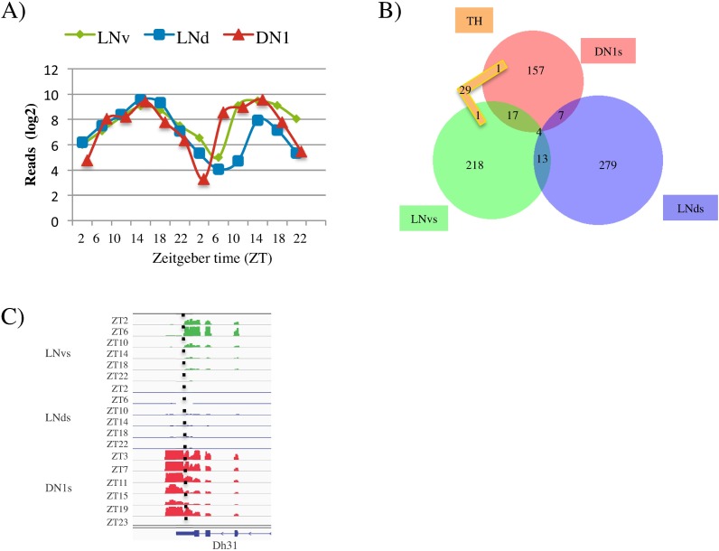
Locomotor activity rhythms are controlled by a network of ~150 circadian neurons within the adult Drosophila brain. They are subdivided based on their anatomical locations and properties. We profiled transcripts "around the clock" from three key groups of circadian neurons with different functions. We also profiled a non-circadian outgroup, dopaminergic (TH) neurons. They have cycling transcripts but fewer than clock neurons as well as low expression and poor cycling of clock gene transcripts. This suggests that TH neurons do not have a canonical circadian clock and that their gene expression cycling is driven by brain systemic cues. The three circadian groups are surprisingly diverse in their cycling transcripts and overall gene expression patterns, which include known and putative novel neuropeptides. Even the overall phase distributions of cycling transcripts are distinct, indicating that different regulatory principles govern transcript oscillations. This surprising cell-type diversity parallels the functional heterogeneity of the different neurons.


Conflict of interest statement

The authors have declared that no competing interests exist.

Figures

Fig 1
Fig 1. Sequencing libraries reflect the transcriptomes of LNds, LNvs, DN1s and TH neurons.
Sequencing results from LNds (blue), LNvs (green), DN1s (red), and TH cells (orange) are shown for a variety of genes. The housekeeping transcript, Act5c, shows similar levels in all four neuronal groups. The two circadian transcripts, tim and cry are found in the three groups of circadian neurons but are expressed at very low levels or not at all in TH cells. The transcript of the neuropeptide sNPF is found in all four subgroups of neurons. The transcripts encoding neuropeptides PDF and ITP are found in the LNvs and LNds, respectively. The tyrosine hydrolyase transcript, ple, is strongly expressed in TH cells and the transcription factor, gl (glass) is expressed specifically in the DN1s. Note that there is some 3’-bias in these libraries; there is generally more signal on the 3’-end of the gene than the 5’-end. Y-axis scale is in reads/million total reads.
Fig 2
Fig 2. Transcripts enriched specifically in one group of circadian neurons.
A) The percentage of transcripts specifically enriched in each of the circadian neuronal groups is represented in a bar graph. LNvs are shown in green. LNds are shown in blue. DN1s are shown in red. The absolute number of enriched genes is indicated in each bar. Results of gene ontology analysis (GO) are included. B, C, and D) Boxplots showing the expression levels of some of the most significantly enriched transcripts in the LNvs (B), LNds (C) and DN1s (D). The purple bar indicates the mean. The asterix denotes those transcripts that show high variability due to cycling transcripts levels.
Fig 3
Fig 3. Known and putative neuropeptide and receptor encoding transcripts found in the three circadian neuronal groups.
A) Table shows neuropeptides listed on the left side and their receptors (if known) on the right side. Those in bold were identified as pro-neuropeptides by NeuroPID (see Methods). Color indicates the level of expression in each group of neurons. For neuropeptides: dark red (>1000 reads/million), red (between 500 and 1000 reads/million) and pink (between 20 and 500 reads/million). Receptor transcripts are generally found at much lower levels: dark red (> 30 reads/million), red (between 10 to 30 reads/million) and pink (5 to 10 reads/million). Those shown in bold were identified via NeuroPID (see Methods). B) The cartoon shows the organization of the circadian neuronal network in Drosophila and the expression pattern of those neuropeptides known to have a role in the circadian system.
Fig 4
Fig 4. Most cycling gene expression is specific to one group of circadian neurons.
In all figures, LNvs are shown in green, LNds are shown in blue, DN1s are shown in read, and TH are shown in orange. A) Timeless (tim) cycles in all three neuronal groups with similar phase and expression level. Transcript levels are represented as reads/million total reads in a log base 2 scale. Two independent six timepoint datasets are concatenated to show cycling. B) Overlap of the high-confidence (HC) cycling transcripts found in the four neuronal groups. Only 4 HC cycling transcripts are in common in the 3 groups of circadian neurons. C) Dh31 is a HC cycling transcript in both the LNvs (green) and DN1s (red) with peak expression in the morning. The dotted line denotes the 3’end of the LNv specific Dh31 isoform. The isoform of Dh31 expressed in DN1s has an extended 3’-UTR. See also Fig 3, S1 Fig and S2 Fig.
Fig 5
Fig 5. Three transcripts that show neuron-specific cycling patterns.
In all figures, LNvs are shown in green, LNds are shown in blue and DN1s are shown in red. A) The transcript encoding the G-protein coupled receptor, CCHA1r, cycles in the LNvs with peak expression in the morning. CCHA1r transcripts are not expressed or expressed at very low levels (<5 average reads/million) in LNds and DN1s. B) The transcript encoding the metabotropic glutamate receptor (mGluRA) cycles robustly in the LNds with peak expression at mid-day. mGluRA is expressed at constant levels in both the LNv and DN1s. C) The transcripts encoding the predicted neuropeptide CG17777 cycle with different phases in the LNvs and DN1s. CG17777 is also expressed in the LNds but does not cycle.
Fig 6
Fig 6. Transcripts cycling in LNvs show bimodal phase distribution.
A) The phase of all the high confidence (HC) cycling transcripts identified in each neuronal group is represented in a histogram (LNvs (green), LNds (blue), DN1s(red)). The transcripts are binned according to their peak expression (phase) and the percentage of all cycling transcripts in that bin is plotted. The shape of the distribution is emphasized by a trendline in the same color as the histogram. The lines indicate the average phase of the core clock genes (vri, pdp1, per, tim) for each of the three neuronal groups. Phase shown was calculated by F24 analysis. B) For every cycling transcript in LNvs, the relative expression in l-LNvs versus s-LNvs was calculated (see Methods). Those transcripts cycling with a morning (ZT3-8) and evening phase (ZT14-ZT19) were binned and the distribution of expression in l-LNvs versus s-LNvs was visualized using a box plot. Transcripts cycling with a morning phase were more likely to be more highly expressed in s-LNvs (relative expression large v. small <1) and transcripts cycling with an evening phase are more likely to have higher expression in l-LNvs (relative expression large v. small >1). These two different distributions are significantly different (p-value: 3x10⁻⁵). C) The phase of all the HC cycling transcripts in dopaminergic (TH) cells. Data representation is the same as in (A).

References

    1. Hardin PE, Panda S. Circadian timekeeping and output mechanisms in animals. Curr Opin Neurobiol. 2013;23(5):724–31. PubMed Central PMCID: PMCPMC3973145. 10.1016/j.conb.2013.02.018 - DOI - PMC - PubMed
    1. Frenkel L, Ceriani MF. Circadian plasticity: from structure to behavior. Int Rev Neurobiol. 2011;99:107–38. 10.1016/B978-0-12-387003-2.00005-7 - DOI - PubMed
    1. Yoshii T, Rieger D, Helfrich-Forster C. Two clocks in the brain: an update of the morning and evening oscillator model in Drosophila. Prog Brain Res. 2012;199:59–82. 10.1016/B978-0-444-59427-3.00027-7 - DOI - PubMed
    1. Tataroglu O, Emery P. Studying circadian rhythms in Drosophila melanogaster. Methods. 2014;68(1):140–50. Epub 2014/01/15. PubMed Central PMCID: PMC4049855. 10.1016/j.ymeth.2014.01.001 - DOI - PMC - PubMed
    1. Renn SC, Park JH, Rosbash M, Hall JC, Taghert PH. A pdf neuropeptide gene mutation and ablation of PDF neurons each cause severe abnormalities of behavioral circadian rhythms in Drosophila. Cell. 1999;99(7):791–802. Epub 2000/01/05. - PubMed

MeSH terms