. 2017 Feb 9;12(2):e0171699.
doi: 10.1371/journal.pone.0171699. eCollection 2017.

Distinct effects of endosomal escape and inhibition of endosomal trafficking on gene delivery via electrotransfection

Affiliations

Distinct effects of endosomal escape and inhibition of endosomal trafficking on gene delivery via electrotransfection

Lisa D Cervia et al. PLoS One. .

Abstract

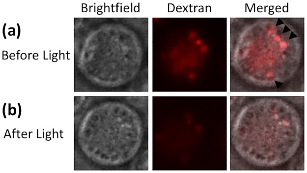
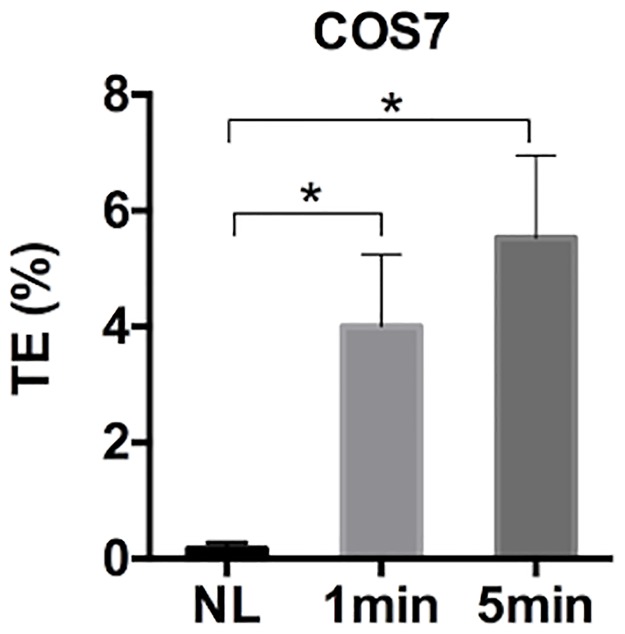
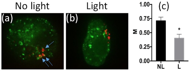
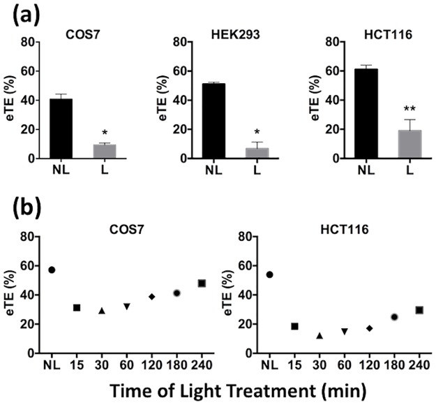
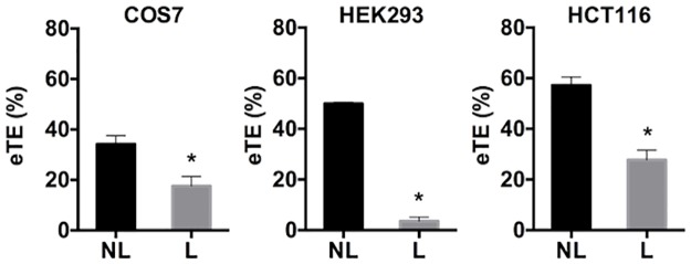
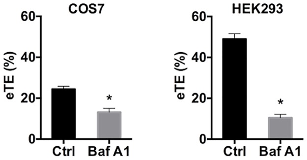
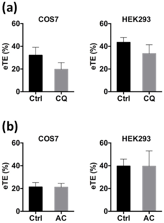
A recent theory suggests that endocytosis is involved in uptake and intracellular transport of electrotransfected plasmid DNA (pDNA). The goal of the current study was to understand if approaches used previously to improve endocytosis of gene delivery vectors could be applied to enhancing electrotransfection efficiency (eTE). Results from the study showed that photochemically induced endosomal escape, which could increase poly-L-lysine (PLL)-mediated gene delivery, decreased eTE. The decrease could not be blocked by treatment of cells with endonuclease inhibitors (aurintricarboxylic acid and zinc ion) or antioxidants (L-glutamine and ascorbic acid). Chemical treatment of cells with an endosomal trafficking inhibitor that blocks endosome progression, bafilomycin A1, resulted in a significant decrease in eTE. However, treatment of cells with lysosomotropic agents (chloroquine and ammonium chloride) had little effects on eTE. These data suggested that endosomes played important roles in protecting and intracellular trafficking of electrotransfected pDNA.


Conflict of interest statement

The authors have declared that no competing interests exist.

Figures

Fig 1
Fig 1. Release of rhodamine B-labeled dextran from endosomes following light treatment.
Intracellular vesicles were preloaded with the photosensitizer. (a) The image of a COS7 cell before light treatment. It was taken at 20 min after the cell was incubated at 37°C with dextran (10,000 MW). The light treatment was performed immediately after the image acquisition. (b) The image of the same cell at 10 min after light treatment. The dextran was punctate within endosomes before light treatment (top panel). However, upon light treatment, dextran diffused out of endosomes, and spread in the cytosol (bottom panel).
Fig 2
Fig 2. Efficiency of cell transfection with Poly-L-lysine (PLL).
Intracellular vesicles were preloaded with the photosensitizer. COS7 cells were transfected with PLL-pDNA complexes for 4 hours at 37°C. Then, the cells were exposed to blue light for 0 (i.e., no light (NL) control), 1 min, and 5 min. The transfection efficiency (TE) was quantified at 24 hours post transfection. (n = 4, *P<0.05, Mann-Whitney U test).
Fig 3
Fig 3. Colocalization of pDNA with Rab5.
(a) Typical image of pDNA and Rab5 distributions in a COS7 cell without light treatment. It was obtained with confocal microscopy. Blue arrows indicate the points of colocalization of rhodamine-labeled pDNA (red) and Rab5-GFP (green). (b) Typical image of pDNA and Rab5 distributions in another COS7 cell after light treatment. The PCI treatment was performed with the light-after protocol, where the cells were treated with light for 2 min at 20 min after electrotransfection. Then, cells were fixed with 2% paraformaldehyde and imaged with a confocal microscope. (c) Quantitative analysis of colocalization. Manders’ colocalization coefficient (M) was calculated for each z-stack of confocal images. The light (L) treated group showed a significant decrease in the colocalization of pDNA with Rab5, compared to the no light (NL) treated group. (n = 10, *P<0.0005, Mann-Whitney U test).
Fig 4
Fig 4. Effects of light-after treatment on eTE.
(a) PCI treatment was performed with the light-after protocol. All cells were exposed to light at 10 min post application of electric field. The treatment resulted in a significant reduction in eTE in COS7, HEK293 and HCT116 cells. (n = 4, *P<0.05, **P<0.01, Mann-Whitney U test.) (b) Dependence of eTE on the time of light treatment. Cells were divided into 7 groups. Cells in each group were exposed to light at a specific time points indicated in the horizontal axis of the plots, or not treated with light (NL). The eTE was quantified at 24 hours post electrotransfection. Each symbol in Panel (b) represents the mean eTE from two repeated experiments.
Fig 5
Fig 5. Effects of light-before treatment on eTE.
PCI treatment was performed with the light-before protocol that prevented pDNA damage by the light treatment or ROS induced by photochemical reactions. However, the eTE still decreased for COS7, HEK293 and HCT116 cell lines after light (L) treatment, compared to the no light (NL) control. (n = 4, *P<0.05, Mann-Whitney U test).
Fig 6
Fig 6. Effects of bafilomycin A1 pretreatment on transgene expression.
COS7 and HEK293 cells were treated with either buffer in the control (Ctrl) or an endocytic trafficking inhibitor, bafilomycin A1 (Baf A1), at 1 μM for 1 hour prior to electrotransfection; and the eTE was quantified at 24 hours post electrotransfection. (n = 4, *P<0.05, Mann-Whitney U test).
Fig 7
Fig 7. Effects of lysosomotropic agents pretreatment on transgene expression.
COS7 and HEK293 cells were treated with either buffer in the control (Ctrl) or a lysosomotropic agent for 4 hours prior to electrotransfection: (a) chloroquine (CQ) at 100 μM, and (b) ammonium chloride (AC) at 10 mM. The treatments had statistically insignificant effects on eTE quantified at 24 hours post electrotransfection. (n = 4, P > 0.05, Mann-Whitney U test).

References

    1. Henshaw JW, Yuan F. Field distribution and DNA transport in solid tumors during electric field-mediated gene delivery. Journal of Pharmaceutical Sciences. 2008. February;97(2):691–711. 10.1002/jps.21000 - DOI - PubMed
    1. Neumann E, Schaefer-Ridder M, Wang Y, Hofschneider PH. Gene transfer into mouse lyoma cells by electroporation in high electric fields. The EMBO Journal. European Molecular Biology Organization; 1982;1(7):841–5. - PMC - PubMed
    1. Weaver JC. Electroporation theory: concepts and mechanisms. Methods in Molecular Biology. 1995;47:1–26. 10.1385/0-89603-310-4:1 - DOI - PubMed
    1. Krassowska W, Filev PD. Modeling electroporation in a single cell. Biophysical Journal. 2007. January 15;92(2):404–17. 10.1529/biophysj.106.094235 - DOI - PMC - PubMed
    1. Zaharoff DA, Henshaw JW, Mossop B, Yuan F. Mechanistic Analysis of Electroporation-Induced Cellular Uptake of Macromolecules. Experimental Biology and Medicine. 2008. January 1;233(1):94–105. 10.3181/0704-RM-113 - DOI - PMC - PubMed

Publication types

MeSH terms