

In Vivo acrylamide exposure may cause severe toxicity to mouse oocytes through its metabolite glycidamide
- PMID: 28182799
- PMCID: PMC5300229
- DOI: 10.1371/journal.pone.0172026
In Vivo acrylamide exposure may cause severe toxicity to mouse oocytes through its metabolite glycidamide
Abstract
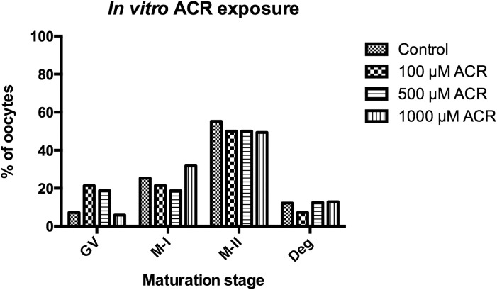
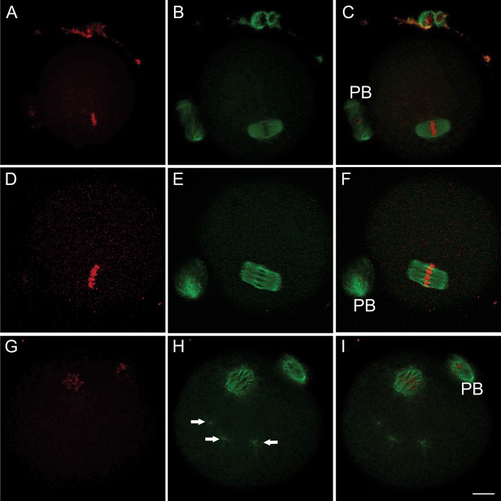
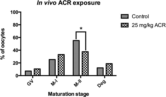
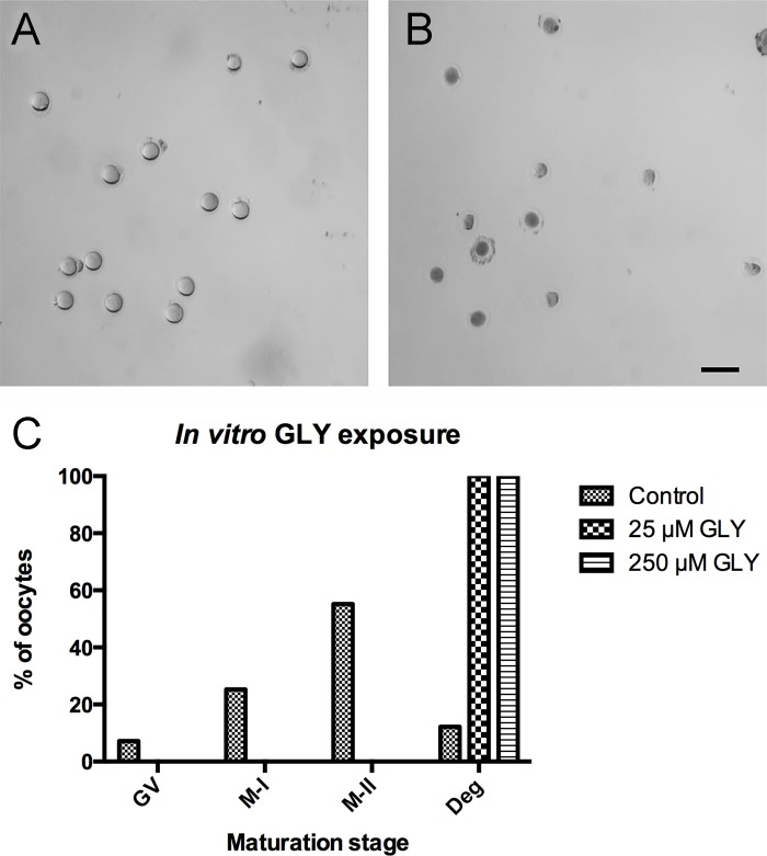
High acrylamide (ACR) content in heat-processed carbohydrate-rich foods, as well as roasted products such as coffee, almonds etc., has been found to be as a risk factor for carcinogenicity and genotoxicity by The World Health Organization. Glycidamide (GLY), the epoxide metabolite of ACR, is processed by the cytochrome P-450 enzyme system and has also been found to be a genotoxic agent. The aim of this study was to determine whether ACR and/or GLY have any detrimental effect on the meiotic cell division of oocytes. For this purpose, germinal vesicle-stage mouse oocytes were treated with 0, 100, 500, or 1000 μM ACR or 0, 25, or 250 μM GLY in vitro. In vivo experiments were performed after an intraperitoneal injection of 25 mg/kg/day ACR of female BALB/c mice for 7 days. The majority of in vitro ACR-treated oocytes reached the metaphase-II stage following 18 hours of incubation, which was not significantly different from the control group. Maturation of the oocytes derived from in vivo ACR-treated mice was impaired significantly. Oocytes, reaching the M-II stage in the in vivo ACR-treated group, were characterized by a decrease in meiotic spindle mass and an increase in chromosomal disruption. In vitro GLY treatment resulted in the degeneration of all oocytes, indicating that ACR toxicity on female germ cells may occur through its metabolite, GLY. Thus, ACR exposure must be considered, together with its metabolite GLY, when female fertility is concerned.
Conflict of interest statement
The authors have declared that no competing interests exist.
Figures
References
-
- Smith E, Prues S, Oehme F. Environmental degradation of acrylamides 2. Effects of environmental (outdoor) exposure. Ecotoxicol Environ Saf. 1997;37: 76–91. - PubMed
-
- NICNAS. Acrylamide—priority existing chemical assessment report. Australia: National Industrial Chemicals Notification and Assessment Scheme (NICNAS); 2002.
-
- Sumner SC, MacNeela JP, Fennell TR. Characterization and quantitation of urinary metabolites of [1, 2, 3-13c] acrylamide in rats and mice using carbon-13 nuclear magnetic resonance spectroscopy. Chem Res Toxicol. 1992;5: 81–89. - PubMed
-
- Doerge DR, Young JF, McDaniel LP, Twaddle NC, Churchwell MI. Toxicokinetics of acrylamide and glycidamide in b6c3f 1 mice. Toxicol Applied Pharm. 2005;202: 258–267. - PubMed
MeSH terms
Substances
LinkOut - more resources
Full Text Sources
Other Literature Sources
