. 2017 Feb 10;16(1):69.
doi: 10.1186/s12936-017-1714-2.

Cellulose filtration of blood from malaria patients for improving ex vivo growth of Plasmodium falciparum parasites

Affiliations

Cellulose filtration of blood from malaria patients for improving ex vivo growth of Plasmodium falciparum parasites

Sixbert I Mkumbaye et al. Malar J. .

Abstract

Background: Establishing in vitro Plasmodium falciparum culture lines from patient parasite isolates can offer deeper understanding of geographic variations of drug sensitivity and mechanisms of malaria pathogenesis and immunity. Cellulose column filtration of blood is an inexpensive, rapid and effective method for the removal of host factors, such as leucocytes and platelets, significantly improving the purification of parasite DNA in a blood sample.

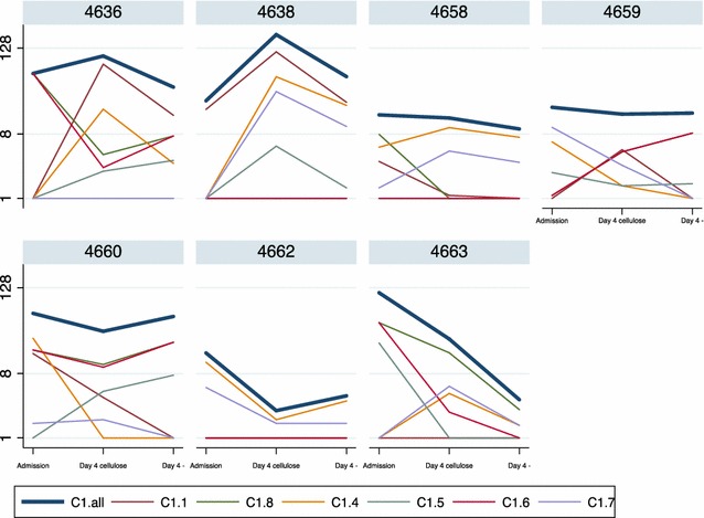
Methods: In this study, the effect of cellulose column filtration of venous blood on the initial in vitro growth of P. falciparum parasite isolates from Tanzanian children admitted to hospital was tested. The parasites were allowed to expand in culture without subcultivation until 5 days after admission or the appearance of dead parasites and parasitaemia was determined daily. To investigate whether the filtration had an effect on clonality, P. falciparum merozoite surface protein 2 genotyping was performed using nested PCR on extracted genomic DNA, and the var gene transcript levels were investigated, using quantitative PCR on extracted RNA, at admission and 4 days of culture.

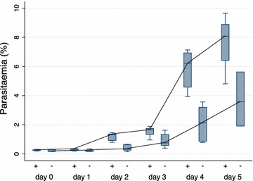
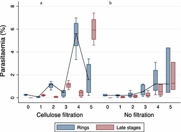
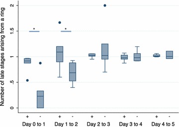
Results: The cellulose-filtered parasites grew to higher parasitaemia faster than non-filtered parasites seemingly due to a higher development ratio of ring stage parasites progressing into the late stages. Cellulose filtration had no apparent effect on clonality or var gene expression; however, evident differences were observed after only 4 days of culture in both the number of clones and transcript levels of var genes compared to the time of admission.

Conclusions: Cellulose column filtration of parasitized blood is a cheap, applicable method for improving cultivation of P. falciparum field isolates for ex vivo based assays; however, when assessing phenotype and genotype of cultured parasites, in general, assumed to represent the in vivo infection, caution is advised.

Keywords: Cellulose column filtration; Ex vivo growth; Plasmodium falciparum.


Figures

Fig. 1
Fig. 1
Parasitaemia of Plasmodium falciparum parasites in culture. Box plots showing the median parasitaemia (%) recorded for every 24 h from day 0 to day 5 of in vitro culture of seven patient parasite isolates either with prior cellulose filtration (+) or no filtration (−). The box plots display median, 1st quartile (Q1), and 3rd quartile (Q3), and the lower whisker indicates the lowest data point above Q1 minus 1.5 times IQR; the upper whisker indicates the highest data point below 1.5 IQR plus Q3. Cellulose filtration was a positive predictor for parasitaemia calculated using generalized least squares (GLS) random-effects regression model (P < 0.001). The growth profile is highlighted with a line through the medians
Fig. 2
Fig. 2
Parasitaemia of ring- and late-stage parasites in a cellulose-filtered and b non-cellulose-filtered parasites. Box plots showing the median parasitaemia (%) of ring- and late-stage parasites recorded for every 24 h from day 0 to day 5 of in vitro culture of seven patient parasite isolates either with prior cellulose filtration (+) or no filtration (−). The box plots display median, 1st quartile (Q1), and 3rd quartile (Q3) and the lower whisker indicates the lowest data point above Q1 minus 1.5 times IQR, the upper whisker indicates the highest data point below 1.5 IQR plus Q3. The growth profile is highlighted with a line only for the ring-stage parasites through the medians
Fig. 3
Fig. 3
Number of ring-stage parasites that develop into late stages. Box plots showing the number of parasites developing from ring- to late-stage parasites, either cellulose-filtered (+) or non-filtered parasites (−) from day 0 to day 5. The lower whisker indicates the lowest data point above Q1 minus 1.5 times IQR, the upper whisker indicates the highest data point below 1.5 IQR plus Q3. Values below Q1 minus 1.5 IQR and above Q3 plus 1.5 IQR are indicated as points. Statistical significance was calculated using Wilcoxon’s signed rank test. *P = 0.02
Fig. 4
Fig. 4
Var transcript levels at admission and after 4 days of culture. Transcript levels (shown as transcript units) of six CIDRα1 var subtypes in P. falciparum parasites at admission, after 4 days in culture of the cellulose-filtered parasites, and after 4 days in culture of the non-filtered parasites. CIDRα1.all is a summated transcript level for all the CIDRα1-domain primers, as described in [14]. C1.all = CIDRα1.all; C1.1 = CIDRα1.1; C1.8 = CIDRα1.8; C1.4 = CIDRα1.4; C1.5 = CIDRα1.5; C1.6 = CIDRα1.6; C1.7 = CIDRα1.7

References

    1. WHO . World malaria report 2015. Geneva: World Health Organization; 2015.
    1. Talundzic E, Okoth SA, Congpuong K, Plucinski MM, Morton L, Goldman IF, et al. Selection and spread of artemisinin-resistant alleles in Thailand prior to the global artemisinin resistance containment campaign. PLoS Pathog. 2015;11:e1004789. doi: 10.1371/journal.ppat.1004789. - DOI - PMC - PubMed
    1. Tun KM, Imwong M, Lwin KM, Win AA, Hlaing TM, Hlaing T, et al. Spread of artemisinin-resistant Plasmodium falciparum in Myanmar: a cross-sectional survey of the K13 molecular marker. Lancet Infect Dis. 2015;15:415–421. doi: 10.1016/S1473-3099(15)70032-0. - DOI - PMC - PubMed
    1. Miller LH, Baruch DI, Marsh K, Doumbo OK. The pathogenic basis of malaria. Nature. 2002;415:673–679. doi: 10.1038/415673a. - DOI - PubMed
    1. Smith JD, Rowe JA, Higgins MK, Lavstsen T. Malaria’s deadly grip: cytoadhesion of Plasmodium falciparum-infected erythrocytes. Cell Microbiol. 2013;15:1976–1983. doi: 10.1111/cmi.12183. - DOI - PMC - PubMed

Publication types

MeSH terms