Review
. 2017 Feb;10(2):171-184.
doi: 10.1016/j.jcmg.2016.12.001.

Cardiac 3D Printing and its Future Directions

Affiliations
Review

Cardiac 3D Printing and its Future Directions

Marija Vukicevic et al. JACC Cardiovasc Imaging. 2017 Feb.

Abstract

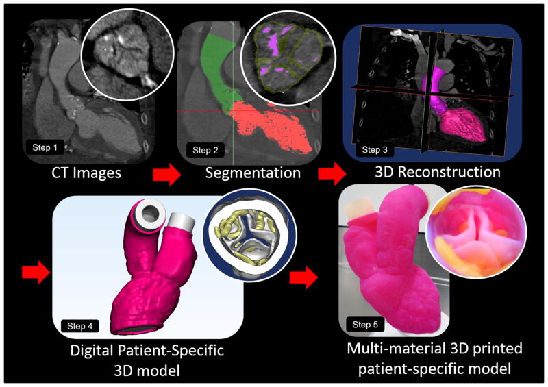
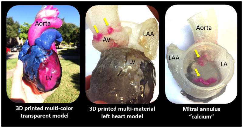
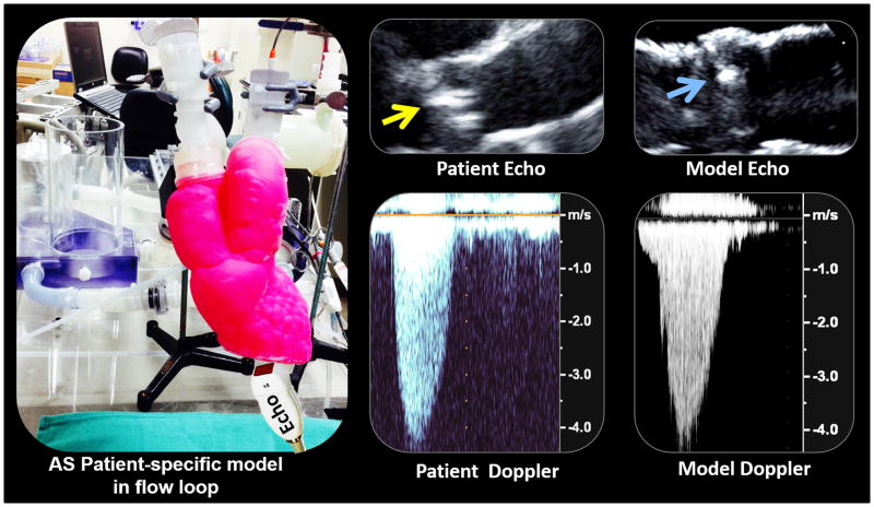
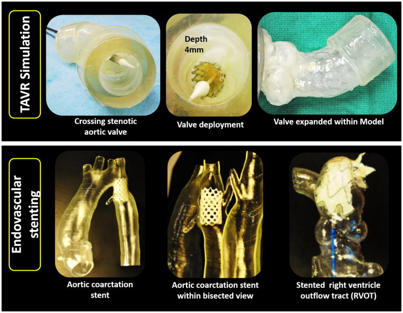
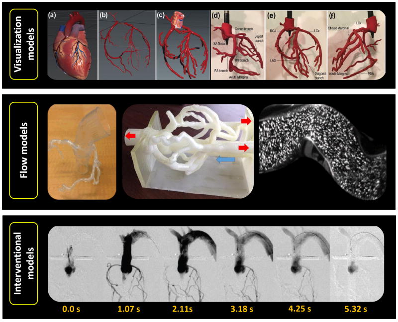
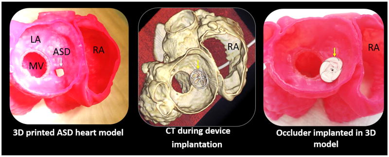
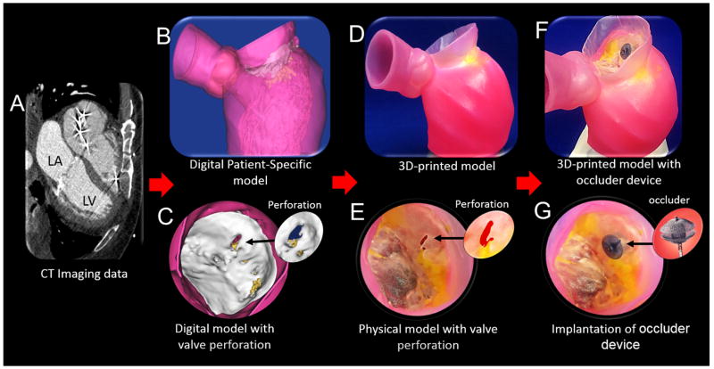
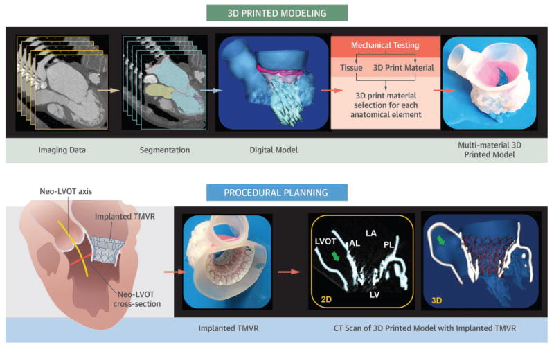
Three-dimensional (3D) printing is at the crossroads of printer and materials engineering, noninvasive diagnostic imaging, computer-aided design, and structural heart intervention. Cardiovascular applications of this technology development include the use of patient-specific 3D models for medical teaching, exploration of valve and vessel function, surgical and catheter-based procedural planning, and early work in designing and refining the latest innovations in percutaneous structural devices. In this review, we discuss the methods and materials being used for 3D printing today. We discuss the basic principles of clinical image segmentation, including coregistration of multiple imaging datasets to create an anatomic model of interest. With applications in congenital heart disease, coronary artery disease, and surgical and catheter-based structural disease, 3D printing is a new tool that is challenging how we image, plan, and carry out cardiovascular interventions.

Keywords: 3D print materials; 3D-printed modeling; aortic valve; congenital heart defects; coronary arteries; mitral valve apparatus.


Figures

Figure 1
Figure 1
3D printed modeling of patient-specific anatomy. Step 1: CT imaging dataset used for image processing. Step 2: Segmentation process and creation of segmentation mask. Step 3: Converting segmentation mask into 3D digital patient-specific model. Step 4: Adjusted digital 3D patient-specific model suitable for implantation in flow loop and medical imaging acquisition. Step 5: 3D printed multi-material patient-specific model.
Figure 2
Figure 2
Multi-material and multi-colored patient-specific 3D printed heart for educational purposes and communication with patients. Yellow arrows indicate regions 3D printed (in pink) to replicate calcium depositions within both the aortic valve (center panel) and mitral valve (right panel).
Figure 3
Figure 3
Functional modeling of patient-specific aortic stenosis. When coupled to a flow loop the echocardiographic image and hemodynamic profile of severe stenosis are replicated. Focal calcification within the echocardiographic image of the patient (yellow arrow) and the patient's model (blue arrow) are indicated. Adapted from Maragiannis et al (9) with permission.
Figure 4
Figure 4
Transcatheter valve and stent implantations within patient-specific models. Bench-top TAVR performed within a model of aortic valve stenosis (upper panel). Endovascular stenting within models of aortic coarctation and a pulmonary artery (lower panel – images courtesy of Dr. Giovanni Biglino).
Figure 5
Figure 5
3D printed coronary models. Visualization models - Rigid and solid structures segmented from clinical data used to visualize paths and geometry of epicardial coronary arteries (top panel (adapted from Jovan et al with permission).(29) Flow models - Hollow structures are used to allow flow of liquids and fluorescent particles to quantitatively asses hemodynamics of coronary vessels (mid panel). Interventional models - Hollow structures facilitating interventional procedures (e.g. angiography), (bottom pane, adapted from Russ et al with permission).(32)
Figure 6
Figure 6
Pre-procedural planning of catheter-based closure of an atrial septal defect (ASD). 3D printed model of an ASD imaged by computer tomorgraphy (CT)(top left); 3D printed model with bench-top implanted septal occluder device (top right); CT scan of septal occluder implantation within the 3D printed model (lower left and right).
Figure 7
Figure 7
3D printed modeling for patient-specific mitral valve repair with a clip and a plug. CT images (A) are used to create a digital model (B) of the mitral valve with a perforation (C). A multi-material patient-specific 3D model (D) was printed to replicate the mitral valve geometry, regional calcium deposition and pathology (E). Images adapted from Little el al (52) with permission.
Central Illustration

References

    1. Farooqi KM, Sengupta PP. Echocardiography and three-dimensional printing: sound ideas to touch a heart. J Am Soc Echocardiogr. 2015;28:398–403. - PubMed
    1. Sumida T, Otawa N, Kamata YU, et al. Custom-made titanium devices as membranes for bone augmentation in implant treatment: Clinical application and the comparison with conventional titanium mesh. J Craniomaxillofac Surg. 2015;43:2183–8. - PubMed
    1. Aranda JL, Jimenez MF, Rodriguez M, Varela G. Tridimensional titanium-printed custom-made prosthesis for sternocostal reconstruction. Eur J Cardiothorac Surg. 2015;48:e92–e94. - PubMed
    1. Ibrahim D, Broilo TL, Heitz C, et al. Dimensional error of selective laser sintering, three-dimensional printing and PolyJet models in the reproduction of mandibular anatomy. J Craniomaxillofac Surg. 2009;37:167–73. - PubMed
    1. Kato K, Ishiguchi T, Maruyama K, Naganawa S, Ishigaki T. Accuracy of plastic replica of aortic aneurysm using 3D-CT data for transluminal stent-grafting: experimental and clinical evaluation. J Comput Assist Tomogr. 2001;25:300–4. - PubMed

Publication types