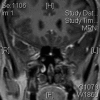
Pituitary tuberculoma with subsequent drug-resistant tuberculous lymphadenopathy: an uncommon presentation of a common disease
- PMID: 28183710
- PMCID: PMC5307273
- DOI: 10.1136/bcr-2016-218330
Pituitary tuberculoma with subsequent drug-resistant tuberculous lymphadenopathy: an uncommon presentation of a common disease
Abstract
We report a case of pituitary tuberculosis which presented as a non-functioning pituitary macroadenoma, and subsequently developed multidrug-resistant tuberculous lymphadenopathy. Pituitary tuberculosis continues to be a rare presentation of tuberculosis, but incidence and prevalence are expected to grow with increasing numbers of multidrug-resistant tuberculosis. Isolated pituitary tuberculosis is rare. Tuberculosis should be considered in the differential diagnosis in evaluation of a sellar mass.
2017 BMJ Publishing Group Ltd.
Conflict of interest statement
Conflicts of Interest: None declared.
Figures


Similar articles
-
[Tuberculous meningitis with pituitary abscess].J Neuroradiol. 2003 Jun;30(3):188-91. J Neuroradiol. 2003. PMID: 12843875 French.
-
Pituitary Tuberculoma.J Coll Physicians Surg Pak. 2018 Jun;28(6):S97-S98. doi: 10.29271/jcpsp.2018.06.S97. J Coll Physicians Surg Pak. 2018. PMID: 29866234
-
Pituitary tuberculoma: a rare cause of sellar mass.Ir J Med Sci. 2018 May;187(2):461-464. doi: 10.1007/s11845-017-1654-4. Epub 2017 Jul 21. Ir J Med Sci. 2018. PMID: 28733940 Review.
-
Intramedullary tuberculoma in a patient with human immunodeficiency virus infection and disseminated multidrug-resistant tuberculosis: case report.Int J Infect Dis. 1998 Jan-Mar;2(3):164-7. doi: 10.1016/s1201-9712(98)90121-7. Int J Infect Dis. 1998. PMID: 9625611 No abstract available.
-
Hypophyseal tuberculoma: direct radiosurgery is contraindicated for a lesion with a thickened pituitary stalk: case report.Neurosurgery. 2000 Mar;46(3):735-8; discussion 738-9. doi: 10.1097/00006123-200003000-00041. Neurosurgery. 2000. PMID: 10719871 Review.
Cited by
-
Idiopathic Granulomatous Hypophysitis with Rapid Onset: A Case Report.Brain Tumor Res Treat. 2019 Apr;7(1):57-61. doi: 10.14791/btrt.2019.7.e22. Brain Tumor Res Treat. 2019. PMID: 31062534 Free PMC article.
-
Primary pituitary tuberculosis.Autops Case Rep. 2020 Dec 8;11:e2020228. doi: 10.4322/acr.2020.228. eCollection 2021. Autops Case Rep. 2020. PMID: 34277492 Free PMC article.
References
-
- Sunil K, Menon R, Goel N. Pituitary tuberculosis. J Assoc Physicians of India 2008;32:453–6. - PubMed
-
- Sharma MC, Arora R, Mahapatra AK et al. . Intrasellar tuberculoma—an enigmatic pituitary infection: a series of 18 cases. Clin Neurol Neurosurg 2000;102:72–7. - PubMed
-
- Dalan R, Leow MK. Pituitary abscess: our experience with a case and a review of the literature. Pituitary 2011;3:299–306. - PubMed
Publication types
MeSH terms
Substances
LinkOut - more resources
Full Text Sources
Other Literature Sources
Medical
